Downloaded 1,162 times
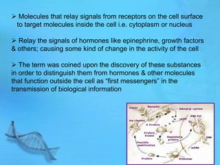
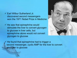
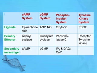
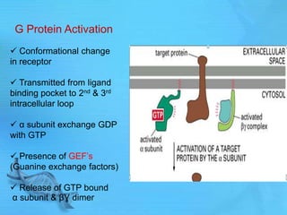
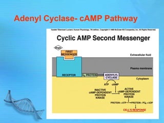


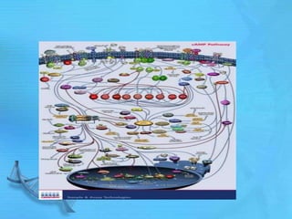


The document discusses second messenger systems. It describes how second messengers relay signals from cell surface receptors to target molecules inside the cell. Some key points discussed include: - Earl Sutherland discovered cyclic AMP (cAMP) as the second messenger for epinephrine and won the Nobel Prize for this work. - Common second messenger systems include those using cAMP, cGMP, phosphatidylinositol, and tyrosine kinases as secondary messengers. - G proteins act as transducers between receptors and effectors and are important drug targets. - cAMP and cGMP have several downstream targets including protein kinases that phosphorylate other proteins and regulate various cellular processes.


