Downloaded 78 times


![ Intrinsic ability of cardiac muscle
Also called ‘inotropism’ or ‘inotropy’
Related to the intracellular [Ca2+]
Inotropic agents
◦ positive: increase contractility
◦ negative: decrease contractility](https://image.slidesharecdn.com/cardiaccontractility-130216085108-phpapp01/85/Cardiac-contractility-10-320.jpg)
![ Chronotropy
◦ rate of contraction
◦ also affected by intracellular [Ca2+]
Dromotropy
◦ rate of impulse conduction
◦ noted particularly at AV node](https://image.slidesharecdn.com/cardiaccontractility-130216085108-phpapp01/85/Cardiac-contractility-11-320.jpg)
![ Increased intracellular [Ca2+]
◦ increased heart rate
◦ cardiac glycosides (e.g. digoxin)
Stimulation of β1-adrenergic receptor
◦ sympathomimetic agents
◦ catecholamines](https://image.slidesharecdn.com/cardiaccontractility-130216085108-phpapp01/85/Cardiac-contractility-12-320.jpg)
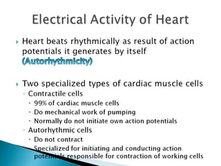
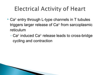


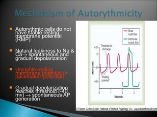


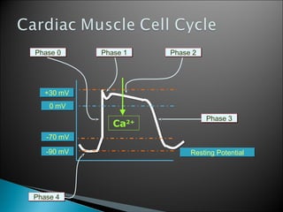
The document discusses cardiac electrophysiology and contractility. It describes the pacemaker potential and automaticity of the sinoatrial node, which allows it to initiate action potentials without external stimulation at a rate of 100 beats per minute. It also discusses the conduction of action potentials through the heart via the atrioventricular node, bundle of His, and Purkinje fibers. Contraction is triggered by increases in intracellular calcium levels. The cardiac action potential is longer than in skeletal muscle, lasting 200-300 milliseconds, which allows time for relaxation between contractions.


