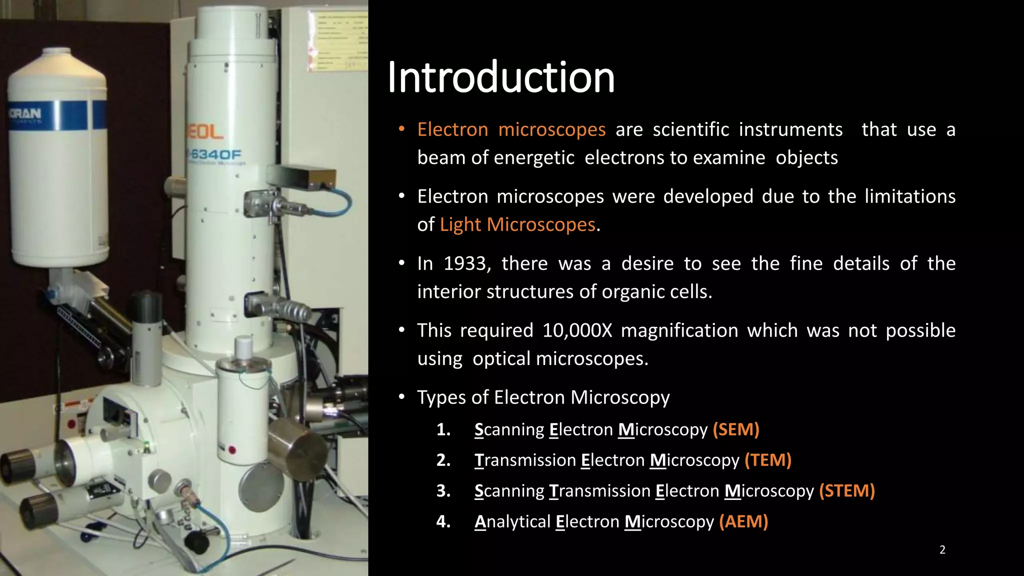
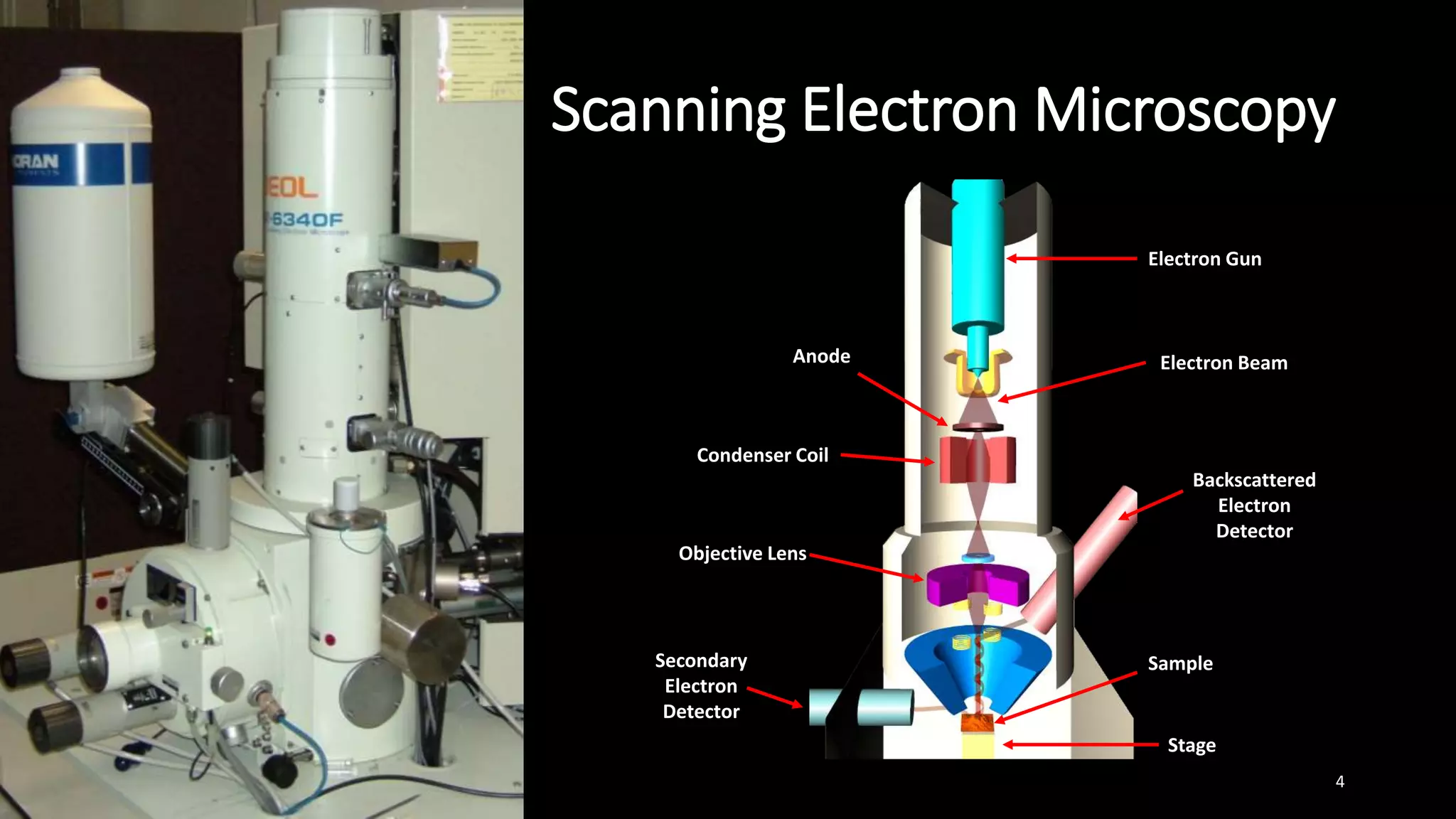
Prepared by : Hamza Suharwardi 1
Introduction
• Electron microscopes are scientific instruments that use a
beam of energetic electrons to examine objects
• Electron microscopes were developed due to the limitations
of Light Microscopes.
• In 1933, there was a desire to see the fine details of the
interior structures of organic cells.
• This required 10,000X magnification which was not possible
using optical microscopes.
• Types of Electron Microscopy
1. Scanning Electron Microscopy (SEM)
2. Transmission Electron Microscopy (TEM)
3. Scanning Transmission Electron Microscopy (STEM)
4. Analytical Electron Microscopy (AEM)
2
Scanning Electron Microscopy
• Scanning Electron Microscopy (SEM) is a type of
microscope that images a sample by scanning it with a
high-energy beam of electron in a raster scan pattern
• The atoms interact with the atoms that make up the
sample producing signals that contain information about
the sample’s surface topography and composition.
• The first scanning electron microscope was debuted in
1938 (Manfred Von Ardenne) with the first commercial
instrument around 1965. It’s late development was due
to the electronics involved in “scanning” the beam of
electrons across the sample .
3
Scanning Electron Microscopy
4
Electron Gun
Electron Beam
Backscattered
Electron
Detector
Anode
Condenser Coil
Objective Lens
Secondary
Electron
Detector
Stage
Sample
Scanning Electron Microscopy
• IFORMATION RETRIEVED
 Topography – The surface features on an object.
 Morphology – The shape and size of the particles
 Composition – The elements and compounds that the
object is composed of and the realative amounts of
them.
 Crystallographic Information – How the atoms are
arranged in the object.
5
Scanning Electron Microscopy
• SURFACE INTERACTIONS:
• The types of signals produced by electron beam are:
1. Secondary Electron
2. Back-scattered Electrons
3. X-rays
4. Light Rays (Cathodoluminescence)
6
Scanning Electron Microscopy
• SECONDRY ELECTRONS:
• The types of signals produced by electron beam are:
1. Secondary Electron
7
Scanning Electron Microscopy
• SURFACE INTERACTIONS:
• The types of signals produced by electron beam are:
1. Secondary Electron
2. Back-scattered Electrons
8
Scanning Electron Microscopy
• SURFACE INTERACTIONS:
• The types of signals produced by electron beam are:
1. Secondary Electron
2. Back-scattered Electrons
3. X-rays
9
Scanning Electron Microscopy
• SURFACE INTERACTIONS:
• The types of signals produced by electron beam are:
1. Secondary Electron
2. Back-scattered Electrons
3. X-rays
• EDX Detector
10
Scanning Electron Microscopy
• SURFACE INTERACTIONS:
• The types of signals produced by electron beam are:
1. Secondary Electron
2. Back-scattered Electrons
3. X-rays
11
Scanning Electron Microscopy
• SURFACE INTERACTIONS:
• The types of signals produced by electron beam are:
1. Secondary Electron
2. Back-scattered Electrons
3. X-rays
4. Light Rays (Cathodoluminescence)
• In SEM , CL detectors display an emission spectrum or an
image of the distribution of cathodoluminescence
emitted by the specimen in real color.
• Very powerful probe for studying nanoscale features and
defects
12
Scanning Electron Microscopy
• SAMPLE PREPARATION:
• Cleaning the surface of the specimen
• Stabilizing the specimen
• Rinsing the specimen
• Dehydrating the specimen
• Drying the specimen
• Mounting the specimen
• Coating the specimen
13
Scanning Electron Microscopy
• VACUUM SYSTEM:
 The electron optical system and specimen chamber
must be kept at high vacuum of 10-3 to10-5 Pa.
 High vacuum minimizes the scattering of electron
beam before reaching the specimen.
 This is important as electron beam will increase the
probe size and reduce the resolution.
 Thus, the components are evacuated by diffusion
pump.
14
• ENVIRONMENTAL SEM (ESEM)
 Samples are observed in low pressure gaseous
environments and high relative humidity (up to 100%).
 ESEM is especially useful for non- metallic and biological
materials.
Coating or carbon or gold is unnecessary.
Good for plastic and elastomers and other uncoated
biological samples
ESEM makes it possible to perform X-ray Micro analysis
on uncoated non conductive samples.
Preferred tool for Forensic Analysis.
15
Scanning Electron Microscopy
• APPLICATIONS:
SEM is one of the most versatile technique and can be
used for:
Image morphology of samples
Image compositional and bonding differences
Examine wet and dry samples while viewing them(only
in ESEM)
Generate X-Rays from the samples for microanalysis
(EDS)
View or map grain orientation, crystallographic
orientation and study information like heterogeneity.
16
Thank you
17
Hamza Suharwardi
Researcher
Slow and steady win the race.
Any questions?
You can find me at:
● hamzaahmed0696@gmail.com
• https://www.researchgate.net/profile/Hamza-Suharwardi-2

SCANNING ELECTRON MICROSCOPY (SEM)

  • 1.
    Prepared by :Hamza Suharwardi 1
  • 2.
    Introduction • Electron microscopesare scientific instruments that use a beam of energetic electrons to examine objects • Electron microscopes were developed due to the limitations of Light Microscopes. • In 1933, there was a desire to see the fine details of the interior structures of organic cells. • This required 10,000X magnification which was not possible using optical microscopes. • Types of Electron Microscopy 1. Scanning Electron Microscopy (SEM) 2. Transmission Electron Microscopy (TEM) 3. Scanning Transmission Electron Microscopy (STEM) 4. Analytical Electron Microscopy (AEM) 2
  • 3.
    Scanning Electron Microscopy •Scanning Electron Microscopy (SEM) is a type of microscope that images a sample by scanning it with a high-energy beam of electron in a raster scan pattern • The atoms interact with the atoms that make up the sample producing signals that contain information about the sample’s surface topography and composition. • The first scanning electron microscope was debuted in 1938 (Manfred Von Ardenne) with the first commercial instrument around 1965. It’s late development was due to the electronics involved in “scanning” the beam of electrons across the sample . 3
  • 4.
    Scanning Electron Microscopy 4 ElectronGun Electron Beam Backscattered Electron Detector Anode Condenser Coil Objective Lens Secondary Electron Detector Stage Sample
  • 5.
    Scanning Electron Microscopy •IFORMATION RETRIEVED  Topography – The surface features on an object.  Morphology – The shape and size of the particles  Composition – The elements and compounds that the object is composed of and the realative amounts of them.  Crystallographic Information – How the atoms are arranged in the object. 5
  • 6.
    Scanning Electron Microscopy •SURFACE INTERACTIONS: • The types of signals produced by electron beam are: 1. Secondary Electron 2. Back-scattered Electrons 3. X-rays 4. Light Rays (Cathodoluminescence) 6
  • 7.
    Scanning Electron Microscopy •SECONDRY ELECTRONS: • The types of signals produced by electron beam are: 1. Secondary Electron 7
  • 8.
    Scanning Electron Microscopy •SURFACE INTERACTIONS: • The types of signals produced by electron beam are: 1. Secondary Electron 2. Back-scattered Electrons 8
  • 9.
    Scanning Electron Microscopy •SURFACE INTERACTIONS: • The types of signals produced by electron beam are: 1. Secondary Electron 2. Back-scattered Electrons 3. X-rays 9
  • 10.
    Scanning Electron Microscopy •SURFACE INTERACTIONS: • The types of signals produced by electron beam are: 1. Secondary Electron 2. Back-scattered Electrons 3. X-rays • EDX Detector 10
  • 11.
    Scanning Electron Microscopy •SURFACE INTERACTIONS: • The types of signals produced by electron beam are: 1. Secondary Electron 2. Back-scattered Electrons 3. X-rays 11
  • 12.
    Scanning Electron Microscopy •SURFACE INTERACTIONS: • The types of signals produced by electron beam are: 1. Secondary Electron 2. Back-scattered Electrons 3. X-rays 4. Light Rays (Cathodoluminescence) • In SEM , CL detectors display an emission spectrum or an image of the distribution of cathodoluminescence emitted by the specimen in real color. • Very powerful probe for studying nanoscale features and defects 12
  • 13.
    Scanning Electron Microscopy •SAMPLE PREPARATION: • Cleaning the surface of the specimen • Stabilizing the specimen • Rinsing the specimen • Dehydrating the specimen • Drying the specimen • Mounting the specimen • Coating the specimen 13
  • 14.
    Scanning Electron Microscopy •VACUUM SYSTEM:  The electron optical system and specimen chamber must be kept at high vacuum of 10-3 to10-5 Pa.  High vacuum minimizes the scattering of electron beam before reaching the specimen.  This is important as electron beam will increase the probe size and reduce the resolution.  Thus, the components are evacuated by diffusion pump. 14
  • 15.
    • ENVIRONMENTAL SEM(ESEM)  Samples are observed in low pressure gaseous environments and high relative humidity (up to 100%).  ESEM is especially useful for non- metallic and biological materials. Coating or carbon or gold is unnecessary. Good for plastic and elastomers and other uncoated biological samples ESEM makes it possible to perform X-ray Micro analysis on uncoated non conductive samples. Preferred tool for Forensic Analysis. 15 Scanning Electron Microscopy
  • 16.
    Scanning Electron Microscopy •APPLICATIONS: SEM is one of the most versatile technique and can be used for: Image morphology of samples Image compositional and bonding differences Examine wet and dry samples while viewing them(only in ESEM) Generate X-Rays from the samples for microanalysis (EDS) View or map grain orientation, crystallographic orientation and study information like heterogeneity. 16
  • 17.
    Thank you 17 Hamza Suharwardi Researcher Slowand steady win the race. Any questions? You can find me at: ● hamzaahmed0696@gmail.com • https://www.researchgate.net/profile/Hamza-Suharwardi-2