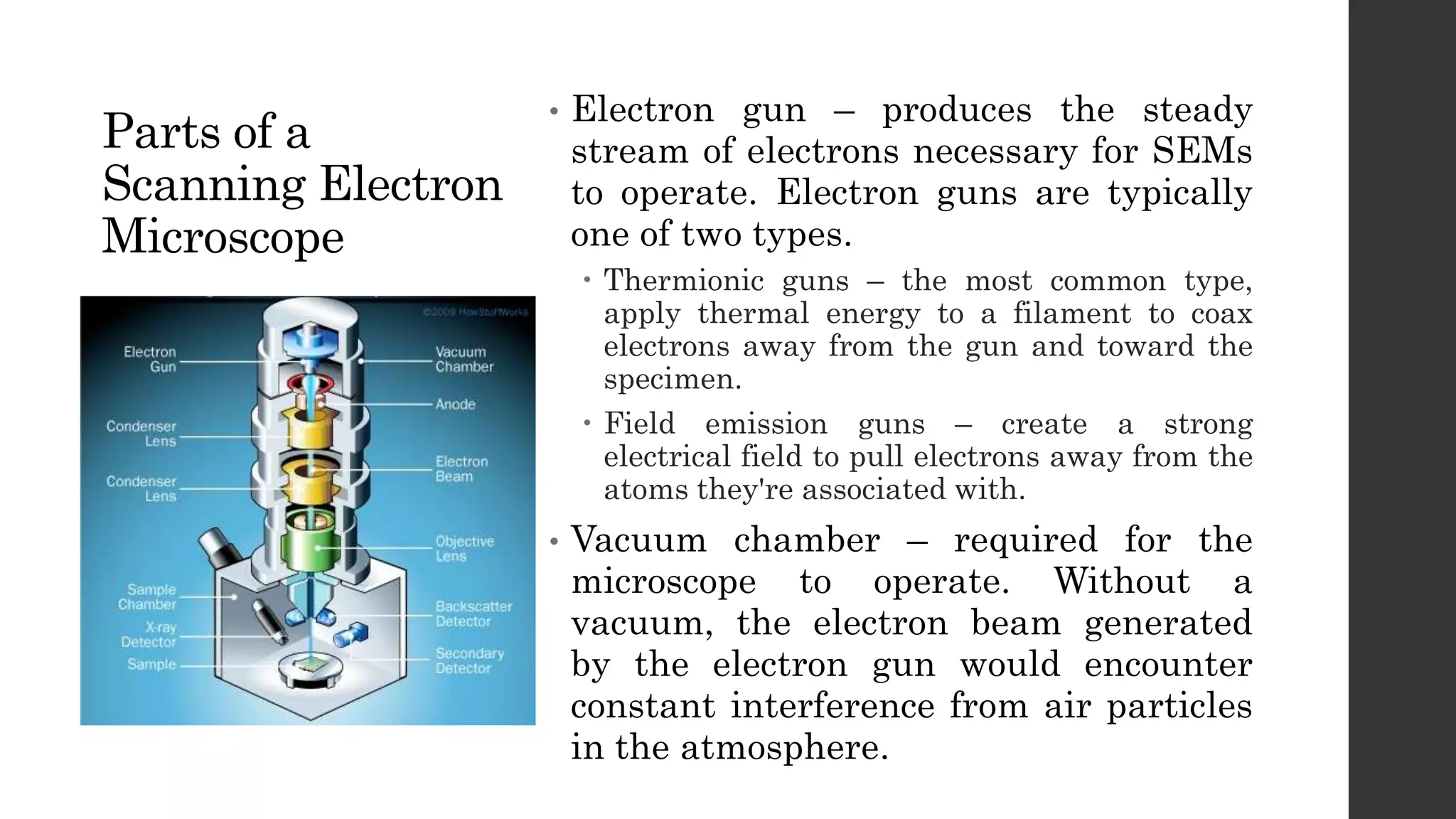
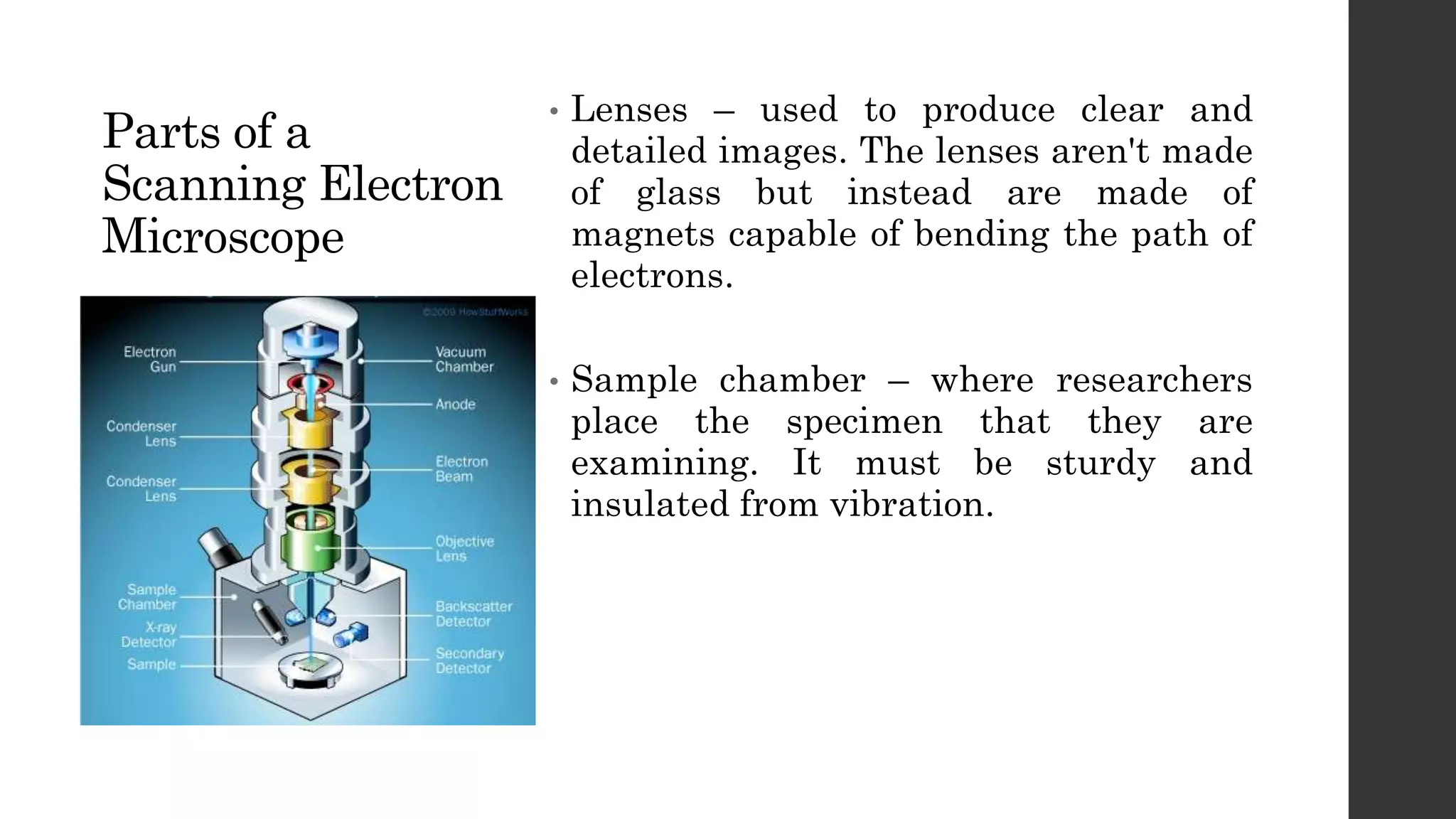
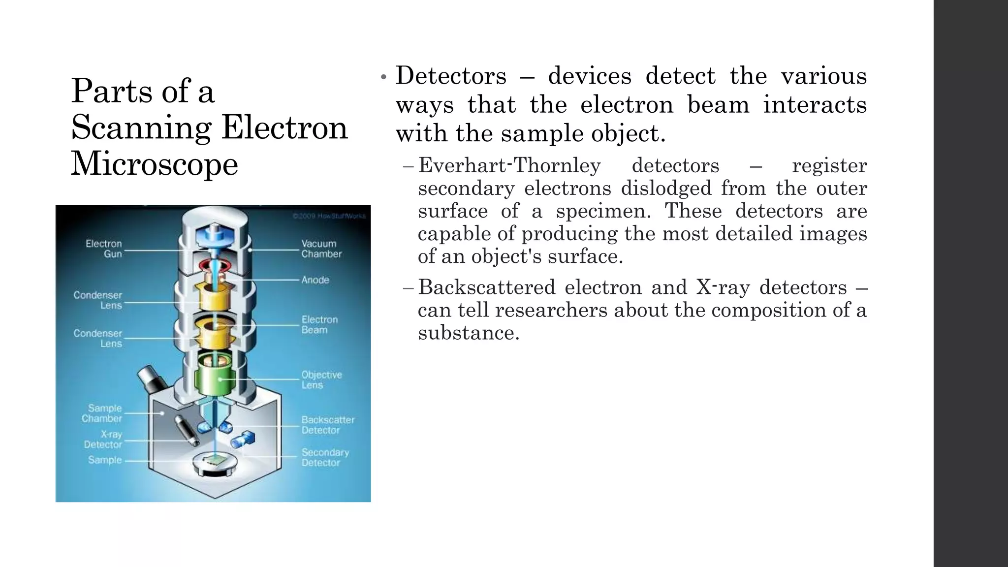
SCANNING
ELECTRON
MICROSCOPE
Electron Microscopes
The electron microscope is a type of microscope
that uses a beam of electrons to create an image of
the specimen.
It is capable of much higher magnifications and
has a greater resolving power than a light
microscope, allowing it to see much smaller objects
in finer detail.
Electron Microscopes
TRANSMISSION ELECTRON
MICROSCOPE
A high energy beam of electrons
is shone through a very thin
sample, and the interactions
between the electrons and the
atoms can be used to observe
features such as the crystal
structure. It can reveal the finest
details of internal structure.
SCANNING ELECTRON
MICROSCOPE
Designed for directly studying
the surfaces of solid objects,
that utilizes a beam of focused
electrons of relatively low
energy as a probe that is
scanned in a regular manner
over the specimen.
TYPES
Scanning Electron Microscope
A Scanning Electron Microscope (SEM) is a
powerful magnification tool that utilizes focused
beams of electrons to obtain information.
The high-resolution, three-dimensional images
produced by SEMs provide topographical,
morphological and compositional information
makes them vital in science and industry.
Scanning Electron Microscope
The Scanning Electron Microscope was developed
by professor Charles Oatley of Cambridge
University's Engineering Department.
Mr. Oatley, together with his students, put
together their first Scanning Electron Microscope
in 1948, and four years later, it was producing
three-dimensional images.
BRIEF HISTORY
Parts of a
Scanning Electron
Microscope
• Electron gun – produces the steady
stream of electrons necessary for SEMs
to operate. Electron guns are typically
one of two types.
 Thermionic guns – the most common type,
apply thermal energy to a filament to coax
electrons away from the gun and toward the
specimen.
 Field emission guns – create a strong
electrical field to pull electrons away from the
atoms they're associated with.
• Vacuum chamber – required for the
microscope to operate. Without a
vacuum, the electron beam generated
by the electron gun would encounter
constant interference from air particles
in the atmosphere.
Parts of a
Scanning Electron
Microscope
• Lenses – used to produce clear and
detailed images. The lenses aren't made
of glass but instead are made of
magnets capable of bending the path of
electrons.
• Sample chamber – where researchers
place the specimen that they are
examining. It must be sturdy and
insulated from vibration.
Parts of a
Scanning Electron
Microscope
• Detectors – devices detect the various
ways that the electron beam interacts
with the sample object.
 Everhart-Thornley detectors – register
secondary electrons dislodged from the outer
surface of a specimen. These detectors are
capable of producing the most detailed images
of an object's surface.
 Backscattered electron and X-ray detectors –
can tell researchers about the composition of a
substance.
Scanning Electron Microscope
Metals require no preparation, as they already
conduct electricity and will respond favorably
when bombarded with electrons.
Non-metals need to be coated by a conductive
material like gold or platinum through a process
called sputter coating. Sputter coating allows a
sample to be grounded, preventing it from being
damaged by the electron beam.
SAMPLE PREPARATION
Scanning Electron Microscope
In addition, preparation traditionally includes the
removal of all water. Water molecules will
vaporize in a vacuum, creating obstacles for the
electron beams and obscuring the clarity of the
image.
SAMPLE PREPARATION
Scanning Electron Microscope
ADVANTAGES
• Has a wide array of applications, detailed
three-dimensional imaging and versatile
information.
• Easy to operate with the proper training.
• Advances in computer technology and
associated software make operation user-
friendly.
• This instrument works fast, often
completing analyses in less than five
minutes.
• It allows the generation of data in digital
form.
• Most samples require minimal preparation
actions.
DISADVANTAGES
• Expensive, large and must be housed in an
area free of any possible electric, magnetic
or vibration interference.
• Maintenance involves keeping a steady
voltage, currents to electromagnetic coils
and circulation of cool water.
• Special training is required to operate.
• The preparation of samples can result in
artifacts.
• Limited to solid, inorganic samples small
enough to fit inside the vacuum chamber
that can handle moderate vacuum
pressure.
• Carries a small risk of radiation exposure
associated with the electrons that scatter
from beneath the sample surface.

Scanning Electron Microscope

  • 1.
  • 2.
    Electron Microscopes The electronmicroscope is a type of microscope that uses a beam of electrons to create an image of the specimen. It is capable of much higher magnifications and has a greater resolving power than a light microscope, allowing it to see much smaller objects in finer detail.
  • 3.
    Electron Microscopes TRANSMISSION ELECTRON MICROSCOPE Ahigh energy beam of electrons is shone through a very thin sample, and the interactions between the electrons and the atoms can be used to observe features such as the crystal structure. It can reveal the finest details of internal structure. SCANNING ELECTRON MICROSCOPE Designed for directly studying the surfaces of solid objects, that utilizes a beam of focused electrons of relatively low energy as a probe that is scanned in a regular manner over the specimen. TYPES
  • 4.
    Scanning Electron Microscope AScanning Electron Microscope (SEM) is a powerful magnification tool that utilizes focused beams of electrons to obtain information. The high-resolution, three-dimensional images produced by SEMs provide topographical, morphological and compositional information makes them vital in science and industry.
  • 5.
    Scanning Electron Microscope TheScanning Electron Microscope was developed by professor Charles Oatley of Cambridge University's Engineering Department. Mr. Oatley, together with his students, put together their first Scanning Electron Microscope in 1948, and four years later, it was producing three-dimensional images. BRIEF HISTORY
  • 6.
    Parts of a ScanningElectron Microscope • Electron gun – produces the steady stream of electrons necessary for SEMs to operate. Electron guns are typically one of two types.  Thermionic guns – the most common type, apply thermal energy to a filament to coax electrons away from the gun and toward the specimen.  Field emission guns – create a strong electrical field to pull electrons away from the atoms they're associated with. • Vacuum chamber – required for the microscope to operate. Without a vacuum, the electron beam generated by the electron gun would encounter constant interference from air particles in the atmosphere.
  • 7.
    Parts of a ScanningElectron Microscope • Lenses – used to produce clear and detailed images. The lenses aren't made of glass but instead are made of magnets capable of bending the path of electrons. • Sample chamber – where researchers place the specimen that they are examining. It must be sturdy and insulated from vibration.
  • 8.
    Parts of a ScanningElectron Microscope • Detectors – devices detect the various ways that the electron beam interacts with the sample object.  Everhart-Thornley detectors – register secondary electrons dislodged from the outer surface of a specimen. These detectors are capable of producing the most detailed images of an object's surface.  Backscattered electron and X-ray detectors – can tell researchers about the composition of a substance.
  • 9.
    Scanning Electron Microscope Metalsrequire no preparation, as they already conduct electricity and will respond favorably when bombarded with electrons. Non-metals need to be coated by a conductive material like gold or platinum through a process called sputter coating. Sputter coating allows a sample to be grounded, preventing it from being damaged by the electron beam. SAMPLE PREPARATION
  • 10.
    Scanning Electron Microscope Inaddition, preparation traditionally includes the removal of all water. Water molecules will vaporize in a vacuum, creating obstacles for the electron beams and obscuring the clarity of the image. SAMPLE PREPARATION
  • 11.
    Scanning Electron Microscope ADVANTAGES •Has a wide array of applications, detailed three-dimensional imaging and versatile information. • Easy to operate with the proper training. • Advances in computer technology and associated software make operation user- friendly. • This instrument works fast, often completing analyses in less than five minutes. • It allows the generation of data in digital form. • Most samples require minimal preparation actions. DISADVANTAGES • Expensive, large and must be housed in an area free of any possible electric, magnetic or vibration interference. • Maintenance involves keeping a steady voltage, currents to electromagnetic coils and circulation of cool water. • Special training is required to operate. • The preparation of samples can result in artifacts. • Limited to solid, inorganic samples small enough to fit inside the vacuum chamber that can handle moderate vacuum pressure. • Carries a small risk of radiation exposure associated with the electrons that scatter from beneath the sample surface.