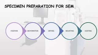
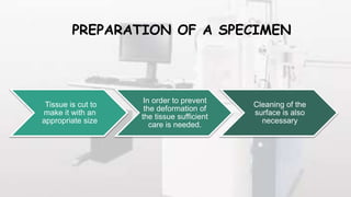
Downloaded 18 times
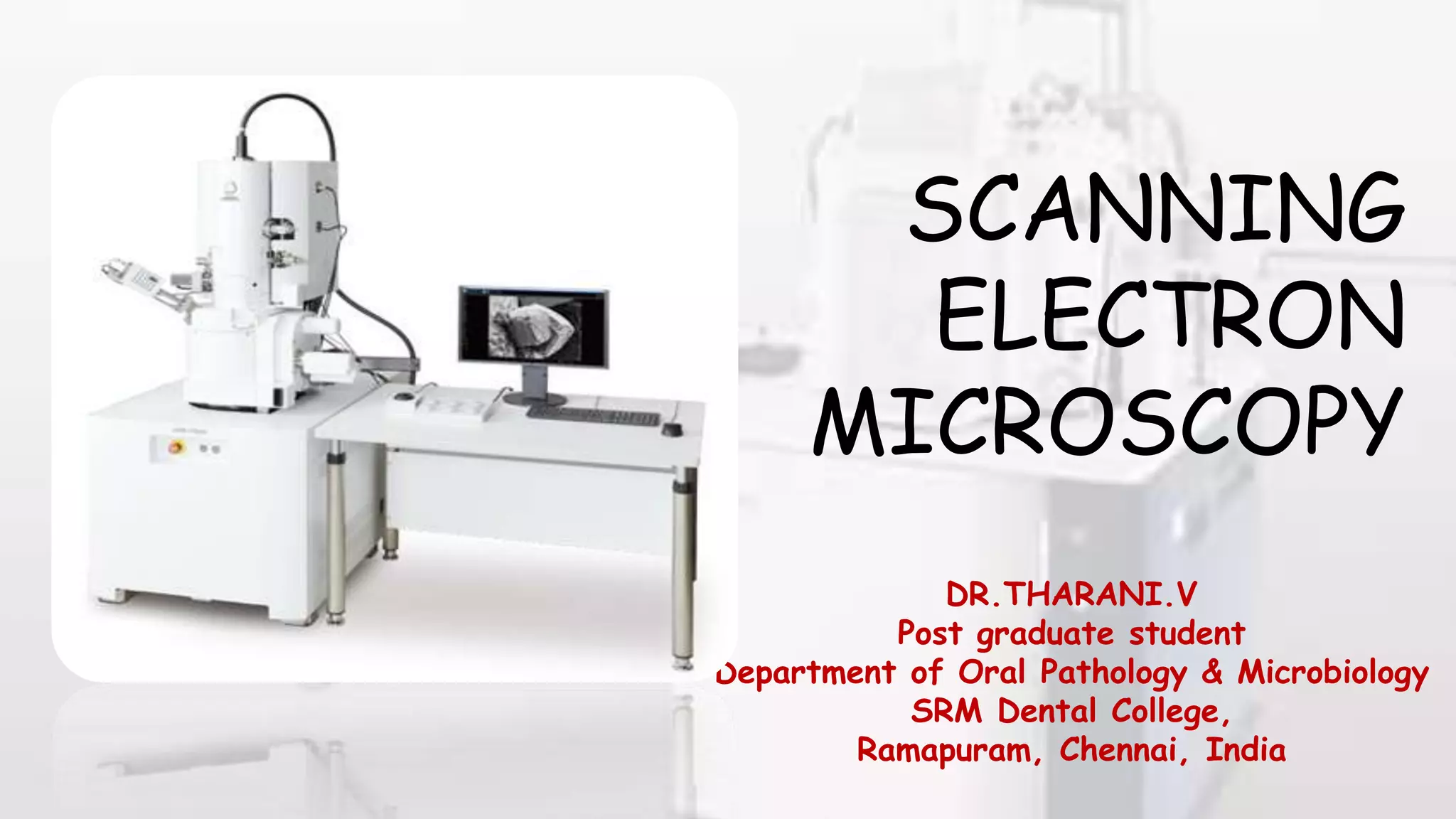
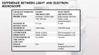
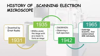
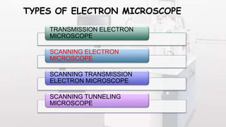
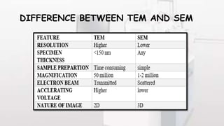
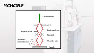

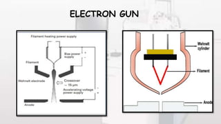
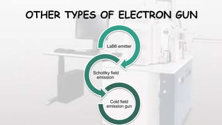
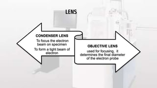
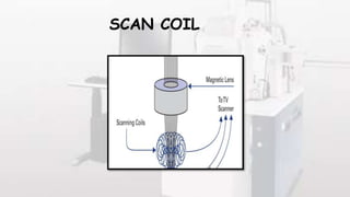
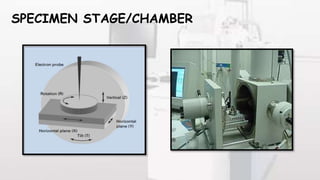
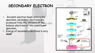
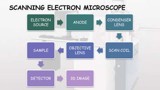
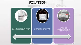
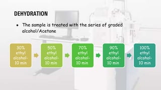
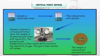
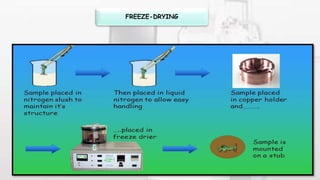
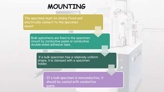
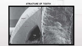
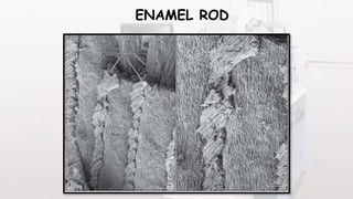


This document provides an overview of scanning electron microscopy (SEM). It discusses the history and development of SEM from the 1930s to present. The key components of an SEM are described, including the electron gun, lenses, scan coils, vacuum system and detectors. SEM provides higher magnification and resolution than light microscopes. Specimen preparation such as fixation, dehydration and coating are outlined. Applications include examining the structure of cells, crystals, and biological and inorganic materials. Advantages are high magnification and digital image capture, while limitations include expense and inability to view non-conductive samples.


