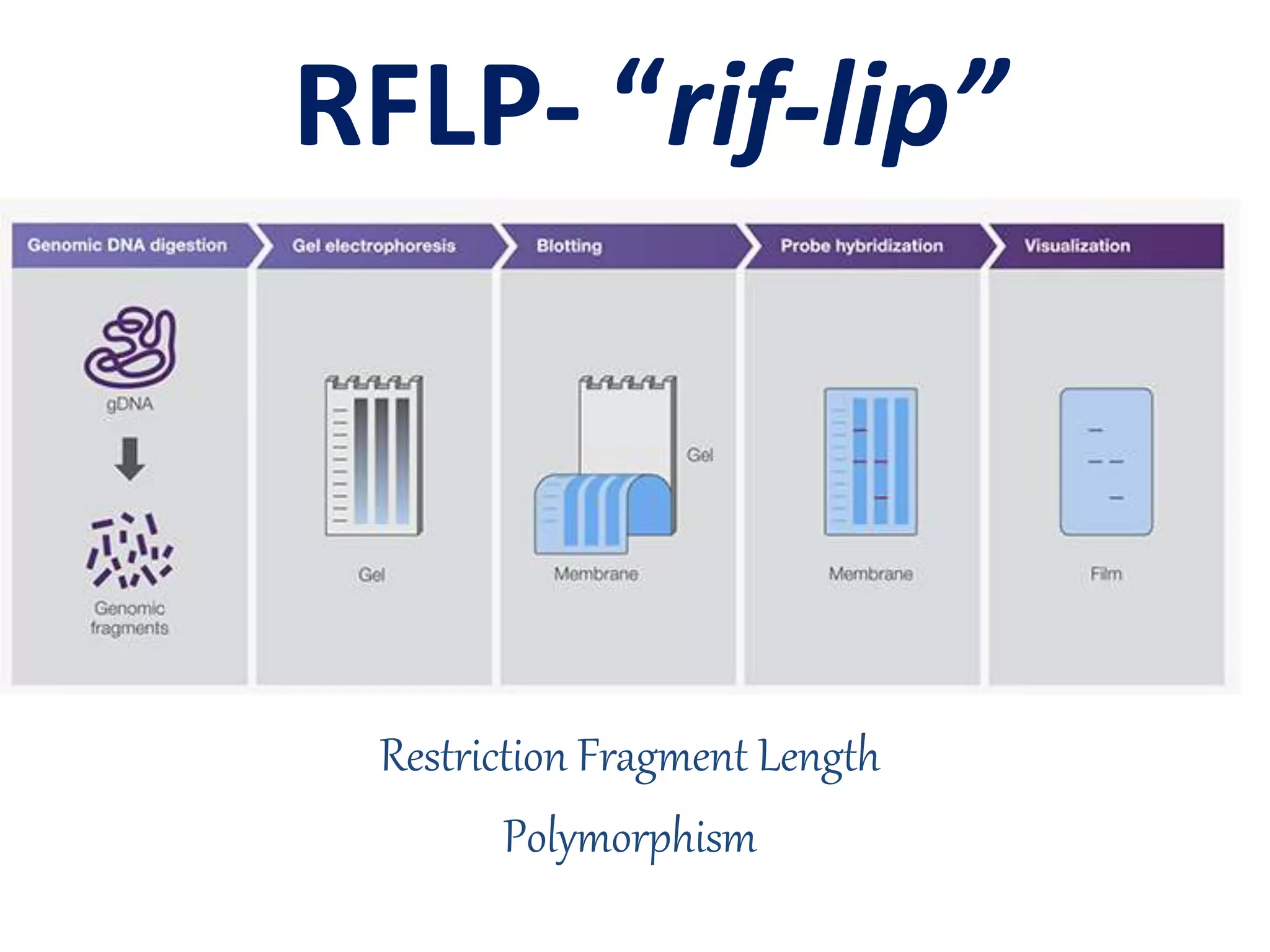
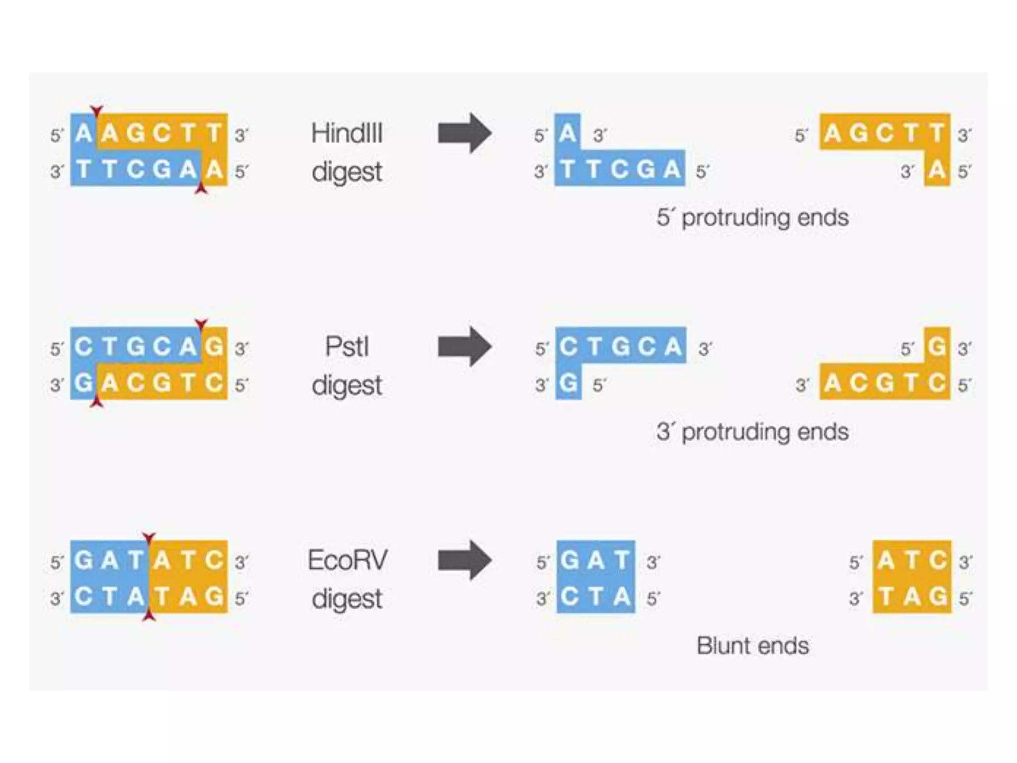
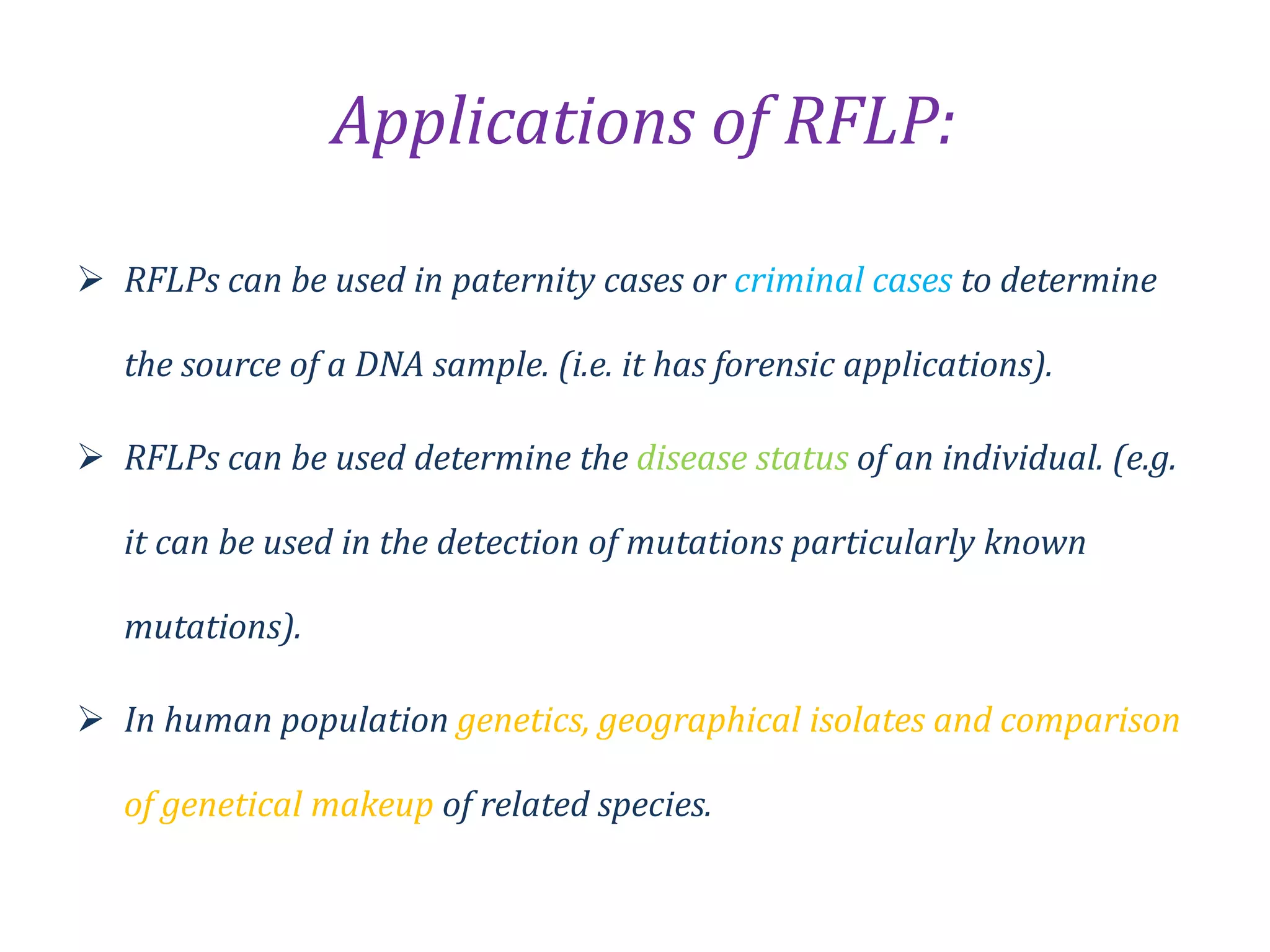
Restriction Fragment Length
Polymorphism
RFLP- “rif-lip”
RFLP’s contribution . . . . .
• Restriction fragment length polymorphism
(RFLP) markers were regarded as the first shot
in the genome revolution, marking the start of
an entirely different era in the biological
sciences.
• RFLP was the most popular approach for
analysis of genetic variation during the entire
1980s.
• RFLP is based DNA fragment length
differences after digesting genomic DNA with
one or more restriction enzymes.
• DNA is digested by one or more restriction
enzymes and separated on an agarose gel.
• The DNA in the gel transferred to
nitrocellulose or nylon membranes.
Intro. . .
• The specific DNA locus with a potential fragment
length difference is characterized by
hybridization to a probe, a radioactively labeled
DNA or ribonucleic acid (RNA) molecule with
sequence similarities to the locus of interest.
• After hybridization, the nonspecific probes must
be washed away leaving only hybridized probes
to the specific locus.
• The membrane is then exposed to a piece of X-
ray film for autoradiography to visualize the DNA
bands.
Technology Advances - Development of RFLP
• Two specific technological advances that set
the foundation for RFLP were —
– The discovery and application
of restriction enzymes and
– The development of DNA hybridisation.
The History of RE. . . .
• The phenomenon was first identified in work done in the laboratories of Salvador
Luria and Giuseppe Bertani in early 1950s.
• It was found that, for a bacteriophage λ that can grow well in one strain
of Escherichia coli
• In the 1960s, it was shown in work done in the laboratories of Werner
Arber and Matthew Meselson that the restriction is caused by an enzymatic
cleavage of the phage DNA, and the enzyme involved was therefore termed a
restriction enzyme.
• The restriction enzymes studied by Arber and Meselson were type I restriction
enzymes, which cleave DNA randomly away from the recognition site.
• In 1970, Hamilton O. Smith, Thomas Kelly and Kent Wilcox isolated and
characterized the first type II restriction enzyme, HindII, from the
bacterium Haemophilus influenzae.
• Restriction enzymes of this type are more useful for laboratory work as they
cleave DNA at the site of their recognition sequence.
• For their work in the discovery and characterization of restriction enzymes, the
1978 Nobel Prize for Physiology or Medicine was awarded to Werner Arber, Daniel
Nathans, and Hamilton O. Smith.
Restriction enzymes- Restriction endonucleases
• Proteins produced by bacteria that cleave DNA
at specific sites along the molecule.
• In the bacterial cell, restriction enzymes
cleave foreign DNA, thus eliminating infecting
organisms.
• Restriction enzymes can be isolated from
bacterial cells and used in the laboratory to
manipulate fragments of DNA.
• They are indispensable tools of . . . . .
The obvious question . . ..
Why the restriction enzymes do
not digest bacterial DNA ??????
“The answer is that the bacteria also harbor a
set of defense weaponry containing so-called
restriction enzyme modification systems.
Usually, organisms that make restriction
enzymes also make a companion modification
enzyme (DNA methyltransferase) that protects
their own DNA from cleavage. These enzymes
recognize the same DNA sequence as the
restriction enzyme they accompany, but
instead of cleaving the sequence, they
disguise it by methylating one of the bases in
each DNA strand.”
The vast nature of RE
• To date, more than 10,000 bacteria species
have been screened for the existence of
restriction enzymes; more than 2,500
restriction enzymes have been found with
more than 250 distinct specificities.
• Occasionally enzymes with novel DNA
sequence specificities are still found, but most
now prove to be duplicates (isoschizomers) of
already discovered specificities.
Isoschizomers ???
• Isoschizomers are pairs of restriction
enzymes specific to the same recognition
sequence.
• For example, SphI (CGTAC/G) and BbuI
(CGTAC/G) are isoschizomersof each other.
• Type I and III enzymes are similar in that both
restriction and methylase activities are carried
out by one large enzyme complex,
• in contrast to the Type II system, in which the
restriction enzyme is independent of its
methylase.
• Type II restriction enzymes also differ from the
other types in that they cleave DNA at specific
sites within the recognition site; the others
cleave DNA randomly.
Types of RE
The palindromes. . .
• Generally speaking, Type II restriction
enzymes recognition sites are palindromes.
• A palindrome read from both sides yields the
same sequence of characters (e.g., 121, IFFI,
ABA).
• However, for a DNA sequence, a palindrome
refers to reading the sequence from both
strands from 5-3. For instance, the EcoR1 site
is 5-GAATTC-3; and its complementary strand
should also read 5-GAATTC-3.
The nomenclature of RE . . .
• Restriction enzymes are named by using the
first letter of the genus name and the first two
letters of the species name from which they
were isolated.
• Often, additional letters are used to designate
the strains from which they were derived, or
the chronological order in which the enzyme
was isolated from the species.
For example . .
• The enzyme EcoRI is produced by Escherichia
coli strain RY13;
• Pst I was isolated from Providencia stuartii;
• Hind III was isolated from Haemophilus
influenza, and
• Not I was isolated from Norcardia otitidis-
caviarum
• Restriction enzymes have recognition
sequences of 4, 6, or 8 base pairs.
4-base pair
Taq 1,
, Hpa II
Msp I
6-bp
cutters
EcoR1,
Hind III,
Bam H1
8-bp
cutters
Not I
Sfi I.
• The odds or frequency of restriction enzymes digesting
DNA depends on their recognition sequences.
“The shorter the recognition sequences, the higher the
cutting frequency.”
• Restriction enzymes with 4-bp recognition sequences
digest DNA at a frequency of one per 44 = 256 bp;
restriction enzymes with 6-bp recognition sequences
digest DNA at a frequency of one per 46 = 4,096 bp;
restriction enzymes with 8-bp recognition sequences
digest DNA at a frequency of one per 48 = 65,536 bp.
The frequency of restriction enzymes
• Three types of ends can be produced by Type
II restriction enzymes including
– 3-overhang (protruding),
– 5-overhang, and
– blunt-ended molecules
• Choose RE wisely –
– some restriction enzymes do not digest DNA
efficiently when the recognition sites are located
close to the end of DNA.
– This is particularly important when incorporating
restriction sites into PCR primers for cloning.
The development of DNA hybridisation
• These hybridisation techniques are used
extensively in the research laboratory for
detecting specific nucleotide sequences in
DNA and RNA and are increasingly being
applied in medicine for diagnosing diseases.
• All of the hybridization techniques started
with a simple hybridization technique called
Southern blot (Southern 1975).
Southern blot
• A Southern blot is a method in molecular biology
of enhancing the result of an agarose gel
electrophoresis by marking specific DNA
sequences.
• The method is named after its inventor, the
British biologist Edwin Southern.
• This caused other blot methods to be named as
plays on Southern’s name (for example, western
blot, northern blot, southwestern blot, etc.).
• All of these blotting techniques require the use of
molecular probes.
Probe
• Agent that is used to detect the presence of a
molecule in the sample.
• For Southern blot, the probe is a DNA sequence
that is used to detect the presence of a
complementary sequence by hybridization with a
DNA sample.
• Probes are needed to screen for a gene of
interest, to determine genomic structure and
gene copy numbers, to analyze gene expression,
or to validate allelic amplification in PCR.
Principle and Molecular Basis of RFLP
• Restriction endonucleases cut DNA wherever
their recognition sequences are encountered
so that changes in the DNA sequence due to
indels, base substitutions, or rearrangements
involving the restriction sites can result in the
gain, loss, or relocation of a restriction site.
• Two approaches are widely used for RFLP
analysis.
Two
approaches
Use of
hybridization
Use of PCR
Southern blot analysis
• Traditionally, fragments were separated using
Southern blot analysis (Southern 1975), in which
genomic DNA is
– digested
– subjected to electrophoresis through an agarose gel
– transferred to a membrane
– visualized by hybridization to specific probes.
PCR
• In recent approaches - PCR replaces the Southern
blot analysis.
• If flanking sequences are known for a locus, the
segment containing the RFLP region is amplified via
PCR.
• If the length polymorphism is caused by a
relatively large (approximately 100 bps depending
on the size of the undigested PCR product) deletion
or insertion, gel electrophoresis of the PCR
products should reveal the size difference.
• However, if the length polymorphism is caused by
base substitution at a restriction site, PCR products
must be digested with a restriction enzyme to
reveal the RFLP.
• With the increasing number of ‘universal’ primers
available in the literature a researcher can target
DNA regions that are either relatively conserved
or rapidly evolving, depending on the amount of
variation observed and the taxonomic level under
examination.
• Also, PCR products can be digested with
restriction enzymes and visualized by simple
staining with ethidium bromide due to the
increased amount of DNA produced by the PCR
method.
• If the size shift is small, polyacrylamide gels or
sequencing gels should be considered rather than
agarose gels.
Inheritance of RFLP Markers
• RFLP markers are inherited in a Mendelian
fashion as codominant markers.-The strength
of RFLP markers.
• Both alleles are expressed in molecular
phenotypes (here, bands on gels).
• In the case of an individual heterozygous for
two allelic RFLP patterns on alternative
chromosomes, the phenotype includes both
of the patterns .
In the example, a base substitutio within the 8 kb fragment leads to the gainin of a new
restriction site. For homozygous AA, one band of 8 kb should be generated; for
homozygous BB, two bands of 3 kb and 5 kb should be generated; for heterozygous AB,
three bands of 8 kb (from allele A), 3 kb and 5 kb (both from allele B) should be
generated.
• RFLP is a non-PCR based method .
• In this Method DNA is digested with restriction
Enzymes.
• RFLP is the co dominant marker.
• RFLP is 1-10 loci detected.
RFLP RAPD
1-3 loci detected.  1-10 loci detected
Can detect allelic variants. Cannot detect allelic variants
Technique comparatively more
reliable.
Technique comparatively less
reliable.
Large quantity of purified DNA
required i.e. 2-10µg.
Quantity of DNA required for
analysis is small 10-50µg.
Different species specific probes are
required.
Same primers with arbitrary sequence
can be used for different species.
Advantages of RFLPs
• Highly robust methodology with good transferability
between laboratories.
• No sequence information required.
• highly recommended for phylogenetic analysis between
related species.
• Well suited for constructing genetic linkage maps.
• Simplicity—given the availability of suitable probes, the
technique can readily be applied to any plant.
Disadvantages of RFLPs
• Large amounts of DNA required.
• Automation not possible.
• Low levels of polymorphism in some species.
• Time consuming, especially with single-copy probes
• Costly.
• Moderately demanding technically.
“It can detect only insertions and deletions of
large sizes, and the gain or loss of restriction
sites. It is unable to detect the vast majority of
point mutations and deletions or insertions
involving small-sized segments because of its
low resolution using agarose gel
electrophoresis.”
Applications of RFLP:
 RFLPs can be used in paternity cases or criminal cases to determine
the source of a DNA sample. (i.e. it has forensic applications).
 RFLPs can be used determine the disease status of an individual. (e.g.
it can be used in the detection of mutations particularly known
mutations).
 In human population genetics, geographical isolates and comparison
of genetical makeup of related species.
RFLP in fisheries. . ..
THANK YOU !!!

Rflp wrt Restriction enzymes and pcr

  • 1.
  • 3.
    RFLP’s contribution .. . . . • Restriction fragment length polymorphism (RFLP) markers were regarded as the first shot in the genome revolution, marking the start of an entirely different era in the biological sciences. • RFLP was the most popular approach for analysis of genetic variation during the entire 1980s.
  • 4.
    • RFLP isbased DNA fragment length differences after digesting genomic DNA with one or more restriction enzymes. • DNA is digested by one or more restriction enzymes and separated on an agarose gel. • The DNA in the gel transferred to nitrocellulose or nylon membranes. Intro. . .
  • 5.
    • The specificDNA locus with a potential fragment length difference is characterized by hybridization to a probe, a radioactively labeled DNA or ribonucleic acid (RNA) molecule with sequence similarities to the locus of interest. • After hybridization, the nonspecific probes must be washed away leaving only hybridized probes to the specific locus. • The membrane is then exposed to a piece of X- ray film for autoradiography to visualize the DNA bands.
  • 7.
    Technology Advances -Development of RFLP • Two specific technological advances that set the foundation for RFLP were — – The discovery and application of restriction enzymes and – The development of DNA hybridisation.
  • 8.
    The History ofRE. . . . • The phenomenon was first identified in work done in the laboratories of Salvador Luria and Giuseppe Bertani in early 1950s. • It was found that, for a bacteriophage λ that can grow well in one strain of Escherichia coli • In the 1960s, it was shown in work done in the laboratories of Werner Arber and Matthew Meselson that the restriction is caused by an enzymatic cleavage of the phage DNA, and the enzyme involved was therefore termed a restriction enzyme. • The restriction enzymes studied by Arber and Meselson were type I restriction enzymes, which cleave DNA randomly away from the recognition site. • In 1970, Hamilton O. Smith, Thomas Kelly and Kent Wilcox isolated and characterized the first type II restriction enzyme, HindII, from the bacterium Haemophilus influenzae. • Restriction enzymes of this type are more useful for laboratory work as they cleave DNA at the site of their recognition sequence. • For their work in the discovery and characterization of restriction enzymes, the 1978 Nobel Prize for Physiology or Medicine was awarded to Werner Arber, Daniel Nathans, and Hamilton O. Smith.
  • 9.
    Restriction enzymes- Restrictionendonucleases • Proteins produced by bacteria that cleave DNA at specific sites along the molecule. • In the bacterial cell, restriction enzymes cleave foreign DNA, thus eliminating infecting organisms. • Restriction enzymes can be isolated from bacterial cells and used in the laboratory to manipulate fragments of DNA. • They are indispensable tools of . . . . .
  • 10.
    The obvious question. . .. Why the restriction enzymes do not digest bacterial DNA ??????
  • 11.
    “The answer isthat the bacteria also harbor a set of defense weaponry containing so-called restriction enzyme modification systems. Usually, organisms that make restriction enzymes also make a companion modification enzyme (DNA methyltransferase) that protects their own DNA from cleavage. These enzymes recognize the same DNA sequence as the restriction enzyme they accompany, but instead of cleaving the sequence, they disguise it by methylating one of the bases in each DNA strand.”
  • 12.
    The vast natureof RE • To date, more than 10,000 bacteria species have been screened for the existence of restriction enzymes; more than 2,500 restriction enzymes have been found with more than 250 distinct specificities. • Occasionally enzymes with novel DNA sequence specificities are still found, but most now prove to be duplicates (isoschizomers) of already discovered specificities.
  • 13.
    Isoschizomers ??? • Isoschizomersare pairs of restriction enzymes specific to the same recognition sequence. • For example, SphI (CGTAC/G) and BbuI (CGTAC/G) are isoschizomersof each other.
  • 14.
    • Type Iand III enzymes are similar in that both restriction and methylase activities are carried out by one large enzyme complex, • in contrast to the Type II system, in which the restriction enzyme is independent of its methylase. • Type II restriction enzymes also differ from the other types in that they cleave DNA at specific sites within the recognition site; the others cleave DNA randomly. Types of RE
  • 17.
    The palindromes. .. • Generally speaking, Type II restriction enzymes recognition sites are palindromes. • A palindrome read from both sides yields the same sequence of characters (e.g., 121, IFFI, ABA). • However, for a DNA sequence, a palindrome refers to reading the sequence from both strands from 5-3. For instance, the EcoR1 site is 5-GAATTC-3; and its complementary strand should also read 5-GAATTC-3.
  • 18.
    The nomenclature ofRE . . . • Restriction enzymes are named by using the first letter of the genus name and the first two letters of the species name from which they were isolated. • Often, additional letters are used to designate the strains from which they were derived, or the chronological order in which the enzyme was isolated from the species.
  • 19.
    For example .. • The enzyme EcoRI is produced by Escherichia coli strain RY13; • Pst I was isolated from Providencia stuartii; • Hind III was isolated from Haemophilus influenza, and • Not I was isolated from Norcardia otitidis- caviarum
  • 20.
    • Restriction enzymeshave recognition sequences of 4, 6, or 8 base pairs. 4-base pair Taq 1, , Hpa II Msp I 6-bp cutters EcoR1, Hind III, Bam H1 8-bp cutters Not I Sfi I.
  • 21.
    • The oddsor frequency of restriction enzymes digesting DNA depends on their recognition sequences. “The shorter the recognition sequences, the higher the cutting frequency.” • Restriction enzymes with 4-bp recognition sequences digest DNA at a frequency of one per 44 = 256 bp; restriction enzymes with 6-bp recognition sequences digest DNA at a frequency of one per 46 = 4,096 bp; restriction enzymes with 8-bp recognition sequences digest DNA at a frequency of one per 48 = 65,536 bp. The frequency of restriction enzymes
  • 22.
    • Three typesof ends can be produced by Type II restriction enzymes including – 3-overhang (protruding), – 5-overhang, and – blunt-ended molecules • Choose RE wisely – – some restriction enzymes do not digest DNA efficiently when the recognition sites are located close to the end of DNA. – This is particularly important when incorporating restriction sites into PCR primers for cloning.
  • 24.
    The development ofDNA hybridisation • These hybridisation techniques are used extensively in the research laboratory for detecting specific nucleotide sequences in DNA and RNA and are increasingly being applied in medicine for diagnosing diseases. • All of the hybridization techniques started with a simple hybridization technique called Southern blot (Southern 1975).
  • 26.
    Southern blot • ASouthern blot is a method in molecular biology of enhancing the result of an agarose gel electrophoresis by marking specific DNA sequences. • The method is named after its inventor, the British biologist Edwin Southern. • This caused other blot methods to be named as plays on Southern’s name (for example, western blot, northern blot, southwestern blot, etc.). • All of these blotting techniques require the use of molecular probes.
  • 27.
    Probe • Agent thatis used to detect the presence of a molecule in the sample. • For Southern blot, the probe is a DNA sequence that is used to detect the presence of a complementary sequence by hybridization with a DNA sample. • Probes are needed to screen for a gene of interest, to determine genomic structure and gene copy numbers, to analyze gene expression, or to validate allelic amplification in PCR.
  • 29.
    Principle and MolecularBasis of RFLP • Restriction endonucleases cut DNA wherever their recognition sequences are encountered so that changes in the DNA sequence due to indels, base substitutions, or rearrangements involving the restriction sites can result in the gain, loss, or relocation of a restriction site.
  • 31.
    • Two approachesare widely used for RFLP analysis. Two approaches Use of hybridization Use of PCR
  • 32.
    Southern blot analysis •Traditionally, fragments were separated using Southern blot analysis (Southern 1975), in which genomic DNA is – digested – subjected to electrophoresis through an agarose gel – transferred to a membrane – visualized by hybridization to specific probes.
  • 35.
    PCR • In recentapproaches - PCR replaces the Southern blot analysis. • If flanking sequences are known for a locus, the segment containing the RFLP region is amplified via PCR. • If the length polymorphism is caused by a relatively large (approximately 100 bps depending on the size of the undigested PCR product) deletion or insertion, gel electrophoresis of the PCR products should reveal the size difference. • However, if the length polymorphism is caused by base substitution at a restriction site, PCR products must be digested with a restriction enzyme to reveal the RFLP.
  • 36.
    • With theincreasing number of ‘universal’ primers available in the literature a researcher can target DNA regions that are either relatively conserved or rapidly evolving, depending on the amount of variation observed and the taxonomic level under examination. • Also, PCR products can be digested with restriction enzymes and visualized by simple staining with ethidium bromide due to the increased amount of DNA produced by the PCR method. • If the size shift is small, polyacrylamide gels or sequencing gels should be considered rather than agarose gels.
  • 37.
    Inheritance of RFLPMarkers • RFLP markers are inherited in a Mendelian fashion as codominant markers.-The strength of RFLP markers. • Both alleles are expressed in molecular phenotypes (here, bands on gels). • In the case of an individual heterozygous for two allelic RFLP patterns on alternative chromosomes, the phenotype includes both of the patterns .
  • 38.
    In the example,a base substitutio within the 8 kb fragment leads to the gainin of a new restriction site. For homozygous AA, one band of 8 kb should be generated; for homozygous BB, two bands of 3 kb and 5 kb should be generated; for heterozygous AB, three bands of 8 kb (from allele A), 3 kb and 5 kb (both from allele B) should be generated.
  • 39.
    • RFLP isa non-PCR based method . • In this Method DNA is digested with restriction Enzymes. • RFLP is the co dominant marker. • RFLP is 1-10 loci detected.
  • 41.
    RFLP RAPD 1-3 locidetected.  1-10 loci detected Can detect allelic variants. Cannot detect allelic variants Technique comparatively more reliable. Technique comparatively less reliable. Large quantity of purified DNA required i.e. 2-10µg. Quantity of DNA required for analysis is small 10-50µg. Different species specific probes are required. Same primers with arbitrary sequence can be used for different species.
  • 42.
    Advantages of RFLPs •Highly robust methodology with good transferability between laboratories. • No sequence information required. • highly recommended for phylogenetic analysis between related species. • Well suited for constructing genetic linkage maps. • Simplicity—given the availability of suitable probes, the technique can readily be applied to any plant.
  • 43.
    Disadvantages of RFLPs •Large amounts of DNA required. • Automation not possible. • Low levels of polymorphism in some species. • Time consuming, especially with single-copy probes • Costly. • Moderately demanding technically.
  • 44.
    “It can detectonly insertions and deletions of large sizes, and the gain or loss of restriction sites. It is unable to detect the vast majority of point mutations and deletions or insertions involving small-sized segments because of its low resolution using agarose gel electrophoresis.”
  • 45.
    Applications of RFLP: RFLPs can be used in paternity cases or criminal cases to determine the source of a DNA sample. (i.e. it has forensic applications).  RFLPs can be used determine the disease status of an individual. (e.g. it can be used in the detection of mutations particularly known mutations).  In human population genetics, geographical isolates and comparison of genetical makeup of related species.
  • 47.
  • 50.