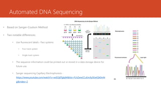
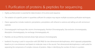
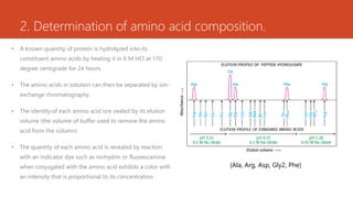
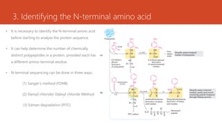
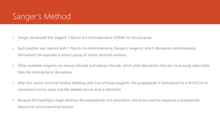
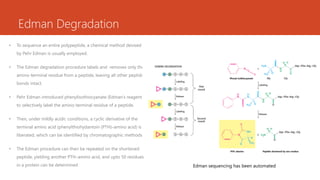
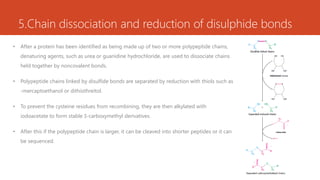
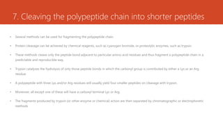
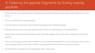
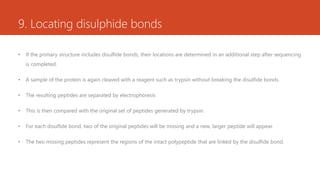
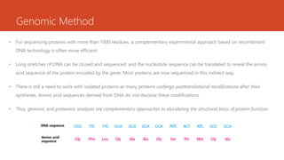
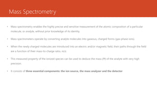
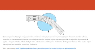
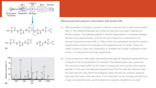
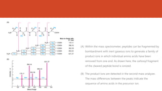
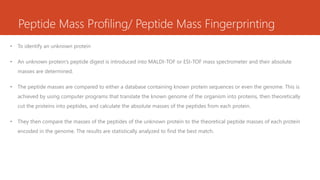
DNA Sequencing
Rima Joseph
Assistant Professor
Department of Zoology
Union Christian College, Aluva
Automated DNA Sequencing
• Based on Sanger-Coulson Method
• Two notable differences:
• Use fluorescent labels –Two systems
• Four track system
• Single track system
• The sequence information could be printed out or stored in a data storage device for
future use.
• Sanger sequencing Capillary Electrophoresis -
https://www.youtube.com/watch?v=wdS3j0TgbjM&list=FLhZwwCCuEmJly5EwtQk0mN
g&index=2
Advantages
• Radioactivity is not used
• Gel processing not needed
• Manual reading of gels not required
• Sequence data is directly fed and stored into a computer
• Extremely fast
Sanger Sequencing - https://www.youtube.com/watch?v=YeHtjO7vlyg&list=FLhZwwCCuEmJly5EwtQk0mNg&index=4
Maxam & Gilbert Sequencing - https://www.youtube.com/watch?v=_B5Dj8PL4E0&list=FLhZwwCCuEmJly5EwtQk0mNg&index=2
Protein Sequencing
Rima Joseph
Assistant Professor
Department of Zoology
Union Christian College, Aluva
Steps in Protein Sequencing
1. Purification of proteins & peptides for sequencing.
2. Determination of amino acid composition.
3. Identifying the N-terminal amino acid
4. C-terminal sequencing
5. In case of large proteins, dissociate chains held together by non-covalent bonds using denaturing agents
6. Breaking disulphide bonds
7. Cleaving the polypeptide chain into shorter peptides
8. Ordering the peptide fragments by finding overlap peptides
9. Locating disulphide bonds
1. Purification of proteins & peptides for sequencing.
• Highly purified protein is essential for determination of its amino acid sequence.
• The isolation of a specific protein in quantities sufficient for analysis may require multiple successive purification techniques.
• Classic approaches includes isoelectric precipitation, precipitation with ethanol or acetone and salting out with ammonium
sulphate.
• Chromatographic techniques like column chromatography, Partition Chromatography, Size exclusion chromatography,
Absorption chromatography, Ion exchange chromatography etc.
• Peptides can be purified by reversed-phase high-pressure chromatography.
• Isoelectric focusing is used in conjunction with SDS PAGE for two-dimensional electrophoresis, which separates polypeptides
based on pI in one dimension and based on molecular mass in the second. Two-dimensional electrophoresis is well suited for
separating the components of complex mixtures of proteins. Helps in identifying the number of chains in a protein.
2. Determination of amino acid composition.
• A known quantity of protein is hydrolyzed into its
constituent amino acids by heating it in 6 M HCl at 110
degree centigrade for 24 hours.
• The amino acids in solution can then be separated by ion-
exchange chromatography.
• The identity of each amino acid isre vealed by its elution
volume (the volume of buffer used to remove the amino
acid from the column)
• The quantity of each amino acid is revealed by reaction
with an indicator dye such as ninhydrin or fluorescamine
when conjugated with the amino acid exhibits a color with
an intensity that is proportional to its concentration.
(Ala, Arg, Asp, Gly2, Phe)
3. Identifying the N-terminal amino acid
• It is necessary to identify the N-terminal amino acid
before starting to analyze the protein sequence.
• It can help determine the number of chemically
distinct polypeptides in a protein, provided each has
a different amino-terminal residue.
• N-terminal sequencing can be done in three ways:
(1) Sanger’s method (FDNB)
(2) Dansyl chloride/ Dabsyl chloride Method
(3) Edman degradation (PITC)
Sanger’s Method
• Sanger developed the reagent 1-fluoro-2,4-dinitrobenzene (FDNB) for this purpose
• Each peptide was reacted with 1-fluoro-2,4-dinitrobenzene (Sanger’s reagent), which derivatizes (dinitrophenyl
derivatives) the exposed α-amino group of amino terminal residues.
• Other available reagents are dansyl chloride and dabsyl chloride, which yield derivatives that are more easily detectable
than the dinitrophenyl derivatives.
• After this amino-terminal residue labelling with one of these reagents, the polypeptide is hydrolyzed (in 6 M HCl) to its
constituent amino acids and the labeled amino acid is identified.
• Because the hydrolysis stage destroys the polypeptide, this procedure cannot be used to sequence a polypeptide
beyond its amino-terminal residue.
Edman Degradation
• To sequence an entire polypeptide, a chemical method devised
by Pehr Edman is usually employed.
• The Edman degradation procedure labels and removes only the
amino-terminal residue from a peptide, leaving all other peptide
bonds intact.
• Pehr Edman introduced phenylisothiocyanate (Edman’s reagent)
to selectively label the amino-terminal residue of a peptide.
• Then, under mildly acidic conditions, a cyclic derivative of the
terminal amino acid (phenylthiohydantoin (PTH)–amino acid) is
liberated, which can be identified by chromatographic methods.
• The Edman procedure can then be repeated on the shortened
peptide, yielding another PTH–amino acid, and upto 50 residues
in a protein can be determined Edman sequencing has been automated
Advantages & Disadvantages of Edman Degradation
Advantages
1. Sensitive method, i.e. a picogram quantity of the
sample is enough.
2. Intact proteins can be directly analyzed, no need for
protein digestion, a small quantity of the protein
only is needed.
3. More reliable sequencing technique.
Disadvantages
1. N-terminal modified proteins cannot be sequenced.
2. Proteins stained with silver nitrate cannot be
sequenced.
3. Slow processing; only 10 amino acids are detected
in 24 hours.
4. Only up to 50 amino acids can be sequenced in a
single reaction.
4. C-terminal sequencing
• This is done with an enzyme known as carboxypeptidase.
• It specifically cleaves the amino acids from the C-terminal end of the polypeptide.
5.Chain dissociation and reduction of disulphide bonds
• After a protein has been identified as being made up of two or more polypeptide chains,
denaturing agents, such as urea or guanidine hydrochloride, are used to dissociate chains
held together by noncovalent bonds.
• Polypeptide chains linked by disulfide bonds are separated by reduction with thiols such as
-mercaptoethanol or dithiothreitol.
• To prevent the cysteine residues from recombining, they are then alkylated with
iodoacetate to form stable S-carboxymethyl derivatives.
• After this if the polypeptide chain is larger, it can be cleaved into shorter peptides or it can
be sequenced.
7. Cleaving the polypeptide chain into shorter peptides
• Several methods can be used for fragmenting the polypeptide chain.
• Protein cleavage can be achieved by chemical reagents, such as cyanogen bromide, or proteolytic enzymes, such as trypsin.
• These methods cleave only the peptide bond adjacent to particular amino acid residues and thus fragment a polypeptide chain in a
predictable and reproducible way.
• Trypsin catalyzes the hydrolysis of only those peptide bonds in which the carbonyl group is contributed by either a Lys or an Arg
residue.
• A polypeptide with three Lys and/or Arg residues will usually yield four smaller peptides on cleavage with trypsin.
• Moreover, all except one of these will have a carboxyl terminal Lys or Arg.
• The fragments produced by trypsin (or other enzyme or chemical) action are then separated by chromatographic or electrophoretic
methods
8. Ordering the peptide fragments by finding overlap
peptides
• At this point, the amino acid sequences of segments of the protein are known, but the order of these segments is not yet
defined.
• This can be obtained from overlap peptides.
• A second enzyme or chemical is used to split the polypeptide chain at different linkages.
• Cyanogen bromide cleaves only those peptide bonds in which the carbonyl group is contributed by Met.
• Because these cyanogen bromide peptides overlap with tryptic peptides, they can be used to establish the order of the
peptides.
• If the second cleavage procedure fails to establish continuity between all peptides from the first cleavage, a third or even a
fourth cleavage method must be used to obtain a set of peptides that can provide the necessary overlap(s).
• The entire amino acid sequence of the polypeptide chain is then known.
9. Locating disulphide bonds
• If the primary structure includes disulfide bonds, their locations are determined in an additional step after sequencing
is completed.
• A sample of the protein is again cleaved with a reagent such as trypsin without breaking the disulfide bonds.
• The resulting peptides are separated by electrophoresis
• This is then compared with the original set of peptides generated by trypsin.
• For each disulfide bond, two of the original peptides will be missing and a new, larger peptide will appear.
• The two missing peptides represent the regions of the intact polypeptide that are linked by the disulfide bond.
Newer Methods of Protein Sequencing
1. Using genomic methods
2. Using Mass Spectrometry
1. Investigating the mass of protein/ peptide using MALDI-MS & ESI-MS
2. De novo protein sequencing using Tandem MS
3. Peptide mass profiling/ Peptide mass fingerprinting to identify an unknown protein
Genomic Method
• For sequencing proteins with more than 1000 residues, a complementary experimental approach based on recombinant
DNA technology is often more efficient.
• Long stretches of DNA can be cloned and sequenced, and the nucleotide sequence can be translated to reveal the amino
acid sequence of the protein encoded by the gene. Most proteins are now sequenced in this indirect way.
• There is still a need to work with isolated proteins as many proteins undergo posttranslational modifications after their
syntheses. Amino acid sequences derived from DNA do not disclose these modifications.
• Thus, genomic and proteomic analyses are complementary approaches to elucidating the structural basis of protein function.
Mass Spectrometry
• Mass spectrometry enables the highly precise and sensitive measurement of the atomic composition of a particular
molecule, or analyte, without prior knowledge of its identity.
• Mass spectrometers operate by converting analyte molecules into gaseous, charged forms (gas-phase ions).
• When the newly charged molecules are introduced into an electric and/or magnetic field, their paths through the field
are a function of their mass-to-charge ratio, m/z.
• This measured property of the ionized species can be used to deduce the mass (M) of the analyte with very high
precision.
• It consists of three essential components: the ion source, the mass analyzer and the detector
Basic components of a simple mass spectrometer. A mixture of molecules is vaporized in an ionized state in the sample chamber S. These
molecules are then accelerated down the flight tube by an electrical potential applied to accelerator grid A. An adjustable electromagnet, E,
applies a magnetic field that deflects the flight of the individual ions until they strike the detector, D. The greater the mass of the ion, the higher
the magnetic field required to focus it onto the detector.
Mass Spectrometry - https://www.youtube.com/watch?v=EzvQzImBuq8&list=FLhZwwCCuEmJly5EwtQk0mNg&index=6
• Ion Source : Conversion of analyte into gas-phase ions (ionization)
2 techniques - matrix-assisted laser desorption/ionization (MALDI) and electrospray ionization (ESI)
• Mass Analyzer: There are a number of different types of mass analyzers. Consider one of the simplest, the time-of-flight (TOF)
mass analyzer – in this ions are accelerated through an elongated chamber under a fixed electrostatic potential. Given two ions
of identical net charge, the smaller ion will require less time to traverse the chamber than will the larger ion. The mass of each
ion can be determined by measuring the time required for each ion to pass through the chamber.
• The molecular mass of a protein can be accurately measured by MALDI-MS. & ESI-MS
• These can also detect changes in mass due to the presence of bound cofactors, bound metal ions, covalent modifications, and
so on.
Proteins are placed in a light-
absorbing matrix. With a short pulse
of laser light, the proteins are ionized
and then desorbed from the matrix
into the vacuum system.
A solution of analytes is passed
through a charged needle that is
kept at a high electrical potential,
dispersing the solution into a fine
mist of charged microdroplets.
Macromolecular ions are thus
introduced nondestructively into the
gas phase.
• MALDI TOF - https://www.youtube.com/watch?v=0jeFpXHZ8W0&list=FLhZwwCCuEmJly5EwtQk0mNg&index=5
• ESI-MS - https://www.youtube.com/watch?v=22dr-3XDNmQ&list=FLhZwwCCuEmJly5EwtQk0mNg&index=4
Tandem Mass Spectrometry (MS/MS)
• Sequence information is extracted using a technique called tandem MS, or MS/MS.
• A solution containing the protein under investigation is first treated with a protease or chemical reagent to hydrolyze it to a
mixture of shorter peptides.
• The mixture is then injected into a device that is essentially two mass spectrometers in tandem
• The first spectrometer separates individual peptides based upon their differences in mass.
• By adjusting the field strength of the first magnet, a single peptide can be directed into a vaccum chamber, the ‘collision cell’
• In this collision cell, the peptide is further fragmented by high-energy impact with a “collision gas” (helium or argon)
• A family of ions is detected; each ion represents a fragment of the original peptide with one or more amino acids removed
from one end. The second mass spectrometer then measures the m/z ratios of all these charged fragments
• This generates one or more sets of peaks. A given set of peaks consists of all the charged fragments that were generated by
breaking the same type of bond (but at different points in the peptide) and are derived from the same side of the bond
breakage, either the carboxyl- or amino-terminal side.
• Each successive peak in a given set has one less amino acid than the peak before. The difference in mass from peak to peak
identifies the amino acid that was lost in each case, thus revealing the sequence of the peptide.
• As the sensitivity and versatility of mass spectrometry continue to increase, it is displacing Edman sequencers for the direct
analysis of protein primary structure.
• Tandem MS - https://www.youtube.com/watch?v=Jc1uC6EbMCs&list=FLhZwwCCuEmJly5EwtQk0mNg
Obtaining protein sequence information with tandem MS.
(a) After proteolytic hydrolysis, a protein solution is injected into a mass spectrometer
(MS-1). The different peptides are sorted so that only one type is selected for
further analysis. The selected peptide is further fragmented in a chamber between
the two mass spectrometers, and m/z for each fragment is measured in the
second mass spectrometer (MS-2). Many of the ions generated during this second
fragmentation result from breakage of the peptide bond, as shown. These are
called b-type or y-type ions, depending on whether the charge is retained on the
amino- or carboxyl-terminal side, respectively.
(b) A typical spectrum with peaks representing the peptide fragments generated from
a sample of one small peptide (10 residues). The labeled peaks are y-type ions.
The successive peaks differ by the mass of a particular amino acid in the original
peptide. In this case, the deduced sequence was Phe–Pro–Gly–Gln–(Ile/Leu)–Asn–
Ala–Asp–(Ile/Leu)–Arg. Note the ambiguity about Ile and Leu residues, because
they have the same molecular mass. In this example, the set of peaks derived from
y-type ions predominates, and the spectrum is greatly simplified as a result.
(A) Within the mass spectrometer, peptides can be fragmented by
bombardment with inert gaseous ions to generate a family of
product ions in which individual amino acids have been
removed from one end. As drawn here, the carboxyl fragment
of the cleaved peptide bond is ionized.
(B) The product ions are detected in the second mass analyzer.
The mass differences between the peaks indicate the
sequence of amino acids in the precursor ion.
Peptide Mass Profiling/ Peptide Mass Fingerprinting
• To identify an unknown protein
• An unknown protein’s peptide digest is introduced into MALDI-TOF or ESI-TOF mass spectrometer and their absolute
masses are determined.
• The peptide masses are compared to either a database containing known protein sequences or even the genome. This is
achieved by using computer programs that translate the known genome of the organism into proteins, then theoretically
cut the proteins into peptides, and calculate the absolute masses of the peptides from each protein.
• They then compare the masses of the peptides of the unknown protein to the theoretical peptide masses of each protein
encoded in the genome. The results are statistically analyzed to find the best match.
References
• Berg, Jeremy M., et al. Biochemistry. W.H. Freeman, 2012.
• Lehninger, Albert. Lehninger Principles of Biochemistry & EBook .-5th Ed. (9781429224161). W.H. Freeman, 2005.
• Murray, Robert K., and Harold A. Harper. Harpers Illustrated Biochemistry. McGraw-Hill, 2003.
• Saraswathy, Nachimuthu, and Ponnusamy Ramalingam. Concepts and Techniques in Genomics and Proteomics.
Woodhead Publishing, 2012.
Automated DNA sequencing ; Protein sequencing

Automated DNA sequencing ; Protein sequencing

  • 1.
    DNA Sequencing Rima Joseph AssistantProfessor Department of Zoology Union Christian College, Aluva
  • 2.
    Automated DNA Sequencing •Based on Sanger-Coulson Method • Two notable differences: • Use fluorescent labels –Two systems • Four track system • Single track system • The sequence information could be printed out or stored in a data storage device for future use. • Sanger sequencing Capillary Electrophoresis - https://www.youtube.com/watch?v=wdS3j0TgbjM&list=FLhZwwCCuEmJly5EwtQk0mN g&index=2
  • 3.
    Advantages • Radioactivity isnot used • Gel processing not needed • Manual reading of gels not required • Sequence data is directly fed and stored into a computer • Extremely fast Sanger Sequencing - https://www.youtube.com/watch?v=YeHtjO7vlyg&list=FLhZwwCCuEmJly5EwtQk0mNg&index=4 Maxam & Gilbert Sequencing - https://www.youtube.com/watch?v=_B5Dj8PL4E0&list=FLhZwwCCuEmJly5EwtQk0mNg&index=2
  • 4.
    Protein Sequencing Rima Joseph AssistantProfessor Department of Zoology Union Christian College, Aluva
  • 5.
    Steps in ProteinSequencing 1. Purification of proteins & peptides for sequencing. 2. Determination of amino acid composition. 3. Identifying the N-terminal amino acid 4. C-terminal sequencing 5. In case of large proteins, dissociate chains held together by non-covalent bonds using denaturing agents 6. Breaking disulphide bonds 7. Cleaving the polypeptide chain into shorter peptides 8. Ordering the peptide fragments by finding overlap peptides 9. Locating disulphide bonds
  • 6.
    1. Purification ofproteins & peptides for sequencing. • Highly purified protein is essential for determination of its amino acid sequence. • The isolation of a specific protein in quantities sufficient for analysis may require multiple successive purification techniques. • Classic approaches includes isoelectric precipitation, precipitation with ethanol or acetone and salting out with ammonium sulphate. • Chromatographic techniques like column chromatography, Partition Chromatography, Size exclusion chromatography, Absorption chromatography, Ion exchange chromatography etc. • Peptides can be purified by reversed-phase high-pressure chromatography. • Isoelectric focusing is used in conjunction with SDS PAGE for two-dimensional electrophoresis, which separates polypeptides based on pI in one dimension and based on molecular mass in the second. Two-dimensional electrophoresis is well suited for separating the components of complex mixtures of proteins. Helps in identifying the number of chains in a protein.
  • 7.
    2. Determination ofamino acid composition. • A known quantity of protein is hydrolyzed into its constituent amino acids by heating it in 6 M HCl at 110 degree centigrade for 24 hours. • The amino acids in solution can then be separated by ion- exchange chromatography. • The identity of each amino acid isre vealed by its elution volume (the volume of buffer used to remove the amino acid from the column) • The quantity of each amino acid is revealed by reaction with an indicator dye such as ninhydrin or fluorescamine when conjugated with the amino acid exhibits a color with an intensity that is proportional to its concentration. (Ala, Arg, Asp, Gly2, Phe)
  • 8.
    3. Identifying theN-terminal amino acid • It is necessary to identify the N-terminal amino acid before starting to analyze the protein sequence. • It can help determine the number of chemically distinct polypeptides in a protein, provided each has a different amino-terminal residue. • N-terminal sequencing can be done in three ways: (1) Sanger’s method (FDNB) (2) Dansyl chloride/ Dabsyl chloride Method (3) Edman degradation (PITC)
  • 9.
    Sanger’s Method • Sangerdeveloped the reagent 1-fluoro-2,4-dinitrobenzene (FDNB) for this purpose • Each peptide was reacted with 1-fluoro-2,4-dinitrobenzene (Sanger’s reagent), which derivatizes (dinitrophenyl derivatives) the exposed α-amino group of amino terminal residues. • Other available reagents are dansyl chloride and dabsyl chloride, which yield derivatives that are more easily detectable than the dinitrophenyl derivatives. • After this amino-terminal residue labelling with one of these reagents, the polypeptide is hydrolyzed (in 6 M HCl) to its constituent amino acids and the labeled amino acid is identified. • Because the hydrolysis stage destroys the polypeptide, this procedure cannot be used to sequence a polypeptide beyond its amino-terminal residue.
  • 10.
    Edman Degradation • Tosequence an entire polypeptide, a chemical method devised by Pehr Edman is usually employed. • The Edman degradation procedure labels and removes only the amino-terminal residue from a peptide, leaving all other peptide bonds intact. • Pehr Edman introduced phenylisothiocyanate (Edman’s reagent) to selectively label the amino-terminal residue of a peptide. • Then, under mildly acidic conditions, a cyclic derivative of the terminal amino acid (phenylthiohydantoin (PTH)–amino acid) is liberated, which can be identified by chromatographic methods. • The Edman procedure can then be repeated on the shortened peptide, yielding another PTH–amino acid, and upto 50 residues in a protein can be determined Edman sequencing has been automated
  • 11.
    Advantages & Disadvantagesof Edman Degradation Advantages 1. Sensitive method, i.e. a picogram quantity of the sample is enough. 2. Intact proteins can be directly analyzed, no need for protein digestion, a small quantity of the protein only is needed. 3. More reliable sequencing technique. Disadvantages 1. N-terminal modified proteins cannot be sequenced. 2. Proteins stained with silver nitrate cannot be sequenced. 3. Slow processing; only 10 amino acids are detected in 24 hours. 4. Only up to 50 amino acids can be sequenced in a single reaction.
  • 12.
    4. C-terminal sequencing •This is done with an enzyme known as carboxypeptidase. • It specifically cleaves the amino acids from the C-terminal end of the polypeptide.
  • 13.
    5.Chain dissociation andreduction of disulphide bonds • After a protein has been identified as being made up of two or more polypeptide chains, denaturing agents, such as urea or guanidine hydrochloride, are used to dissociate chains held together by noncovalent bonds. • Polypeptide chains linked by disulfide bonds are separated by reduction with thiols such as -mercaptoethanol or dithiothreitol. • To prevent the cysteine residues from recombining, they are then alkylated with iodoacetate to form stable S-carboxymethyl derivatives. • After this if the polypeptide chain is larger, it can be cleaved into shorter peptides or it can be sequenced.
  • 14.
    7. Cleaving thepolypeptide chain into shorter peptides • Several methods can be used for fragmenting the polypeptide chain. • Protein cleavage can be achieved by chemical reagents, such as cyanogen bromide, or proteolytic enzymes, such as trypsin. • These methods cleave only the peptide bond adjacent to particular amino acid residues and thus fragment a polypeptide chain in a predictable and reproducible way. • Trypsin catalyzes the hydrolysis of only those peptide bonds in which the carbonyl group is contributed by either a Lys or an Arg residue. • A polypeptide with three Lys and/or Arg residues will usually yield four smaller peptides on cleavage with trypsin. • Moreover, all except one of these will have a carboxyl terminal Lys or Arg. • The fragments produced by trypsin (or other enzyme or chemical) action are then separated by chromatographic or electrophoretic methods
  • 16.
    8. Ordering thepeptide fragments by finding overlap peptides • At this point, the amino acid sequences of segments of the protein are known, but the order of these segments is not yet defined. • This can be obtained from overlap peptides. • A second enzyme or chemical is used to split the polypeptide chain at different linkages. • Cyanogen bromide cleaves only those peptide bonds in which the carbonyl group is contributed by Met. • Because these cyanogen bromide peptides overlap with tryptic peptides, they can be used to establish the order of the peptides. • If the second cleavage procedure fails to establish continuity between all peptides from the first cleavage, a third or even a fourth cleavage method must be used to obtain a set of peptides that can provide the necessary overlap(s). • The entire amino acid sequence of the polypeptide chain is then known.
  • 17.
    9. Locating disulphidebonds • If the primary structure includes disulfide bonds, their locations are determined in an additional step after sequencing is completed. • A sample of the protein is again cleaved with a reagent such as trypsin without breaking the disulfide bonds. • The resulting peptides are separated by electrophoresis • This is then compared with the original set of peptides generated by trypsin. • For each disulfide bond, two of the original peptides will be missing and a new, larger peptide will appear. • The two missing peptides represent the regions of the intact polypeptide that are linked by the disulfide bond.
  • 18.
    Newer Methods ofProtein Sequencing 1. Using genomic methods 2. Using Mass Spectrometry 1. Investigating the mass of protein/ peptide using MALDI-MS & ESI-MS 2. De novo protein sequencing using Tandem MS 3. Peptide mass profiling/ Peptide mass fingerprinting to identify an unknown protein
  • 19.
    Genomic Method • Forsequencing proteins with more than 1000 residues, a complementary experimental approach based on recombinant DNA technology is often more efficient. • Long stretches of DNA can be cloned and sequenced, and the nucleotide sequence can be translated to reveal the amino acid sequence of the protein encoded by the gene. Most proteins are now sequenced in this indirect way. • There is still a need to work with isolated proteins as many proteins undergo posttranslational modifications after their syntheses. Amino acid sequences derived from DNA do not disclose these modifications. • Thus, genomic and proteomic analyses are complementary approaches to elucidating the structural basis of protein function.
  • 20.
    Mass Spectrometry • Massspectrometry enables the highly precise and sensitive measurement of the atomic composition of a particular molecule, or analyte, without prior knowledge of its identity. • Mass spectrometers operate by converting analyte molecules into gaseous, charged forms (gas-phase ions). • When the newly charged molecules are introduced into an electric and/or magnetic field, their paths through the field are a function of their mass-to-charge ratio, m/z. • This measured property of the ionized species can be used to deduce the mass (M) of the analyte with very high precision. • It consists of three essential components: the ion source, the mass analyzer and the detector
  • 21.
    Basic components ofa simple mass spectrometer. A mixture of molecules is vaporized in an ionized state in the sample chamber S. These molecules are then accelerated down the flight tube by an electrical potential applied to accelerator grid A. An adjustable electromagnet, E, applies a magnetic field that deflects the flight of the individual ions until they strike the detector, D. The greater the mass of the ion, the higher the magnetic field required to focus it onto the detector. Mass Spectrometry - https://www.youtube.com/watch?v=EzvQzImBuq8&list=FLhZwwCCuEmJly5EwtQk0mNg&index=6
  • 22.
    • Ion Source: Conversion of analyte into gas-phase ions (ionization) 2 techniques - matrix-assisted laser desorption/ionization (MALDI) and electrospray ionization (ESI) • Mass Analyzer: There are a number of different types of mass analyzers. Consider one of the simplest, the time-of-flight (TOF) mass analyzer – in this ions are accelerated through an elongated chamber under a fixed electrostatic potential. Given two ions of identical net charge, the smaller ion will require less time to traverse the chamber than will the larger ion. The mass of each ion can be determined by measuring the time required for each ion to pass through the chamber. • The molecular mass of a protein can be accurately measured by MALDI-MS. & ESI-MS • These can also detect changes in mass due to the presence of bound cofactors, bound metal ions, covalent modifications, and so on. Proteins are placed in a light- absorbing matrix. With a short pulse of laser light, the proteins are ionized and then desorbed from the matrix into the vacuum system. A solution of analytes is passed through a charged needle that is kept at a high electrical potential, dispersing the solution into a fine mist of charged microdroplets. Macromolecular ions are thus introduced nondestructively into the gas phase.
  • 23.
    • MALDI TOF- https://www.youtube.com/watch?v=0jeFpXHZ8W0&list=FLhZwwCCuEmJly5EwtQk0mNg&index=5 • ESI-MS - https://www.youtube.com/watch?v=22dr-3XDNmQ&list=FLhZwwCCuEmJly5EwtQk0mNg&index=4
  • 24.
    Tandem Mass Spectrometry(MS/MS) • Sequence information is extracted using a technique called tandem MS, or MS/MS. • A solution containing the protein under investigation is first treated with a protease or chemical reagent to hydrolyze it to a mixture of shorter peptides. • The mixture is then injected into a device that is essentially two mass spectrometers in tandem • The first spectrometer separates individual peptides based upon their differences in mass. • By adjusting the field strength of the first magnet, a single peptide can be directed into a vaccum chamber, the ‘collision cell’ • In this collision cell, the peptide is further fragmented by high-energy impact with a “collision gas” (helium or argon) • A family of ions is detected; each ion represents a fragment of the original peptide with one or more amino acids removed from one end. The second mass spectrometer then measures the m/z ratios of all these charged fragments
  • 25.
    • This generatesone or more sets of peaks. A given set of peaks consists of all the charged fragments that were generated by breaking the same type of bond (but at different points in the peptide) and are derived from the same side of the bond breakage, either the carboxyl- or amino-terminal side. • Each successive peak in a given set has one less amino acid than the peak before. The difference in mass from peak to peak identifies the amino acid that was lost in each case, thus revealing the sequence of the peptide. • As the sensitivity and versatility of mass spectrometry continue to increase, it is displacing Edman sequencers for the direct analysis of protein primary structure. • Tandem MS - https://www.youtube.com/watch?v=Jc1uC6EbMCs&list=FLhZwwCCuEmJly5EwtQk0mNg
  • 26.
    Obtaining protein sequenceinformation with tandem MS. (a) After proteolytic hydrolysis, a protein solution is injected into a mass spectrometer (MS-1). The different peptides are sorted so that only one type is selected for further analysis. The selected peptide is further fragmented in a chamber between the two mass spectrometers, and m/z for each fragment is measured in the second mass spectrometer (MS-2). Many of the ions generated during this second fragmentation result from breakage of the peptide bond, as shown. These are called b-type or y-type ions, depending on whether the charge is retained on the amino- or carboxyl-terminal side, respectively. (b) A typical spectrum with peaks representing the peptide fragments generated from a sample of one small peptide (10 residues). The labeled peaks are y-type ions. The successive peaks differ by the mass of a particular amino acid in the original peptide. In this case, the deduced sequence was Phe–Pro–Gly–Gln–(Ile/Leu)–Asn– Ala–Asp–(Ile/Leu)–Arg. Note the ambiguity about Ile and Leu residues, because they have the same molecular mass. In this example, the set of peaks derived from y-type ions predominates, and the spectrum is greatly simplified as a result.
  • 27.
    (A) Within themass spectrometer, peptides can be fragmented by bombardment with inert gaseous ions to generate a family of product ions in which individual amino acids have been removed from one end. As drawn here, the carboxyl fragment of the cleaved peptide bond is ionized. (B) The product ions are detected in the second mass analyzer. The mass differences between the peaks indicate the sequence of amino acids in the precursor ion.
  • 28.
    Peptide Mass Profiling/Peptide Mass Fingerprinting • To identify an unknown protein • An unknown protein’s peptide digest is introduced into MALDI-TOF or ESI-TOF mass spectrometer and their absolute masses are determined. • The peptide masses are compared to either a database containing known protein sequences or even the genome. This is achieved by using computer programs that translate the known genome of the organism into proteins, then theoretically cut the proteins into peptides, and calculate the absolute masses of the peptides from each protein. • They then compare the masses of the peptides of the unknown protein to the theoretical peptide masses of each protein encoded in the genome. The results are statistically analyzed to find the best match.
  • 29.
    References • Berg, JeremyM., et al. Biochemistry. W.H. Freeman, 2012. • Lehninger, Albert. Lehninger Principles of Biochemistry & EBook .-5th Ed. (9781429224161). W.H. Freeman, 2005. • Murray, Robert K., and Harold A. Harper. Harpers Illustrated Biochemistry. McGraw-Hill, 2003. • Saraswathy, Nachimuthu, and Ponnusamy Ramalingam. Concepts and Techniques in Genomics and Proteomics. Woodhead Publishing, 2012.