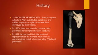
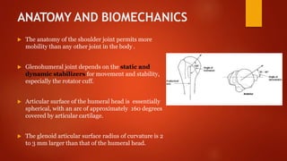
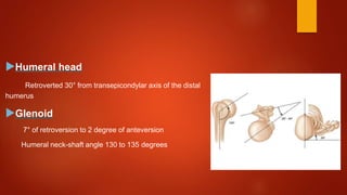
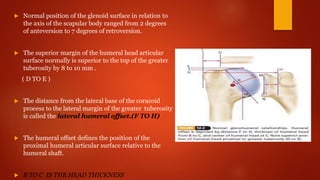
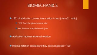
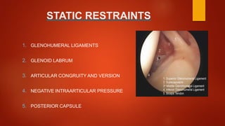
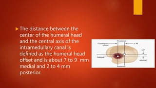
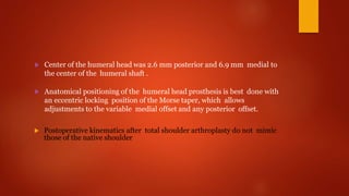
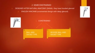
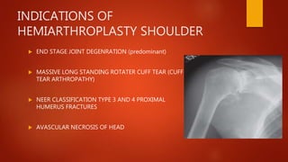
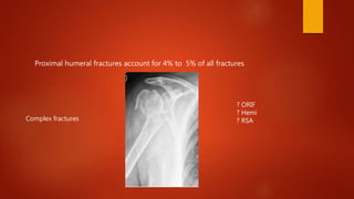
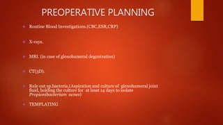
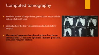
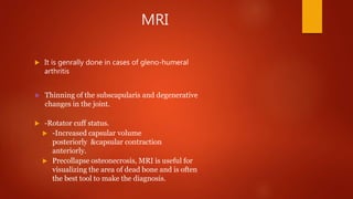
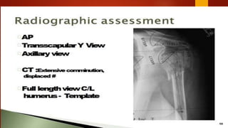
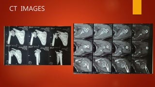
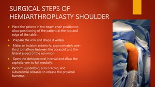
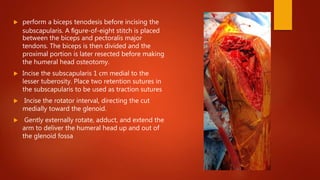
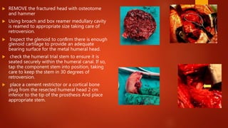
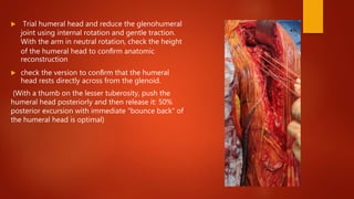
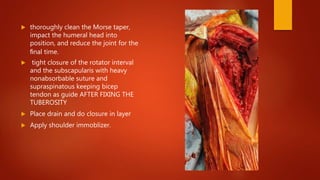
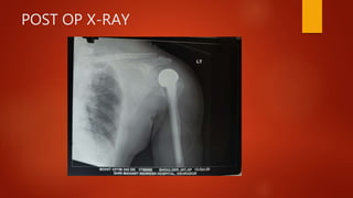
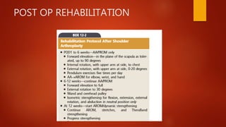
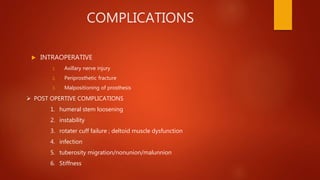
HEMIARTHROPLASTY
SHOULDER
DR ANSHUL SETHI
PG ORTHOPAEDICS
Outline
 HISTORY.
 ANATOMY & BIOMECHANICS.
 PROSTHESIS DEIGNS.
 PROSTHESIS & ITS FEATURES.
 INDICATIONS
 CONTRAINDICATIONS
 OBJECTIVE
 CASE DISCUSSION
 SURGICAL APPROACH.
 COMPLICATIONS.
History
 1st SHOULDER ARTHROPLASTY French surgeon,
Jules Emil Péan, substituted a platinum and
rubber implant for a gleno-humeral joint
destroyed by tuberculosis.
 In 1950s, Neer introduced a humeral head
prosthesis for complex shoulder fractures.
 In 1951, he reported his initial results of
replacement of the humeral head with an
unconstrained cobalt-chromium alloy (Vitallium)
prosthesis.
ANATOMY AND BIOMECHANICS
 The anatomy of the shoulder joint permits more
mobility than any other joint in the body .
 Glenohumeral joint depends on the static and
dynamic stabilizers for movement and stability,
especially the rotator cuff.
 Articular surface of the humeral head is essentially
spherical, with an arc of approximately 160 degrees
covered by articular cartilage.
 The glenoid articular surface radius of curvature is 2
to 3 mm larger than that of the humeral head.
Humeral head
Retroverted 30° from transepicondylar axis of the distal
humerus
Glenoid
7° of retroversion to 2 degree of anteversion
Humeral neck-shaft angle 130 to 135 degrees
 Normal position of the glenoid surface in relation to
the axis of the scapular body ranged from 2 degrees
of anteversion to 7 degrees of retroversion.
 The superior margin of the humeral head articular
surface normally is superior to the top of the greater
tuberosity by 8 to 10 mm .
( D TO E )
 The distance from the lateral base of the coracoid
process to the lateral margin of the greater tuberosity
is called the lateral humeral offset.(F TO H)
 The humeral offset defines the position of the
proximal humeral articular surface relative to the
humeral shaft.
 B TO C IS THE HEAD THICKNESS
BIOMECHANICS
180° of abduction comes from motion in two joints (2:1 ratio)
 120° from the glenohumeral joint
 60° from the scapulothoracic joint
Abduction requires external rotation
Internal rotation contracture they can not abduct > 120
STATIC RESTRAINTS
1. GLENOHUMERAL LIGAMENTS
2. GLENOID LABRUM
3. ARTICULAR CONGRUITY AND VERSION
4. NEGATIVE INTRAARTICULAR PRESSURE
5. POSTERIOR CAPSULE
Dynamic restraints
 Rotator cuff muscles
 Rotator interval
 Biceps long head
 Periscapular muscles
 The distance between the
center of the humeral head
and the central axis of the
intramedullary canal is
defined as the humeral head
offset and is about 7 to 9 mm
medial and 2 to 4 mm
posterior.
 Increasing the humeral head thickness by 5 mm has been shown to
reduce the range of motion at the glenohumeral joint by 20 to 30
degrees, whereas decreasing the thickness by 5 mm can diminish
motion.
 Replacement of the anatomical humeral head size and
position aims to restore normal shoulder biomechanics.
PROSTHESIS DESIGN
 Based on NEER II implant many modular designs were developed to
impove implant fixation and durability .
 Modularity allows a better fit for individual patients because various stem
and head sizes can be “mixed and matched” to an individual’s anatomy.
 Most stems are made of cobalt-chrome alloy, have proximal porous
ingrowth coating, and have proximal fins.
(A fin is a thin component or appendage attached to a larger body or structure)
for rotational stability.
 Center of the humeral head was 2.6 mm posterior and 6.9 mm medial to
the center of the humeral shaft .
 Anatomical positioning of the humeral head prosthesis is best done with
an eccentric locking position of the Morse taper, which allows
adjustments to the variable medial offset and any posterior offset.
 Postoperative kinematics after total shoulder arthroplasty do not mimic
those of the native shoulder
 Prosthetic head should be within 4 mm of the
original humeral head thickness.(to prevent
overstuffing of joint)
 Most stems can be inserted with a press-fit or
cemented technique.
 Glenoid part should have an increased radius of
curvature compared with the humeral head (2 to 6
mm larger) to allow translation during movement
and to decrease edge loading in order to prevent
rocking –horse effect (damage to articular cartilage
or glenoid loosening in TSA)
ROCKING HORSE EFFECT
 Eccentric loading of the glenoid caused by
superior migration of the humeral
component has been cited as a cause of
glenoid loosening.
 Association between glenoid component
loosening and irreparable rotator cuff
tears.
DEVELOPMENT IS SHOULDER
ARTHROPLASTY PROSTHESIS
1. UNCONSTRAINED
1. NEER II (DESIGNED TO REPRODUCE NORMAL ANATOMY)
2. CUP ARTHROPLASTY (CAN BE OF METAL ,SILASTIC CUP
3. BIPOLAR (MORE MOTION WITH LESS STRESS ON GLENOID)
NEER TYPE (LESS CONSTRAINT AND BETTER SOFT TISSUE BALANCING)
BIPOLAR (DEAL WITH EXTENSIVE ROTATER CUFF DEFICIENCY)
2. SEMICONSTRAINED
1. DESIGNED AFTER NATURAL ANATOMY (DANA)- they have hooded glenoid
2. ENGLISH-MACNAB (uncemented design with deep glenoid)
1. CONSTRAINED
BALL AND
SOCKET TYPE
REVERSE BALL AND
SOCKET TYPE
INDICATIONS OF
HEMIARTHROPLASTY SHOULDER
 END STAGE JOINT DEGENRATION (predominant)
 MASSIVE LONG STANDING ROTATER CUFF TEAR (CUFF
TEAR ARTHROPATHY)
 NEER CLASSIFICATION TYPE 3 AND 4 PROXIMAL
HUMERUS FRACTURES
 AVASCULAR NECROSIS OF HEAD
MATSEN ET AL Listed INSICATIONS OF
hemi arthroplasty
1. Humeral joint surface is rough but cartillegnous surface of glenoid is intact
2. Insufficient bone to support glenoid component
3. Fixed proximal migration of humeral head as in rotater cuff arthropathy
4. Remote joint infection
5. Heavy occupational demands
CONTRAINDICATIONS
1. Recent sepsis
2. Neuropathic joint
3. Deltoid muscle deficient
4. Paralytic disorder
Proximal humeral fractures account for 4% to 5% of all fractures
? ORIF
? Hemi
? RSAComplex fractures
PREOPERATIVE PLANNING
 Routine Blood Investigations.(CBC,ESR,CRP)
 X-rays.
 MRI. (in case of glenohumeral degentration)
 CT(3D).
 Rule out sp.bacteria.(Aspiration and culture of glenohumeral joint
fluid, holding the culture for at least 14 days to isolate
Propionibacterium acnes)
 TEMPLATING
Computed tomography
 Excellent picture of the patient’s glenoid bone stock and the
pattern of glenoid wear.
 precisely show the bony deformities and defects before
surgery.
 The role of preoperative planning based on three-
dimensional CT scans to optimize implant position,
size, and range of motion
MRI
 It is genrally done in cases of gleno-humeral
arthritis
 Thinning of the subscapularis and degenerative
changes in the joint.
 -Rotator cuff status.
 -Increased capsular volume
posteriorly &capsular contraction
anteriorly.
 Precollapse osteonecrosis, MRI is useful for
visualizing the area of dead bone and is often
the best tool to make the diagnosis.
2
OBJECTIVE
 ABOLISH PAIN arising from the pathological joint
 Restore functional range of motion
CASE
 PATIENT MOHIT BISHT 35 YEAR OLD MALE PRESENTED TO EMERGENCY
WITH A/H/O RTA (SLIP FROM BIKE) FOLLWED BY COMPLAIN OF
1. PAIN , SWELLING AND DEFORMITY IN LEFT SHOULDER
2. INABILITY TO MOVE LEFT SHOULDER
DIAGNOSED AS
FRACTURE PROXIMAL HUMERUS LEFT SIDE
(NEER CLASSIFICATION TYPE 3 )
A.O CLASSIFICATION (11 C)
PRE OP X RAY
CT IMAGES
PATIENT MANAGED WITH
 MODULAR BIPOLAR CEMENTED HEMIARTHROPLASTY LEFT SHOULDER
OTHER TYPE OF HEMIARTHROPLASTY :
1. MODIFIED HEMIARTHROPLASTY: INTERPOSITION ARTHROPLASTY AND
GLENOIDPLASTY (REAM AND RUN)
2. RESURFACING HEMIARTHROPLASTY
SURGICAL STEPS OF
HEMIARTHROPLASTY SHOULDER
 Place the patient in the beach chair position to
allow positioning of the patient at the top and
edge of the table .
 Prepare the arm and drape it widely
 Make an incision anteriorly, approximately one-
third to halfway between the coracoid and the
lateral aspect of the acromion
 Open the deltopectoral interval and allow the
cephalic vein to fall medially.
 Perform subdeltoid, subcoracoid, and
subacromial releases to release the proximal
humerus
 perform a biceps tenodesis before incising the
subscapularis. A figure-of-eight stitch is placed
between the biceps and pectoralis major
tendons. The biceps is then divided and the
proximal portion is later resected before making
the humeral head osteotomy.
 Incise the subscapularis 1 cm medial to the
lesser tuberosity. Place two retention sutures in
the subscapularis to be used as traction sutures
 Incise the rotator interval, directing the cut
medially toward the glenoid.
 Gently externally rotate, adduct, and extend the
arm to deliver the humeral head up and out of
the glenoid fossa
 REMOVE the fractured head with osteotome
and hammer
 Using broach and box reamer medullary cavity
is reamed to appropriate size taking care of
retroversion.
 Inspect the glenoid to confirm there is enough
glenoid cartilage to provide an adequate
bearing surface for the metal humeral head.
 check the humeral trial stem to ensure it is
seated securely within the humeral canal. If so,
tap the component stem into position, taking
care to keep the stem in 30 degrees of
retroversion.
 place a cement restrictor or a cortical bone
plug from the resected humeral head 2 cm
inferior to the tip of the prosthesis And place
appropriate stem.
 Trial humeral head and reduce the glenohumeral
joint using internal rotation and gentle traction.
With the arm in neutral rotation, check the height
of the humeral head to confirm anatomic
reconstruction
 check the version to confirm that the humeral
head rests directly across from the glenoid.
(With a thumb on the lesser tuberosity, push the
humeral head posteriorly and then release it: 50%
posterior excursion with immediate “bounce back” of
the humeral head is optimal)
 thoroughly clean the Morse taper,
impact the humeral head into
position, and reduce the joint for the
final time.
 tight closure of the rotator interval
and the subscapularis with heavy
nonabsorbable suture and
supraspinatous keeping bicep
tendon as guide AFTER FIXING THE
TUBEROSITY
 Place drain and do closure in layer
 Apply shoulder immoblizer.
POST OP X-RAY
POST OP REHABILITATION
COMPLICATIONS
 INTRAOPERATIVE
1. Axillary nerve injury
2. Periprosthetic fracture
3. Malpositioning of prosthesis
 POST OPERTIVE COMPLICATIONS
1. humeral stem loosening
2. instability
3. rotater cuff failure ; deltoid muscle dysfunction
4. infection
5. tuberosity migration/nonunion/malunnion
6. Stiffness
STUDIES COMPARING ORIF VS HEMI
 Bastian and Hertel : ORIF vs Hemi
 similar functional results & patient satisfaction.
 ORIF with preservation of the humeral head should be considered
when an adequate reduction and stable conditions for
revascularization can be obtained.
 Hemiarthroplasty is a viable alternative for patients with osteopenic
bone and/or a comminuted fracture.
 Bastian JD, Hertel R. Osteosynthesis and hemiarthroplasty of fractures of the
proximal humerus:
 outcomes in a consecutive case series. J Shoulder Elbow Surg. 2009;18:216-9
BoileauP, WalchG, KrishnanSG. Tuberosity
osteosynthesisandhemiarthroplastyforfour-part
fracturesof theproximalhumerus.TechShoulder
ElbowSurg. 2000;1:96-109.
 Krishnan et al. : using ‘Gothic arch’ technique reported 88% of the
tuberosities healed anatomically. The mean active anterior elevation was
129˚, and pain scores averaged 1.2 points on a 10-point scale.
KrishnanSG, BennionPW, ReineckJR,Burkhead WZ.
Hemiarthroplastyforproximalhumeralfracture: restorationof theGothic
arch.OrthopClinNorthAm. 2008;39:441-50,
THANK YOU

Hemiarthroplasty shlulder

  • 1.
  • 2.
    Outline  HISTORY.  ANATOMY& BIOMECHANICS.  PROSTHESIS DEIGNS.  PROSTHESIS & ITS FEATURES.  INDICATIONS  CONTRAINDICATIONS  OBJECTIVE  CASE DISCUSSION  SURGICAL APPROACH.  COMPLICATIONS.
  • 3.
    History  1st SHOULDERARTHROPLASTY French surgeon, Jules Emil Péan, substituted a platinum and rubber implant for a gleno-humeral joint destroyed by tuberculosis.  In 1950s, Neer introduced a humeral head prosthesis for complex shoulder fractures.  In 1951, he reported his initial results of replacement of the humeral head with an unconstrained cobalt-chromium alloy (Vitallium) prosthesis.
  • 4.
    ANATOMY AND BIOMECHANICS The anatomy of the shoulder joint permits more mobility than any other joint in the body .  Glenohumeral joint depends on the static and dynamic stabilizers for movement and stability, especially the rotator cuff.  Articular surface of the humeral head is essentially spherical, with an arc of approximately 160 degrees covered by articular cartilage.  The glenoid articular surface radius of curvature is 2 to 3 mm larger than that of the humeral head.
  • 5.
    Humeral head Retroverted 30°from transepicondylar axis of the distal humerus Glenoid 7° of retroversion to 2 degree of anteversion Humeral neck-shaft angle 130 to 135 degrees
  • 6.
     Normal positionof the glenoid surface in relation to the axis of the scapular body ranged from 2 degrees of anteversion to 7 degrees of retroversion.  The superior margin of the humeral head articular surface normally is superior to the top of the greater tuberosity by 8 to 10 mm . ( D TO E )  The distance from the lateral base of the coracoid process to the lateral margin of the greater tuberosity is called the lateral humeral offset.(F TO H)  The humeral offset defines the position of the proximal humeral articular surface relative to the humeral shaft.  B TO C IS THE HEAD THICKNESS
  • 7.
    BIOMECHANICS 180° of abductioncomes from motion in two joints (2:1 ratio)  120° from the glenohumeral joint  60° from the scapulothoracic joint Abduction requires external rotation Internal rotation contracture they can not abduct > 120
  • 8.
    STATIC RESTRAINTS 1. GLENOHUMERALLIGAMENTS 2. GLENOID LABRUM 3. ARTICULAR CONGRUITY AND VERSION 4. NEGATIVE INTRAARTICULAR PRESSURE 5. POSTERIOR CAPSULE
  • 9.
    Dynamic restraints  Rotatorcuff muscles  Rotator interval  Biceps long head  Periscapular muscles
  • 10.
     The distancebetween the center of the humeral head and the central axis of the intramedullary canal is defined as the humeral head offset and is about 7 to 9 mm medial and 2 to 4 mm posterior.
  • 12.
     Increasing thehumeral head thickness by 5 mm has been shown to reduce the range of motion at the glenohumeral joint by 20 to 30 degrees, whereas decreasing the thickness by 5 mm can diminish motion.  Replacement of the anatomical humeral head size and position aims to restore normal shoulder biomechanics.
  • 13.
    PROSTHESIS DESIGN  Basedon NEER II implant many modular designs were developed to impove implant fixation and durability .  Modularity allows a better fit for individual patients because various stem and head sizes can be “mixed and matched” to an individual’s anatomy.  Most stems are made of cobalt-chrome alloy, have proximal porous ingrowth coating, and have proximal fins. (A fin is a thin component or appendage attached to a larger body or structure) for rotational stability.
  • 14.
     Center ofthe humeral head was 2.6 mm posterior and 6.9 mm medial to the center of the humeral shaft .  Anatomical positioning of the humeral head prosthesis is best done with an eccentric locking position of the Morse taper, which allows adjustments to the variable medial offset and any posterior offset.  Postoperative kinematics after total shoulder arthroplasty do not mimic those of the native shoulder
  • 15.
     Prosthetic headshould be within 4 mm of the original humeral head thickness.(to prevent overstuffing of joint)  Most stems can be inserted with a press-fit or cemented technique.  Glenoid part should have an increased radius of curvature compared with the humeral head (2 to 6 mm larger) to allow translation during movement and to decrease edge loading in order to prevent rocking –horse effect (damage to articular cartilage or glenoid loosening in TSA)
  • 16.
    ROCKING HORSE EFFECT Eccentric loading of the glenoid caused by superior migration of the humeral component has been cited as a cause of glenoid loosening.  Association between glenoid component loosening and irreparable rotator cuff tears.
  • 17.
    DEVELOPMENT IS SHOULDER ARTHROPLASTYPROSTHESIS 1. UNCONSTRAINED 1. NEER II (DESIGNED TO REPRODUCE NORMAL ANATOMY) 2. CUP ARTHROPLASTY (CAN BE OF METAL ,SILASTIC CUP 3. BIPOLAR (MORE MOTION WITH LESS STRESS ON GLENOID) NEER TYPE (LESS CONSTRAINT AND BETTER SOFT TISSUE BALANCING) BIPOLAR (DEAL WITH EXTENSIVE ROTATER CUFF DEFICIENCY)
  • 18.
    2. SEMICONSTRAINED 1. DESIGNEDAFTER NATURAL ANATOMY (DANA)- they have hooded glenoid 2. ENGLISH-MACNAB (uncemented design with deep glenoid) 1. CONSTRAINED BALL AND SOCKET TYPE REVERSE BALL AND SOCKET TYPE
  • 19.
    INDICATIONS OF HEMIARTHROPLASTY SHOULDER END STAGE JOINT DEGENRATION (predominant)  MASSIVE LONG STANDING ROTATER CUFF TEAR (CUFF TEAR ARTHROPATHY)  NEER CLASSIFICATION TYPE 3 AND 4 PROXIMAL HUMERUS FRACTURES  AVASCULAR NECROSIS OF HEAD
  • 20.
    MATSEN ET ALListed INSICATIONS OF hemi arthroplasty 1. Humeral joint surface is rough but cartillegnous surface of glenoid is intact 2. Insufficient bone to support glenoid component 3. Fixed proximal migration of humeral head as in rotater cuff arthropathy 4. Remote joint infection 5. Heavy occupational demands
  • 21.
    CONTRAINDICATIONS 1. Recent sepsis 2.Neuropathic joint 3. Deltoid muscle deficient 4. Paralytic disorder
  • 22.
    Proximal humeral fracturesaccount for 4% to 5% of all fractures ? ORIF ? Hemi ? RSAComplex fractures
  • 23.
    PREOPERATIVE PLANNING  RoutineBlood Investigations.(CBC,ESR,CRP)  X-rays.  MRI. (in case of glenohumeral degentration)  CT(3D).  Rule out sp.bacteria.(Aspiration and culture of glenohumeral joint fluid, holding the culture for at least 14 days to isolate Propionibacterium acnes)  TEMPLATING
  • 24.
    Computed tomography  Excellentpicture of the patient’s glenoid bone stock and the pattern of glenoid wear.  precisely show the bony deformities and defects before surgery.  The role of preoperative planning based on three- dimensional CT scans to optimize implant position, size, and range of motion
  • 25.
    MRI  It isgenrally done in cases of gleno-humeral arthritis  Thinning of the subscapularis and degenerative changes in the joint.  -Rotator cuff status.  -Increased capsular volume posteriorly &capsular contraction anteriorly.  Precollapse osteonecrosis, MRI is useful for visualizing the area of dead bone and is often the best tool to make the diagnosis.
  • 27.
  • 28.
    OBJECTIVE  ABOLISH PAINarising from the pathological joint  Restore functional range of motion
  • 30.
    CASE  PATIENT MOHITBISHT 35 YEAR OLD MALE PRESENTED TO EMERGENCY WITH A/H/O RTA (SLIP FROM BIKE) FOLLWED BY COMPLAIN OF 1. PAIN , SWELLING AND DEFORMITY IN LEFT SHOULDER 2. INABILITY TO MOVE LEFT SHOULDER DIAGNOSED AS FRACTURE PROXIMAL HUMERUS LEFT SIDE (NEER CLASSIFICATION TYPE 3 ) A.O CLASSIFICATION (11 C)
  • 31.
  • 32.
  • 33.
    PATIENT MANAGED WITH MODULAR BIPOLAR CEMENTED HEMIARTHROPLASTY LEFT SHOULDER OTHER TYPE OF HEMIARTHROPLASTY : 1. MODIFIED HEMIARTHROPLASTY: INTERPOSITION ARTHROPLASTY AND GLENOIDPLASTY (REAM AND RUN) 2. RESURFACING HEMIARTHROPLASTY
  • 34.
    SURGICAL STEPS OF HEMIARTHROPLASTYSHOULDER  Place the patient in the beach chair position to allow positioning of the patient at the top and edge of the table .  Prepare the arm and drape it widely  Make an incision anteriorly, approximately one- third to halfway between the coracoid and the lateral aspect of the acromion  Open the deltopectoral interval and allow the cephalic vein to fall medially.  Perform subdeltoid, subcoracoid, and subacromial releases to release the proximal humerus
  • 35.
     perform abiceps tenodesis before incising the subscapularis. A figure-of-eight stitch is placed between the biceps and pectoralis major tendons. The biceps is then divided and the proximal portion is later resected before making the humeral head osteotomy.  Incise the subscapularis 1 cm medial to the lesser tuberosity. Place two retention sutures in the subscapularis to be used as traction sutures  Incise the rotator interval, directing the cut medially toward the glenoid.  Gently externally rotate, adduct, and extend the arm to deliver the humeral head up and out of the glenoid fossa
  • 36.
     REMOVE thefractured head with osteotome and hammer  Using broach and box reamer medullary cavity is reamed to appropriate size taking care of retroversion.  Inspect the glenoid to confirm there is enough glenoid cartilage to provide an adequate bearing surface for the metal humeral head.  check the humeral trial stem to ensure it is seated securely within the humeral canal. If so, tap the component stem into position, taking care to keep the stem in 30 degrees of retroversion.  place a cement restrictor or a cortical bone plug from the resected humeral head 2 cm inferior to the tip of the prosthesis And place appropriate stem.
  • 38.
     Trial humeralhead and reduce the glenohumeral joint using internal rotation and gentle traction. With the arm in neutral rotation, check the height of the humeral head to confirm anatomic reconstruction  check the version to confirm that the humeral head rests directly across from the glenoid. (With a thumb on the lesser tuberosity, push the humeral head posteriorly and then release it: 50% posterior excursion with immediate “bounce back” of the humeral head is optimal)
  • 40.
     thoroughly cleanthe Morse taper, impact the humeral head into position, and reduce the joint for the final time.  tight closure of the rotator interval and the subscapularis with heavy nonabsorbable suture and supraspinatous keeping bicep tendon as guide AFTER FIXING THE TUBEROSITY  Place drain and do closure in layer  Apply shoulder immoblizer.
  • 41.
  • 42.
  • 43.
    COMPLICATIONS  INTRAOPERATIVE 1. Axillarynerve injury 2. Periprosthetic fracture 3. Malpositioning of prosthesis  POST OPERTIVE COMPLICATIONS 1. humeral stem loosening 2. instability 3. rotater cuff failure ; deltoid muscle dysfunction 4. infection 5. tuberosity migration/nonunion/malunnion 6. Stiffness
  • 44.
    STUDIES COMPARING ORIFVS HEMI  Bastian and Hertel : ORIF vs Hemi  similar functional results & patient satisfaction.  ORIF with preservation of the humeral head should be considered when an adequate reduction and stable conditions for revascularization can be obtained.  Hemiarthroplasty is a viable alternative for patients with osteopenic bone and/or a comminuted fracture.  Bastian JD, Hertel R. Osteosynthesis and hemiarthroplasty of fractures of the proximal humerus:  outcomes in a consecutive case series. J Shoulder Elbow Surg. 2009;18:216-9
  • 45.
    BoileauP, WalchG, KrishnanSG.Tuberosity osteosynthesisandhemiarthroplastyforfour-part fracturesof theproximalhumerus.TechShoulder ElbowSurg. 2000;1:96-109.
  • 46.
     Krishnan etal. : using ‘Gothic arch’ technique reported 88% of the tuberosities healed anatomically. The mean active anterior elevation was 129˚, and pain scores averaged 1.2 points on a 10-point scale. KrishnanSG, BennionPW, ReineckJR,Burkhead WZ. Hemiarthroplastyforproximalhumeralfracture: restorationof theGothic arch.OrthopClinNorthAm. 2008;39:441-50,
  • 47.