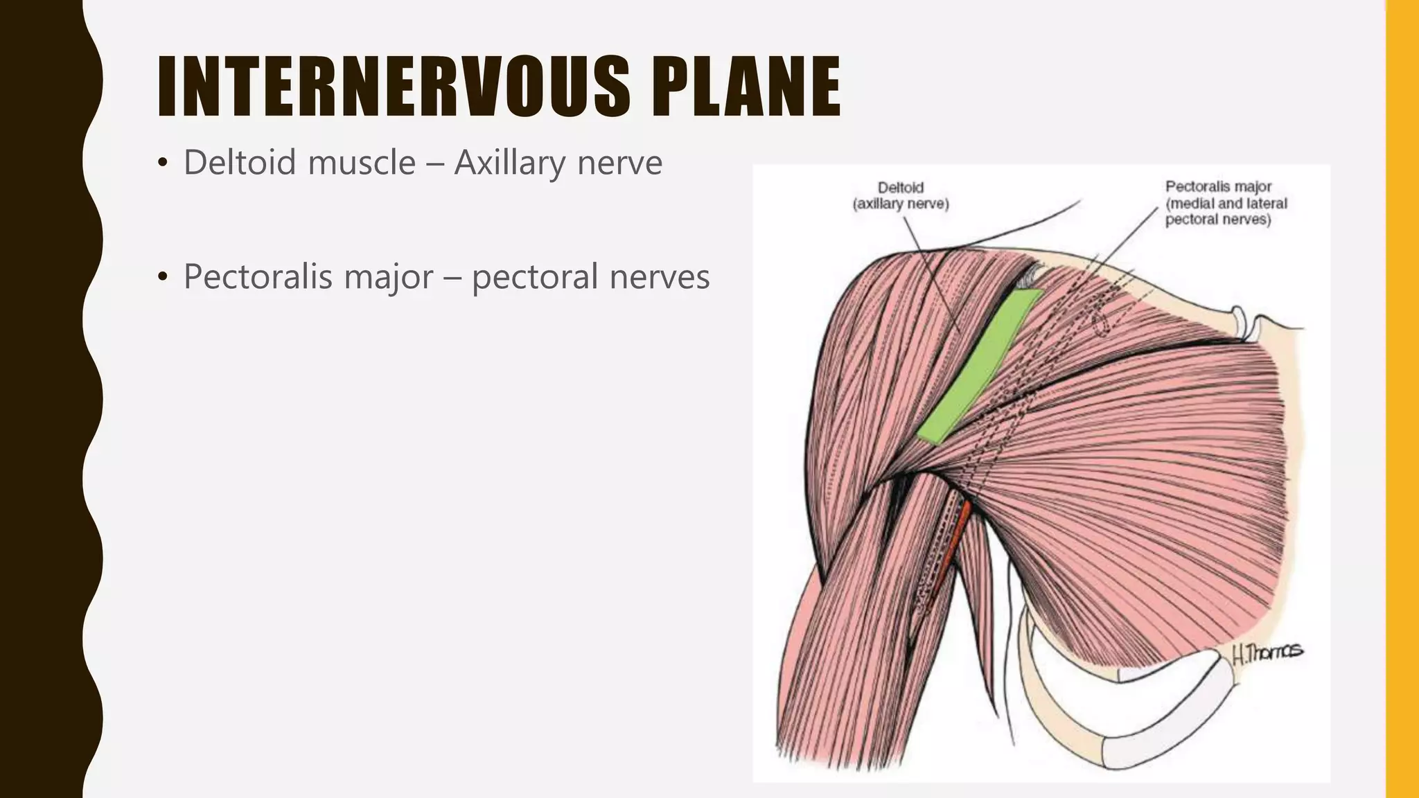
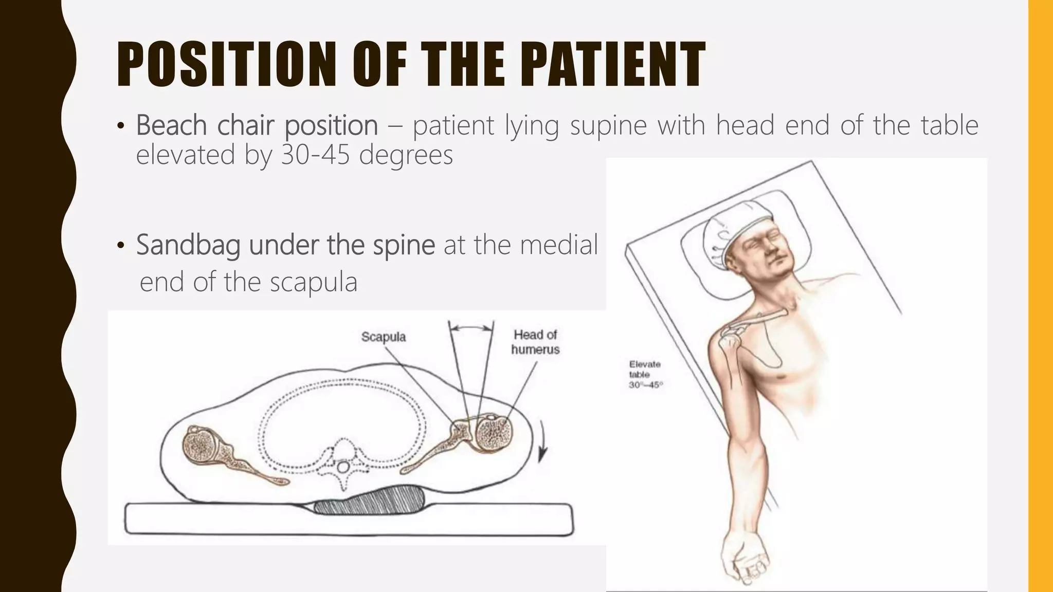
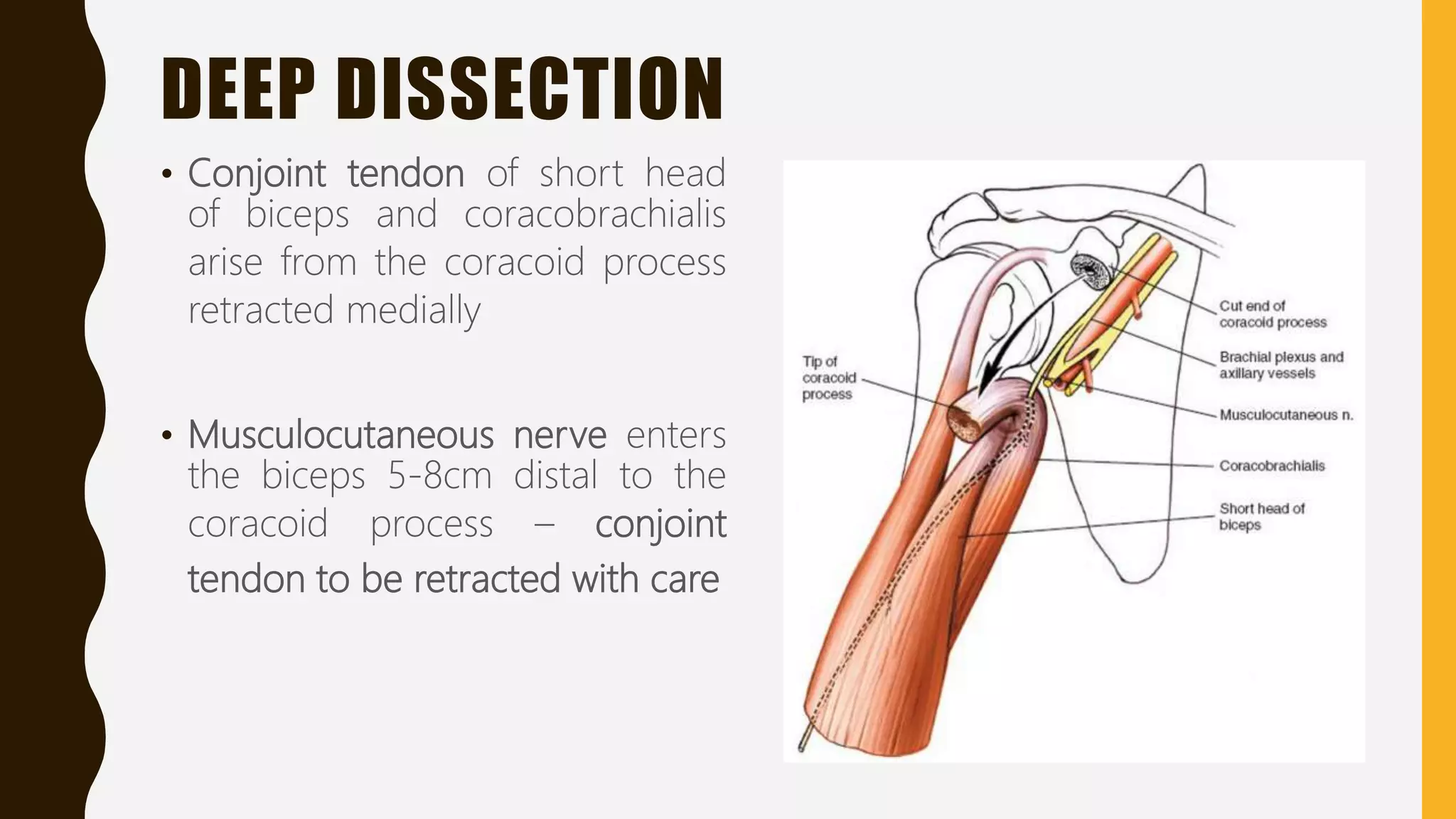
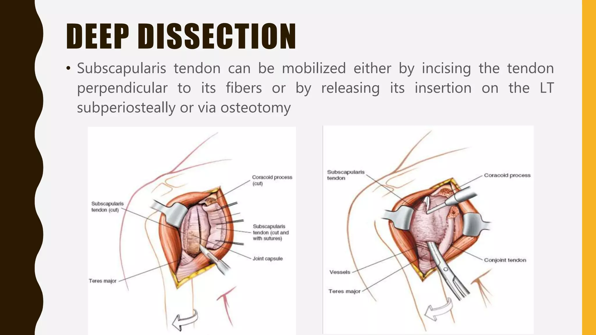
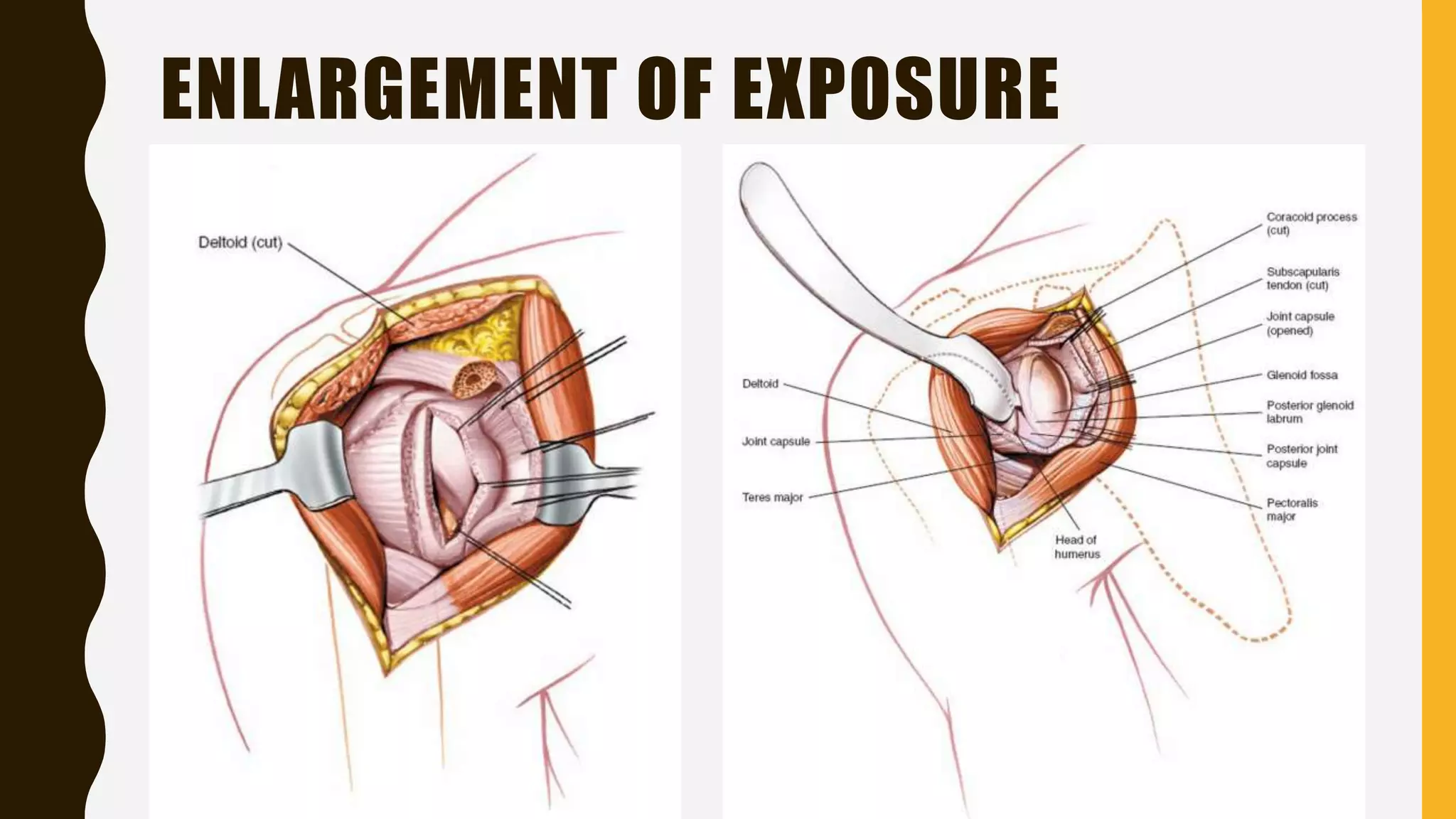
ANTERIOR
APPROACH TO
THE SHOULDER
P R E S E N T E D BY : D R . S A C H I N . M .
2 N D Y E A R P G T, D E P T O F O R T H O PA E D I C S , S M C H
M O D E R ATO R : D R . A . K . S I PA N I
P R O F & H O D, D E P T O F O R T H O PA E D I C S , S M C H
INTRODUCTION
• Also known as Deltopectoral approach
• Fairly extensile exposure – gives access to the anterior, medial and
lateral aspects of the shoulder
• Can extend distally to include the anterior approach to the humerus.
INDICATIONS
• Shoulder arthroplasty
• Proximal humerus fractures
• Reconstruction of recurrent dislocations
• Injury to long head of the biceps – repair
• Septic glenohumeral joint – drainage
• Biopsy and excision of tumerus
INTERNERVOUS PLANE
• Deltoid muscle – Axillary nerve
• Pectoralis major – pectoral nerves
POSITION OF THE PATIENT
• Beach chair position – patient lying supine with head end of the table
elevated by 30-45 degrees
• Sandbag under the spine at the medial
end of the scapula
LANDMARKS
• Coracoid process
– Lies 2.5 cm distal to the point of maximum concavity of the distal clavicle
– Directed anterolaterally and covered by Pectoralis major
• Deltopectoral groove
– Look for cephalic vein which runs in this groove
– Easily seen in thin patients, but difficult in obese individuals
INCISION
• 10 - to 15 cm linear incision is made along the deltopectoral groove
• Begins at tip of coracoid process
SUPERFICIAL DISSECTION
• Superficial skin vessels can
bleed significantly – cauterize
• Deltopectoral fascia is
encountered first
• Cephalic vein is the landmark to
identify the deltopectoral
interval
SUPERFICIAL DISSECTION
• Mobilize the cephalic vein either medially or laterally
• Deltoid fibers are retracted laterally and fibers of pectoralis major are
retracted medially
DEEP DISSECTION
• Conjoint tendon of short head
of biceps and coracobrachialis
arise from the coracoid process
retracted medially
• Musculocutaneous nerve enters
the biceps 5-8cm distal to the
coracoid process – conjoint
tendon to be retracted with care
DEEP DISSECTION
• Fascia lateral to the conjoint tendon is cut to expose the subscapularis
tendon
• External rotation of the shoulder makes the subscapularis tendon taut
and pulls it away from the axillary nerve, which travels through the
quadrangular space
DEEP DISSECTION
• Subscapularis tendon can be mobilized either by incising the tendon
perpendicular to its fibers or by releasing its insertion on the LT
subperiosteally or via osteotomy
DEEP DISSECTION
• Capsule is incised to gain access into the joint
ENLARGEMENT OF EXPOSURE
• Extend the skin incision proximally along the clavicle and distally along
the deltopectoral groove
• Release deltoid either from its origin from the clavicle or from its
insertion on the humerus
• Partial detachment of pectoralis major tendon from its insertion
• Use of suitable retractors – Bankart skid
• Internal and external rotation of shoulder to expose the different areas
of the joint
ENLARGEMENT OF EXPOSURE
DANGERS
• Musculocutaneous nerve – neurapraxia if conjoint tendon is retracted
vigorously
• Cephalic vein – has to be preserved, works as landmark in case of
reversion surgeries
– Can be ligated if injured
• Axillary nerve – can be injured while incising the subscapularis tendon
– Can be avoided by external rotation of the shoulder
• Anterior circumflex humeral artery – runs anteriorly around the
proximal humerus proximal to pectoralis major tendon
THANK YOU

Anterior approach to the shoulder - Dr. Sachin M

  • 1.
    ANTERIOR APPROACH TO THE SHOULDER PR E S E N T E D BY : D R . S A C H I N . M . 2 N D Y E A R P G T, D E P T O F O R T H O PA E D I C S , S M C H M O D E R ATO R : D R . A . K . S I PA N I P R O F & H O D, D E P T O F O R T H O PA E D I C S , S M C H
  • 2.
    INTRODUCTION • Also knownas Deltopectoral approach • Fairly extensile exposure – gives access to the anterior, medial and lateral aspects of the shoulder • Can extend distally to include the anterior approach to the humerus.
  • 3.
    INDICATIONS • Shoulder arthroplasty •Proximal humerus fractures • Reconstruction of recurrent dislocations • Injury to long head of the biceps – repair • Septic glenohumeral joint – drainage • Biopsy and excision of tumerus
  • 4.
    INTERNERVOUS PLANE • Deltoidmuscle – Axillary nerve • Pectoralis major – pectoral nerves
  • 5.
    POSITION OF THEPATIENT • Beach chair position – patient lying supine with head end of the table elevated by 30-45 degrees • Sandbag under the spine at the medial end of the scapula
  • 6.
    LANDMARKS • Coracoid process –Lies 2.5 cm distal to the point of maximum concavity of the distal clavicle – Directed anterolaterally and covered by Pectoralis major • Deltopectoral groove – Look for cephalic vein which runs in this groove – Easily seen in thin patients, but difficult in obese individuals
  • 7.
    INCISION • 10 -to 15 cm linear incision is made along the deltopectoral groove • Begins at tip of coracoid process
  • 8.
    SUPERFICIAL DISSECTION • Superficialskin vessels can bleed significantly – cauterize • Deltopectoral fascia is encountered first • Cephalic vein is the landmark to identify the deltopectoral interval
  • 9.
    SUPERFICIAL DISSECTION • Mobilizethe cephalic vein either medially or laterally • Deltoid fibers are retracted laterally and fibers of pectoralis major are retracted medially
  • 10.
    DEEP DISSECTION • Conjointtendon of short head of biceps and coracobrachialis arise from the coracoid process retracted medially • Musculocutaneous nerve enters the biceps 5-8cm distal to the coracoid process – conjoint tendon to be retracted with care
  • 11.
    DEEP DISSECTION • Fascialateral to the conjoint tendon is cut to expose the subscapularis tendon • External rotation of the shoulder makes the subscapularis tendon taut and pulls it away from the axillary nerve, which travels through the quadrangular space
  • 12.
    DEEP DISSECTION • Subscapularistendon can be mobilized either by incising the tendon perpendicular to its fibers or by releasing its insertion on the LT subperiosteally or via osteotomy
  • 13.
    DEEP DISSECTION • Capsuleis incised to gain access into the joint
  • 14.
    ENLARGEMENT OF EXPOSURE •Extend the skin incision proximally along the clavicle and distally along the deltopectoral groove • Release deltoid either from its origin from the clavicle or from its insertion on the humerus • Partial detachment of pectoralis major tendon from its insertion • Use of suitable retractors – Bankart skid • Internal and external rotation of shoulder to expose the different areas of the joint
  • 15.
  • 16.
    DANGERS • Musculocutaneous nerve– neurapraxia if conjoint tendon is retracted vigorously • Cephalic vein – has to be preserved, works as landmark in case of reversion surgeries – Can be ligated if injured • Axillary nerve – can be injured while incising the subscapularis tendon – Can be avoided by external rotation of the shoulder • Anterior circumflex humeral artery – runs anteriorly around the proximal humerus proximal to pectoralis major tendon
  • 17.