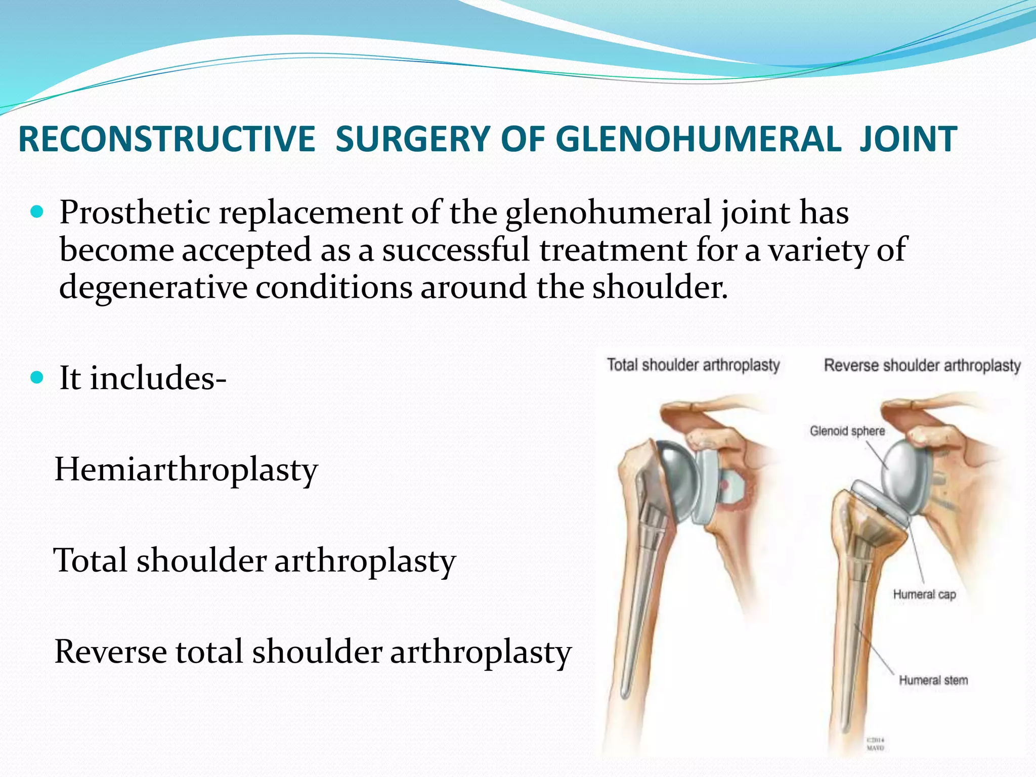
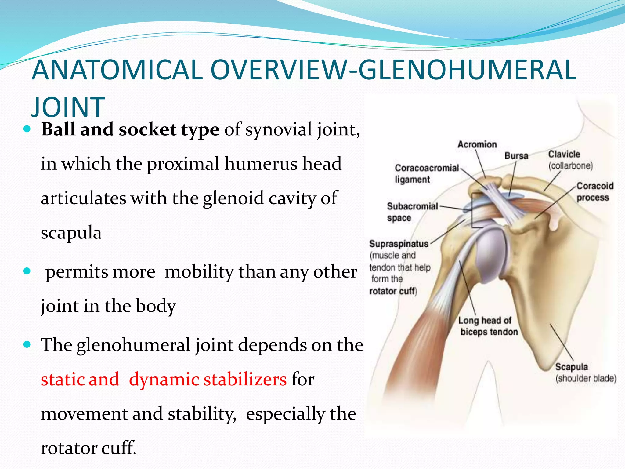
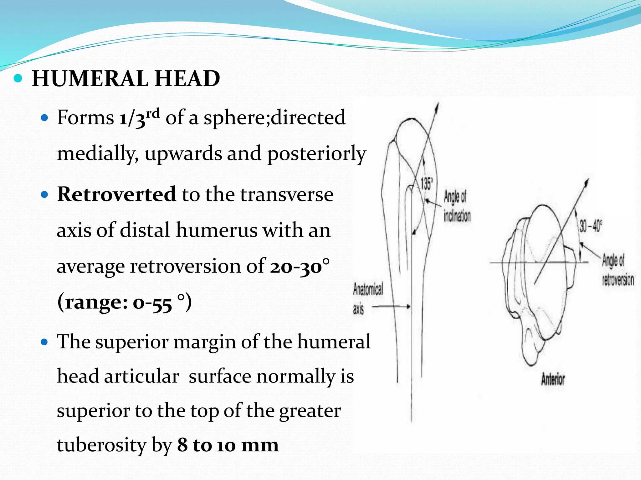
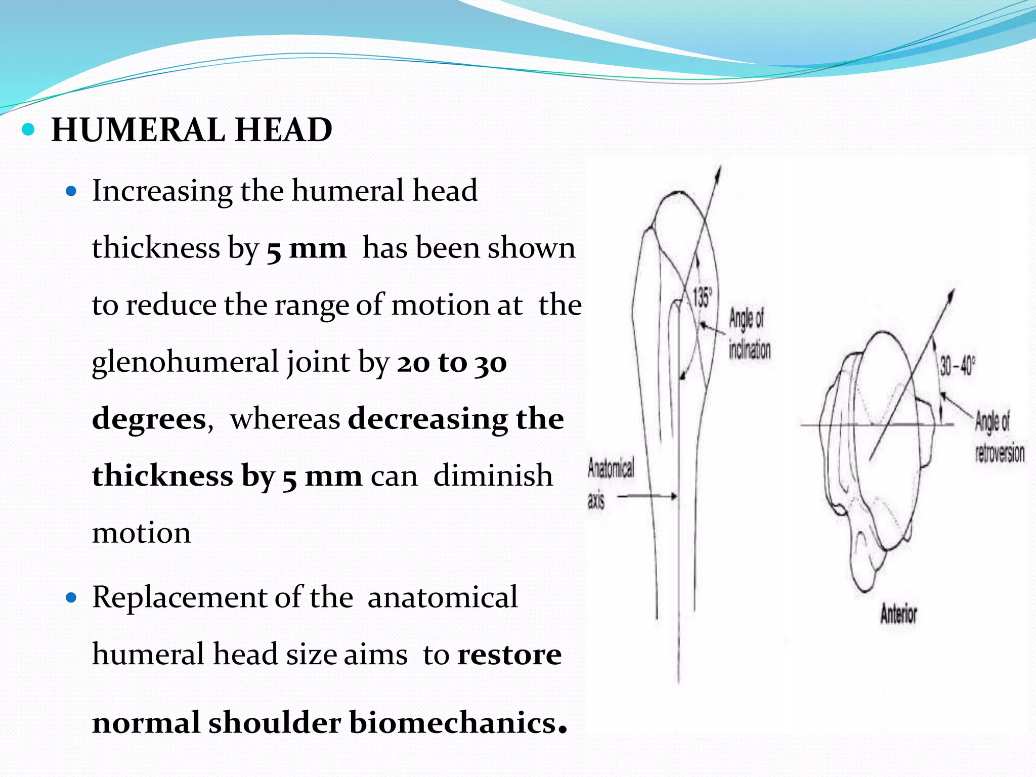
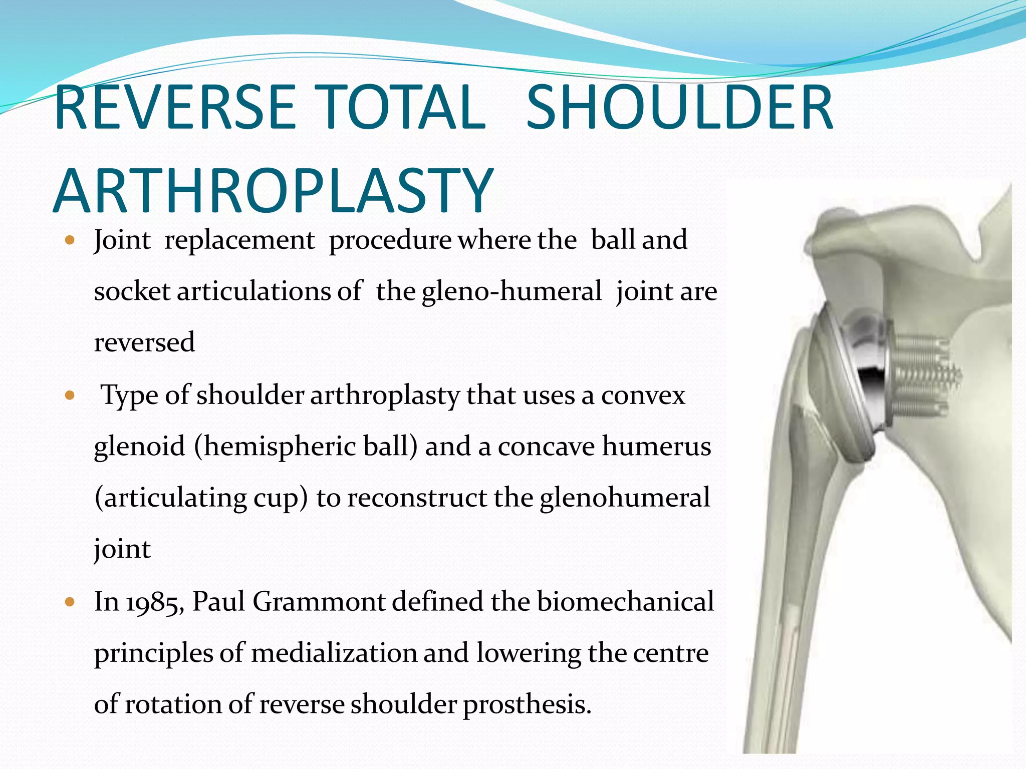
DR. BIPUL BORTHAKUR
PROFESSOR,
DEPTT. OF ORTHOPAEDICS, SMCH
RECONSTRUCTIVE SURGERY OF GLENOHUMERAL JOINT
 Prosthetic replacement of the glenohumeral joint has
become accepted as a successful treatment for a variety of
degenerative conditions around the shoulder.
 It includes-
Hemiarthroplasty
Total shoulder arthroplasty
Reverse total shoulder arthroplasty
 Introduction of reverse total shoulder arthroplasty
 on 1987 (in Europe)
 on 2004 (in the United States)
 Indications have expanded to include several shoulder
conditions, many of which involve dysfunction of rotator
cuff
 Current controversies:
 optimal baseplate positioning
 humeral neck-shaft angle (135○ vs. 155○)
 glenosphere placement (medial, lateral or bony increased
offset RTSA)
 subscapularis repair
ANATOMICAL OVERVIEW-GLENOHUMERAL
JOINT
 Ball and socket type of synovial joint,
in which the proximal humerus head
articulates with the glenoid cavity of
scapula
 permits more mobility than any other
joint in the body
 The glenohumeral joint depends on the
static and dynamic stabilizers for
movement and stability, especially the
rotator cuff.
 HUMERAL HEAD
 Forms 1/3rd of a sphere;directed
medially, upwards and posteriorly
 Retroverted to the transverse
axis of distal humerus with an
average retroversion of 20-30°
(range: 0-55 °)
 The superior margin of the humeral
head articular surface normally is
superior to the top of the greater
tuberosity by 8 to 10 mm
 HUMERAL HEAD
 Increasing the humeral head
thickness by 5 mm has been shown
to reduce the range of motion at the
glenohumeral joint by 20 to 30
degrees, whereas decreasing the
thickness by 5 mm can diminish
motion
 Replacement of the anatomical
humeral head size aims to restore
normal shoulder biomechanics.
 GLENOID CAVITY
 Pear shaped and shallow, 1/4th the size of
head; Represents about a quarter of
humeral surface
 Surface directed outward and
anteriorly
 Normal position of the glenoid surface in
relation to the axis of the scapular body
ranged from 2 ° of anteversion to 7 ° of
retroversion
 GLENOID LABRUM
 Deepens glenoid cavity; Triangular
in cross section
 On superior aspect, tendon of long
head of biceps adheres to labrum
 The labrum passes over the glenoid
notch and forms an osseofibrous
tunnel through which a synovial
extension is created that might
communicate with subscapularis
bursa
 CAPSULE: Fibrous bag of tissue which
provides a sealed sac around gleno-
humeral joint
FACTORS THAT CONTRIBUTE TO MOBILITY OF THE
SHOULDER JOINT
 Type of joint : ball and socket type
 Bony Surfaces: Shallow glenoid cavity and large
Humeral head; there is a 1:4 disproportion in surfaces
 Laxity of the joint capsule
FACTORS THAT CONTRIBUTE TO STABILITY
 Rotator cuff muscles
 Glenoid labrum
 Ligaments
 Biceps tendon
Indications for Shoulder
Arthroplasty
 Osteoarthritis- most
common indication
 Rheumatoid arthritis
 Rotator cuff tear arthropathy
 Humeral head osteonecrosis
 Avascular necrosis
 Instability arthritis
 Post-traumatic arthritis
 Post infectious
 Severe proximal humeral
fractures
Contraindications
 Infection-Local or systemic
 Poor Compliance
 Paralysis with complete loss of rotator cuff and deltoid
function
 Neuropathic arthropathy
 Irreparable rotator cuff tear is a contraindication for
glenoid resurfacing
 Inadequate bonedensity to appropriately support the new
prosthesis
COMPLICATIONS OF SHOULDER
ARTHROPLASTY
Intraoperative Complications Postoperative Complications
 Humeral fracture
 Glenoid fracture
 Vascular injury
 Nerve Injury
 Infection
 Haematoma
 Thromboembolism
 Early instability
 Deltoid muscle dysfunction
Infection
 Prevention
 IV antibiotics before and after
surgery
 Treatment
 Early revision with component
exchange and debridement
Haematoma
 Prevention
 Haemostasis
 Drain
 Management
 Early Evacuation Surgery
Thrombo-embolism
Deep venous thrombosis
 for shoulder arthroplasties-0.5%
 for hip arthroplasties- 1.5%
 for knee arthroplasties- 2.7%
Pulmonary embolism
 for shoulder arthroplasties- 0.2%
 for hip arthroplasties-0.4%
 for knee arthroplasties-0.4%
 Increasing age, trauma, and cancer were risk factors after shoulder
arthroplasties
Early Instability
Component malposition
 Humeral- version, size
 Glenoid- version, size, Offset
Soft tissue
 Posterior Capsular Laxity
 Subscapularis failure
Management
 Address Bone Loss
 Component revision
 soft tissue Balancing
 Reverse geometry arthroplasty
REVERSE TOTAL SHOULDER
ARTHROPLASTY
 Joint replacement procedure where the ball and
socket articulations of the gleno-humeral joint are
reversed
 Type of shoulder arthroplasty that uses a convex
glenoid (hemispheric ball) and a concave humerus
(articulating cup) to reconstruct the glenohumeral
joint
 In 1985, Paul Grammont defined the biomechanical
principles of medialization and lowering the centre
of rotation of reverse shoulder prosthesis.
BIOMECHANICS
The RP design of Grammont combines
 A large hemispherical glenoid component
(36 or 42 mm diameter) that has a
medialized center of rotation and secure
screw fixation to the glenoid
 A cemented humeral component that has a
more horizontally aligned (155°)
metaphyseal neck
 A reverse hemispherical polyethylene cup
that perfectly conforms to the glenosphere
 This design allows for more range
of motion than reverse ball and
socket designs of the past
 With proper placement of this
prosthesis, the center of humeral
rotation is made more distal,
placing the deltoid muscle at
greater tension, and medialized,
which recruits more deltoid muscle
fibers for elevation and increases
the deltoid lever arm.
 These factors increase the deltoid’s
biomechanical ability to generate
rotational torque, elevating the
arm on a new stable fulcrum (the
glenosphere).
 The medialized center of rotation
decreases torsional forces on
glenosphere fixation, decreasing
the tendency of the glenoid
component to loosen.
INDICATIONS
The common characteristic of these indications is-
biomechanical fulcrum for elevation is lost.
 Primary osteoarthritis of the shoulder
 Cuff tear arthropathy
 massive irreparable cuff tear with pseudoparalysis
 Secondary osteoarthritis or osteonecrosis who have a
‘‘decompensated’’ massive cuff deficiency/ tear (‘‘Cuff
Tear Arthritis,’’ CTA)
 Failed shoulder arthroplasty
 Absence of tuberosities ( failed hemiarthroplasty for
fracture/ nonunion)
 Absence of cuff ( failed hemiarthroplasty for cuff-tear
arthroplasty)
 Instability
 Severe Proximal humeral fracture sequelae
 Proximal humeral malunion or non union
 Reimplantation for deep periprosthetic infection
 Reconstruction after tumor removal
CONTRAINDICATIONS
 Articular surface–tuberosity relationships are normal
and the rotator cuff is intact and functional.
 Deltoid loss/ inactivity
 Excessive glenoid bone loss that would not allow
secure implantation of glenoid component
RELATIVE CONTRAINDICATIONS
 Previous infection
 Age < 65 years
 Surgeon inexperience
PROSTHETIC COMPONENTS
HUMERAL COMPONENTS
1.Metaphysis: Upper cup-shaped part of
the prosthetic humeral stem along with
metaphyseal offset extension( 9 mm)
2. Diaphysis: Lower portion of the
humeral stem
3. Polyethylene Cup/ inserts: Becomes
the socket of the new ball & socketjoint.
They are available in increasing diameter
of 6,9 and 12 mm
PROSTHETIC COMPONENTSGLENOID COMPONENTS
1. Metaglene/ base plate: Specially
coated circularmetal plate that is
attached to the glenoid fossa with
2compression and 2 lockedscrews.
The superiorand inferior holes in
the baseplate are threaded and
angled 20 superiorly and inferiorly,
respectively. The anterior and
posterior screw holes are not
threaded.
PROSTHETIC COMPONENTS
GLENOID COMPONENTS
2.Glenosphere: Half globe
cobalt-chrome piece that is
attached to the metaglene via
a Morse taper and
countersunk set screw placing
the center of rotation at the
level of the glenoid/baseplate
interface.
PREOPERATIVE IMAGING
 XRAY
 True AP with arm in neutral rotation(Grashey view)
 Axillary lateral
 Scapular lateral
PREOPERATIVE IMAGING
 CT SCAN of the shoulder including the entire scapula with 2 mm
cuts so that fatty atrophy of the cuff can be examined and accurate 2-
dimensional coronal reconstructions can be obtained.
 MRI of shoulder joint
SURGICAL PROCEDURE
 The most common surgical approach is known as
the 'deltopectoral approach’
 Incision- made just superior and medial to
coracoid process, extended between the deltoid and
pectoralis major
 Subscapularis is tenotomized 1 cm from its
insertion. Release of the subscapularis continues
inferiorly, releasing the inferior muscular fibers and
inferior capsule from the humerus while an
assistant provides gentle progressive external
rotation to the arm. The superior 1 cm of the
pectoralis insertion is also occasionally released to
assist in ease of exposure
Disadvantages:
Some have reported higher ratesof
dislocation
 The cephalic vein is retracted laterally with the
deltoid and the deltopectoral interval fully
dissected from the clavicle to the insertion of
the pectoralis major on the proximal humerus
 The conjoint tendon is gently retracted
medially
 The interval between the cuff and acromion
dissected sharply or with electrocautery
 The posterior cuff and tuberosity is freed from
the overlying deltoid sharply or with
electrocautery, taking care not to injure the
axillary nerve inferiorly.
HUMERAL PREPARATION
 The humerus is readily dislocated anteriorly
and superiorly for humeral preparation
 The highest, most lateral point on the
humeral head is identified as a reference
point
 A hole is created with an awl; the head
cutting guide is inserted into the humeral
shaft via this hole, and retroversion is
determined using a pin set at the desired
degree between 0 and 20° of retroversion.
 A minimal head cut is made parallel to
the undersurface of the neck cutting
guide
 The guide is removed and the cut
completed with either the saw or an
osteotome
 Then the metaphyseal ‘‘cheese grater’’
reamer is used to remove remaining
cancellous bone from the proximal
humerus to allow metaphyseal
component placement
 Distal metaphyseal reaming is then
performed with a long conical reamer,
inserting the reamer so that the height
landmark is level with the highest part of
the humeral cut
 Diaphyseal reaming is then performed
sequentially
 Preparing the canal more aggressively risks
fracturing the humerus as well as not
having an adequate cement mantle.
 The humeral trial is then assembled and
retroversion rod set to 0 to 20°
GLENOID PREPARATION
 A circumferential capsulotomy and labral excision are
performed.
 Origin of the long head of the triceps is released from
the inferior glenoid
 Glenoid osteophytes are removed to reveal the true
glenoid anatomic shape and more correctly identify the
base of glenoid bone that is most solid for baseplate
(metaglene) placement
 The central hole guide is assembled and placed with the
handle inferior for the deltopectoral approach
 The inferior most edge of the guide is placed
against the inferior most edge of the glenoid. This
ensures that the metaglene is placed against the
glenoid as inferior as possible.
 Based on preoperative CT scanning the guide is
angled inferiorly to create an inferior tilt to the
reamed surface of 0–15°
 The central hole is then drilled with a 6 mm bit,
which has a self-stop.
 The guide is then removed, followed by placing
the flat glenoid reamer
 Once a uniformly flat surface has been
created, a 1–2 mm groove is created
circumferentially, to allow placement
of the baseplate flush against the
bone.
 The reamer is removed and the
glenoid central hole drill guide placed,
followed by over-drilling of the central
hole with a 7.5 mm drill with a self-
stop
 The cancellous bone graft is impacted
in the post hole
 The baseplate inserter is
assembled and 8.0 mm baseplate
central post is impacted into 7.5
mm drilled hole for a press-fit
into the scapula.
 There is an ‘‘up’’ and ‘‘down’’
marker on the inserter with a
vertical line that allows
orientation of the baseplate so
that inferior drill hole is aligned
with inferior pillar of the scapular
neck.
 The positioning of the superior
and inferior locking screws is
most critical.
 The baseplate should be very well
fixed to the bone. This requires at
least 3 of the 4 screws having good
purchase.
 The glenosphere of choice (36 or
42 mm) is then guided to the
metaglene using a screwdriver
 The glenosphere is firmly seated onto
the Morse taper of the metaglene
 Final glenosphere fixation is secured by
tightening the center set screw, which
threads into a mating thread on the
inside of the center peg of the
metaglene
 Care is taken throughout these steps to
ensure that soft tissue is completely
cleared from the periphery of the
glenoid
GLENOHUMERAL TRIALING
 A trial reduction is performed. The humeral
component should rotate nicely without signs of
instability.
 Gentle traction is applied to the arm to perform a
‘‘shuck’’ maneuver. There should be less than 1 mm of
diastasis between the humeral cup and glenosphere
during this maneuver to ensure proper stability
 If the shoulder cannot be reduced with a 6 mm
insert, then the trial is removed, adhesions lysed, and
additional proximal humeral bone resected if
necessary.
HUMERAL COMPONENT IMPLANTATION
 Once it is determined that the shoulder will
reduce, the proximal humeral component is
cemented. A cement restrictor is inserted and
the humeral shaft meticulously dried.
 Press fit placement of the humeral component
is not recommended at this time
 After the humeral component is well fixed with
cement, the trial insert is placed into the
metaphysis and the shoulder reduced. If the
tension is adequate, the shoulder is
redislocated and the final polyethylene liner
impacted into the metaphyseal cup
FINAL REDUCTION
 After the shoulder is reduced with final implants in
position, the subscapularis is repaired via 3 to 4 tendon to
tendon sutures, occasionally augmenting the repair with
transosseous sutures if the remaining tendon tag on the
humerus is insufficient.
 The deltopectoral interval is closed over a drain, with a
running absorbable suture.
 The skin is closed in routine fashion.
COMPLICATIONS
 Scapular Notching-most
frequentcomplication
 Instability
 Dislocation
 Acromial stress fractures,
scapular spine fractures
 Infection
 Glenoid loosening
 Humeral loosening
 Peri prosthetic Humerus
fractures
 Neuropraxia
 Heterotopic ossification
SCAPULAR NOTCHING
 Erosion of the inferiorscapular neck
related to impingement by the medial
rim of the polyethylene humeral cup
 10-96% of reverse shoulder
arthroplasties
NEROT CLASSIFICATION
 Grade I: confined to the scapular pillar
 Grade II: notch outline contacts lower
screw
 Grade III: notch over the lower screw
 Grade IV: notch extends to base plate
 Inferior placement of the baseplate with
inferior tilt are the two most important
factors to decrease notching
TECHNIQUES TO DECREASE
NOTCHING
 Placement of the baseplate in an inferior position
with inferior tilt of about 10-15°
 Increase of glenosphere size and lateral offset
 Placement of initial drill hole 11.5 mm superior to
the inferior glenoid rim to optimise baseplate
positioning
 Use of a more varus stem with a neck-shaft angle of
135° rather than 155° and a lateralised glenosphere
 Longer scapular neck
Instability
 It occurs primarily in extension
and adduction
 approximately 5%
 Irreparable subscapularis
tendon has been reported to
correlate with a higher
dislocation rate
Acromial stress fractures
 Acromial stress fractures are
thought to be caused by increased
stress placed on the posterior
aspect of the acromion
 by the configuration of the
prosthesis and resultant increased
deltoid tension.
 Scapular spine fractures are more
common in osteoporotic patients
and are associated with baseplate
relying on center screw for fixation
POST OPERATIVE REHABILITATION
PROTOCOL
 POD1 to 6 weeks— Active Assisted/Passive ROM only
 Forward elevation—in the plane of the scapula as tolerated, up to
90 degrees
 Internal rotation, with upper arm at side, to chest
 External rotation, with upper arm at side, 0-20 degrees
 Pendulum exercises five times per day
 Active Assisted → Active ROM for elbow, wrist, and hand
POST OPERATIVE REHABILITATION
PROTOCOL
 6-12 weeks—continue Active Assisted/Passive ROM
 Forward elevation to full
 External rotation to 30 degrees
 Wand and overhead pulley
 Isometric strengthening for flexion, extension, external
rotation, and abduction in neutral position only
POST OPERATIVE REHABILITATION
PROTOCOL
 At 12 weeks— start Active ROM/dynamic
strengthening
 Continue Active ROM, stretches, and TheraBand
strengthening
 Progress strengthening
 Progress to home program
“satyaṃ ca yena nirataṃ rogaṃ vidhūtaṃ, anveṣitaṃ ca
savidhiṃ ārogyamasya
gūḍhaṃ nigūḍhaṃ auṣadhyarūpam, dhanvantariṃ ca
satataṃ praṇamāmi nityaṃ”
“One who wiped out all the diseases, truly and constantly,
One who discovered the guidelines for (good) health, One
who uncovered the hidden nature of medicinal plants, I,
forever, bow to the God Dhanwantari.”

Seminar recent advances reverse shoulder arthroplasty

  • 1.
  • 2.
    RECONSTRUCTIVE SURGERY OFGLENOHUMERAL JOINT  Prosthetic replacement of the glenohumeral joint has become accepted as a successful treatment for a variety of degenerative conditions around the shoulder.  It includes- Hemiarthroplasty Total shoulder arthroplasty Reverse total shoulder arthroplasty
  • 3.
     Introduction ofreverse total shoulder arthroplasty  on 1987 (in Europe)  on 2004 (in the United States)  Indications have expanded to include several shoulder conditions, many of which involve dysfunction of rotator cuff  Current controversies:  optimal baseplate positioning  humeral neck-shaft angle (135○ vs. 155○)  glenosphere placement (medial, lateral or bony increased offset RTSA)  subscapularis repair
  • 4.
    ANATOMICAL OVERVIEW-GLENOHUMERAL JOINT  Balland socket type of synovial joint, in which the proximal humerus head articulates with the glenoid cavity of scapula  permits more mobility than any other joint in the body  The glenohumeral joint depends on the static and dynamic stabilizers for movement and stability, especially the rotator cuff.
  • 5.
     HUMERAL HEAD Forms 1/3rd of a sphere;directed medially, upwards and posteriorly  Retroverted to the transverse axis of distal humerus with an average retroversion of 20-30° (range: 0-55 °)  The superior margin of the humeral head articular surface normally is superior to the top of the greater tuberosity by 8 to 10 mm
  • 6.
     HUMERAL HEAD Increasing the humeral head thickness by 5 mm has been shown to reduce the range of motion at the glenohumeral joint by 20 to 30 degrees, whereas decreasing the thickness by 5 mm can diminish motion  Replacement of the anatomical humeral head size aims to restore normal shoulder biomechanics.
  • 7.
     GLENOID CAVITY Pear shaped and shallow, 1/4th the size of head; Represents about a quarter of humeral surface  Surface directed outward and anteriorly  Normal position of the glenoid surface in relation to the axis of the scapular body ranged from 2 ° of anteversion to 7 ° of retroversion
  • 8.
     GLENOID LABRUM Deepens glenoid cavity; Triangular in cross section  On superior aspect, tendon of long head of biceps adheres to labrum  The labrum passes over the glenoid notch and forms an osseofibrous tunnel through which a synovial extension is created that might communicate with subscapularis bursa  CAPSULE: Fibrous bag of tissue which provides a sealed sac around gleno- humeral joint
  • 9.
    FACTORS THAT CONTRIBUTETO MOBILITY OF THE SHOULDER JOINT  Type of joint : ball and socket type  Bony Surfaces: Shallow glenoid cavity and large Humeral head; there is a 1:4 disproportion in surfaces  Laxity of the joint capsule FACTORS THAT CONTRIBUTE TO STABILITY  Rotator cuff muscles  Glenoid labrum  Ligaments  Biceps tendon
  • 10.
    Indications for Shoulder Arthroplasty Osteoarthritis- most common indication  Rheumatoid arthritis  Rotator cuff tear arthropathy  Humeral head osteonecrosis  Avascular necrosis  Instability arthritis  Post-traumatic arthritis  Post infectious  Severe proximal humeral fractures
  • 11.
    Contraindications  Infection-Local orsystemic  Poor Compliance  Paralysis with complete loss of rotator cuff and deltoid function  Neuropathic arthropathy  Irreparable rotator cuff tear is a contraindication for glenoid resurfacing  Inadequate bonedensity to appropriately support the new prosthesis
  • 12.
    COMPLICATIONS OF SHOULDER ARTHROPLASTY IntraoperativeComplications Postoperative Complications  Humeral fracture  Glenoid fracture  Vascular injury  Nerve Injury  Infection  Haematoma  Thromboembolism  Early instability  Deltoid muscle dysfunction
  • 13.
    Infection  Prevention  IVantibiotics before and after surgery  Treatment  Early revision with component exchange and debridement
  • 14.
    Haematoma  Prevention  Haemostasis Drain  Management  Early Evacuation Surgery
  • 15.
    Thrombo-embolism Deep venous thrombosis for shoulder arthroplasties-0.5%  for hip arthroplasties- 1.5%  for knee arthroplasties- 2.7% Pulmonary embolism  for shoulder arthroplasties- 0.2%  for hip arthroplasties-0.4%  for knee arthroplasties-0.4%  Increasing age, trauma, and cancer were risk factors after shoulder arthroplasties
  • 16.
    Early Instability Component malposition Humeral- version, size  Glenoid- version, size, Offset Soft tissue  Posterior Capsular Laxity  Subscapularis failure Management  Address Bone Loss  Component revision  soft tissue Balancing  Reverse geometry arthroplasty
  • 17.
    REVERSE TOTAL SHOULDER ARTHROPLASTY Joint replacement procedure where the ball and socket articulations of the gleno-humeral joint are reversed  Type of shoulder arthroplasty that uses a convex glenoid (hemispheric ball) and a concave humerus (articulating cup) to reconstruct the glenohumeral joint  In 1985, Paul Grammont defined the biomechanical principles of medialization and lowering the centre of rotation of reverse shoulder prosthesis.
  • 18.
    BIOMECHANICS The RP designof Grammont combines  A large hemispherical glenoid component (36 or 42 mm diameter) that has a medialized center of rotation and secure screw fixation to the glenoid  A cemented humeral component that has a more horizontally aligned (155°) metaphyseal neck  A reverse hemispherical polyethylene cup that perfectly conforms to the glenosphere
  • 19.
     This designallows for more range of motion than reverse ball and socket designs of the past  With proper placement of this prosthesis, the center of humeral rotation is made more distal, placing the deltoid muscle at greater tension, and medialized, which recruits more deltoid muscle fibers for elevation and increases the deltoid lever arm.
  • 20.
     These factorsincrease the deltoid’s biomechanical ability to generate rotational torque, elevating the arm on a new stable fulcrum (the glenosphere).  The medialized center of rotation decreases torsional forces on glenosphere fixation, decreasing the tendency of the glenoid component to loosen.
  • 21.
    INDICATIONS The common characteristicof these indications is- biomechanical fulcrum for elevation is lost.  Primary osteoarthritis of the shoulder  Cuff tear arthropathy  massive irreparable cuff tear with pseudoparalysis  Secondary osteoarthritis or osteonecrosis who have a ‘‘decompensated’’ massive cuff deficiency/ tear (‘‘Cuff Tear Arthritis,’’ CTA)
  • 22.
     Failed shoulderarthroplasty  Absence of tuberosities ( failed hemiarthroplasty for fracture/ nonunion)  Absence of cuff ( failed hemiarthroplasty for cuff-tear arthroplasty)  Instability  Severe Proximal humeral fracture sequelae  Proximal humeral malunion or non union  Reimplantation for deep periprosthetic infection  Reconstruction after tumor removal
  • 23.
    CONTRAINDICATIONS  Articular surface–tuberosityrelationships are normal and the rotator cuff is intact and functional.  Deltoid loss/ inactivity  Excessive glenoid bone loss that would not allow secure implantation of glenoid component RELATIVE CONTRAINDICATIONS  Previous infection  Age < 65 years  Surgeon inexperience
  • 24.
    PROSTHETIC COMPONENTS HUMERAL COMPONENTS 1.Metaphysis:Upper cup-shaped part of the prosthetic humeral stem along with metaphyseal offset extension( 9 mm) 2. Diaphysis: Lower portion of the humeral stem 3. Polyethylene Cup/ inserts: Becomes the socket of the new ball & socketjoint. They are available in increasing diameter of 6,9 and 12 mm
  • 25.
    PROSTHETIC COMPONENTSGLENOID COMPONENTS 1.Metaglene/ base plate: Specially coated circularmetal plate that is attached to the glenoid fossa with 2compression and 2 lockedscrews. The superiorand inferior holes in the baseplate are threaded and angled 20 superiorly and inferiorly, respectively. The anterior and posterior screw holes are not threaded.
  • 26.
    PROSTHETIC COMPONENTS GLENOID COMPONENTS 2.Glenosphere:Half globe cobalt-chrome piece that is attached to the metaglene via a Morse taper and countersunk set screw placing the center of rotation at the level of the glenoid/baseplate interface.
  • 27.
    PREOPERATIVE IMAGING  XRAY True AP with arm in neutral rotation(Grashey view)  Axillary lateral  Scapular lateral
  • 28.
    PREOPERATIVE IMAGING  CTSCAN of the shoulder including the entire scapula with 2 mm cuts so that fatty atrophy of the cuff can be examined and accurate 2- dimensional coronal reconstructions can be obtained.
  • 29.
     MRI ofshoulder joint
  • 30.
    SURGICAL PROCEDURE  Themost common surgical approach is known as the 'deltopectoral approach’  Incision- made just superior and medial to coracoid process, extended between the deltoid and pectoralis major  Subscapularis is tenotomized 1 cm from its insertion. Release of the subscapularis continues inferiorly, releasing the inferior muscular fibers and inferior capsule from the humerus while an assistant provides gentle progressive external rotation to the arm. The superior 1 cm of the pectoralis insertion is also occasionally released to assist in ease of exposure
  • 31.
    Disadvantages: Some have reportedhigher ratesof dislocation  The cephalic vein is retracted laterally with the deltoid and the deltopectoral interval fully dissected from the clavicle to the insertion of the pectoralis major on the proximal humerus  The conjoint tendon is gently retracted medially  The interval between the cuff and acromion dissected sharply or with electrocautery  The posterior cuff and tuberosity is freed from the overlying deltoid sharply or with electrocautery, taking care not to injure the axillary nerve inferiorly.
  • 32.
    HUMERAL PREPARATION  Thehumerus is readily dislocated anteriorly and superiorly for humeral preparation  The highest, most lateral point on the humeral head is identified as a reference point  A hole is created with an awl; the head cutting guide is inserted into the humeral shaft via this hole, and retroversion is determined using a pin set at the desired degree between 0 and 20° of retroversion.
  • 33.
     A minimalhead cut is made parallel to the undersurface of the neck cutting guide  The guide is removed and the cut completed with either the saw or an osteotome  Then the metaphyseal ‘‘cheese grater’’ reamer is used to remove remaining cancellous bone from the proximal humerus to allow metaphyseal component placement
  • 34.
     Distal metaphysealreaming is then performed with a long conical reamer, inserting the reamer so that the height landmark is level with the highest part of the humeral cut  Diaphyseal reaming is then performed sequentially  Preparing the canal more aggressively risks fracturing the humerus as well as not having an adequate cement mantle.  The humeral trial is then assembled and retroversion rod set to 0 to 20°
  • 35.
    GLENOID PREPARATION  Acircumferential capsulotomy and labral excision are performed.  Origin of the long head of the triceps is released from the inferior glenoid  Glenoid osteophytes are removed to reveal the true glenoid anatomic shape and more correctly identify the base of glenoid bone that is most solid for baseplate (metaglene) placement  The central hole guide is assembled and placed with the handle inferior for the deltopectoral approach
  • 36.
     The inferiormost edge of the guide is placed against the inferior most edge of the glenoid. This ensures that the metaglene is placed against the glenoid as inferior as possible.  Based on preoperative CT scanning the guide is angled inferiorly to create an inferior tilt to the reamed surface of 0–15°  The central hole is then drilled with a 6 mm bit, which has a self-stop.  The guide is then removed, followed by placing the flat glenoid reamer
  • 37.
     Once auniformly flat surface has been created, a 1–2 mm groove is created circumferentially, to allow placement of the baseplate flush against the bone.  The reamer is removed and the glenoid central hole drill guide placed, followed by over-drilling of the central hole with a 7.5 mm drill with a self- stop  The cancellous bone graft is impacted in the post hole
  • 38.
     The baseplateinserter is assembled and 8.0 mm baseplate central post is impacted into 7.5 mm drilled hole for a press-fit into the scapula.  There is an ‘‘up’’ and ‘‘down’’ marker on the inserter with a vertical line that allows orientation of the baseplate so that inferior drill hole is aligned with inferior pillar of the scapular neck.
  • 39.
     The positioningof the superior and inferior locking screws is most critical.  The baseplate should be very well fixed to the bone. This requires at least 3 of the 4 screws having good purchase.  The glenosphere of choice (36 or 42 mm) is then guided to the metaglene using a screwdriver
  • 40.
     The glenosphereis firmly seated onto the Morse taper of the metaglene  Final glenosphere fixation is secured by tightening the center set screw, which threads into a mating thread on the inside of the center peg of the metaglene  Care is taken throughout these steps to ensure that soft tissue is completely cleared from the periphery of the glenoid
  • 41.
    GLENOHUMERAL TRIALING  Atrial reduction is performed. The humeral component should rotate nicely without signs of instability.  Gentle traction is applied to the arm to perform a ‘‘shuck’’ maneuver. There should be less than 1 mm of diastasis between the humeral cup and glenosphere during this maneuver to ensure proper stability  If the shoulder cannot be reduced with a 6 mm insert, then the trial is removed, adhesions lysed, and additional proximal humeral bone resected if necessary.
  • 42.
    HUMERAL COMPONENT IMPLANTATION Once it is determined that the shoulder will reduce, the proximal humeral component is cemented. A cement restrictor is inserted and the humeral shaft meticulously dried.  Press fit placement of the humeral component is not recommended at this time  After the humeral component is well fixed with cement, the trial insert is placed into the metaphysis and the shoulder reduced. If the tension is adequate, the shoulder is redislocated and the final polyethylene liner impacted into the metaphyseal cup
  • 43.
    FINAL REDUCTION  Afterthe shoulder is reduced with final implants in position, the subscapularis is repaired via 3 to 4 tendon to tendon sutures, occasionally augmenting the repair with transosseous sutures if the remaining tendon tag on the humerus is insufficient.  The deltopectoral interval is closed over a drain, with a running absorbable suture.  The skin is closed in routine fashion.
  • 44.
    COMPLICATIONS  Scapular Notching-most frequentcomplication Instability  Dislocation  Acromial stress fractures, scapular spine fractures  Infection  Glenoid loosening  Humeral loosening  Peri prosthetic Humerus fractures  Neuropraxia  Heterotopic ossification
  • 45.
    SCAPULAR NOTCHING  Erosionof the inferiorscapular neck related to impingement by the medial rim of the polyethylene humeral cup  10-96% of reverse shoulder arthroplasties
  • 46.
    NEROT CLASSIFICATION  GradeI: confined to the scapular pillar  Grade II: notch outline contacts lower screw  Grade III: notch over the lower screw  Grade IV: notch extends to base plate  Inferior placement of the baseplate with inferior tilt are the two most important factors to decrease notching
  • 47.
    TECHNIQUES TO DECREASE NOTCHING Placement of the baseplate in an inferior position with inferior tilt of about 10-15°  Increase of glenosphere size and lateral offset  Placement of initial drill hole 11.5 mm superior to the inferior glenoid rim to optimise baseplate positioning  Use of a more varus stem with a neck-shaft angle of 135° rather than 155° and a lateralised glenosphere  Longer scapular neck
  • 48.
    Instability  It occursprimarily in extension and adduction  approximately 5%  Irreparable subscapularis tendon has been reported to correlate with a higher dislocation rate
  • 49.
    Acromial stress fractures Acromial stress fractures are thought to be caused by increased stress placed on the posterior aspect of the acromion  by the configuration of the prosthesis and resultant increased deltoid tension.  Scapular spine fractures are more common in osteoporotic patients and are associated with baseplate relying on center screw for fixation
  • 50.
    POST OPERATIVE REHABILITATION PROTOCOL POD1 to 6 weeks— Active Assisted/Passive ROM only  Forward elevation—in the plane of the scapula as tolerated, up to 90 degrees  Internal rotation, with upper arm at side, to chest  External rotation, with upper arm at side, 0-20 degrees  Pendulum exercises five times per day  Active Assisted → Active ROM for elbow, wrist, and hand
  • 51.
    POST OPERATIVE REHABILITATION PROTOCOL 6-12 weeks—continue Active Assisted/Passive ROM  Forward elevation to full  External rotation to 30 degrees  Wand and overhead pulley  Isometric strengthening for flexion, extension, external rotation, and abduction in neutral position only
  • 52.
    POST OPERATIVE REHABILITATION PROTOCOL At 12 weeks— start Active ROM/dynamic strengthening  Continue Active ROM, stretches, and TheraBand strengthening  Progress strengthening  Progress to home program
  • 53.
    “satyaṃ ca yenanirataṃ rogaṃ vidhūtaṃ, anveṣitaṃ ca savidhiṃ ārogyamasya gūḍhaṃ nigūḍhaṃ auṣadhyarūpam, dhanvantariṃ ca satataṃ praṇamāmi nityaṃ” “One who wiped out all the diseases, truly and constantly, One who discovered the guidelines for (good) health, One who uncovered the hidden nature of medicinal plants, I, forever, bow to the God Dhanwantari.”