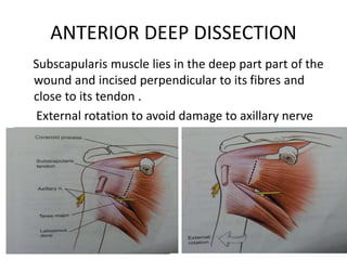
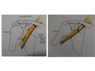
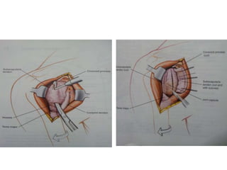
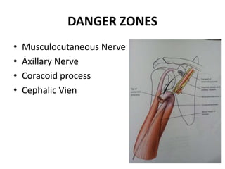
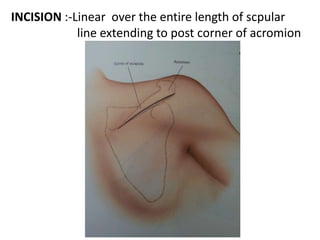
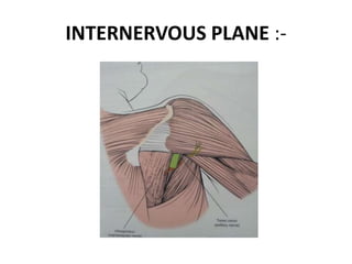
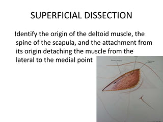
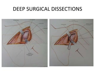
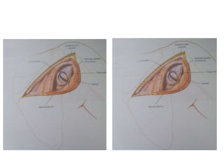
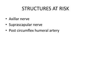
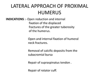
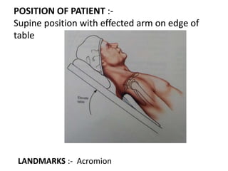
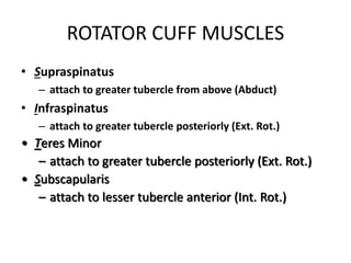
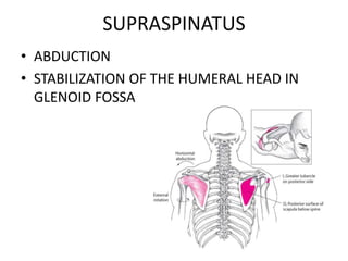
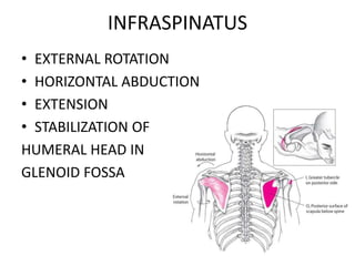
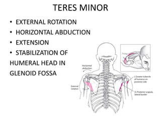
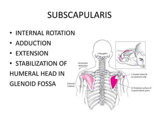
APPROACHES TO SHOULDER JOINT
&
STABILITY OF SHOULDER JOINT
PRESENTATION BY - DR. PRADEEPKUMAR. T
MODRATOR- DR. VINEET THOMAS ABRAHAM
ANTERIOR APPROACH
LATERAL APPROACH
POSTERIOR APPROACH
ANTERIOR APPROACH
INDICATION :-
POSITION OF PATIENT
Coracoid
Process,
Clavicle & Deltopectoral
groove
ANTERIOR INSICION :-10-15cm
straight incision along the D/P groove.
AXILLARY INSICION :-
Supine and abduct the shoulder
to 90 degree.
Vertical insicion 8-10cm
long starting at mid
point of ant axillary fold
extending posteriorly in
to axilla.
TRUE INTERNERVOUS PLANE
Deltoid (axillary) and Pec Major
(pectoral nerve)
ANTERIOR SUPERFICIAL DISSECTION
Develop the groove b/w the fascia overlying
pectoralis Major and deltoid
ANTERIOR DEEP DISSECTION
Subscapularis muscle lies in the deep part part of the
wound and incised perpendicular to its fibres and
close to its tendon .
External rotation to avoid damage to axillary nerve
DANGER ZONES
• Musculocutaneous Nerve
• Axillary Nerve
• Coracoid process
• Cephalic Vien
ADVANTAGES :-
The approach is through an internervous plane
b/w deltoid and pectoralis major Incision can be
expanded proximally or distally
DISADVANTAGES :-
The approach is clearly the best and it
has no significant disadvantages
POSTERIOR APPROACH OF SHOULDER
JOINT
INDICATIONS :-Repairs in cases of recurrent
posterior dislocation or
subluxation of the shoulder
Glenoid osteotomy
Biopsy and excision of tumors
Removal of loose bodies in
Drainage of sepsis
Fractures of the scapula neck,
particularly those in association
with fractured clavicles
Posterior fracture dislocations of
the proximal humerus
POSITION OF THE PATIENT :-
LANDMARKS :- Acromion and spine of scapula
INCISION :-Linear over the entire length of scpular
line extending to post corner of acromion
INTERNERVOUS PLANE :-
SUPERFICIAL DISSECTION
Identify the origin of the deltoid muscle, the
spine of the scapula, and the attachment from
its origin detaching the muscle from the
lateral to the medial point
DEEP SURGICAL DISSECTIONS
STRUCTURES AT RISK
• Axillar nerve
• Suprascapular nerve
• Post circumflex humeral artery
LATERAL APPROACH OF PROXIMAL
HUMERUS
INDICATIONS :- Open reduction and internal
fixation of the displaced
fractures of the greater tuberosity
of the humerus.
Open and internal fixation of humeral
neck fractures.
Removal of calcific deposits from the
subacromial bursa
Repair of supraspinatus tendon .
Repair of rotator cuff.
POSITION OF PATIENT :-
Supine position with effected arm on edge of
table
LANDMARKS :- Acromion
INCISION :-
5cm longitudinal incision from tip of
acromion to lateral aspect of arm
INTERNERVOUS PLANE :- No internervous plane
SUPERFICIAL SURGICAL EXPOSURE
Split the deltoid muscle in the line of fibres
from the acromion downwards for 5cm and
apply a stay sutures at inferior apex.
DEEP SURGICAL EXPOSURES
STRUCTURES AT RISK
AXILLARY NERVE
EXTENSILE MEASURES :-
Proximal Extension -incision superiorly and
medially across the acromion
ROTATOR CUFF MUSCLES
• Supraspinatus
– attach to greater tubercle from above (Abduct)
• Infraspinatus
– attach to greater tubercle posteriorly (Ext. Rot.)
• Teres Minor
– attach to greater tubercle posteriorly (Ext. Rot.)
• Subscapularis
– attach to lesser tubercle anterior (Int. Rot.)
ROTATOR CUFF MUSCLES
• Not very large
• Must possess strength & muscular endurance
• Conducting repetitious overhead activities
(throwing, swimming, & pitching) with poor
technique, muscle fatigue, or inadequate warm-
up & conditioning leads to failure of rotator cuff
muscle group in dynamically stabilizing humeral
head in glenoid cavity
• Leads to further rotator cuff problems such as
tendinitis & rotator cuff impingement within
subacromial space
SUPRASPINATUS
• ABDUCTION
• STABILIZATION OF THE HUMERAL HEAD IN
GLENOID FOSSA
INFRASPINATUS
• EXTERNAL ROTATION
• HORIZONTAL ABDUCTION
• EXTENSION
• STABILIZATION OF
HUMERAL HEAD IN
GLENOID FOSSA
TERES MINOR
• EXTERNAL ROTATION
• HORIZONTAL ABDUCTION
• EXTENSION
• STABILIZATION OF
HUMERAL HEAD IN
GLENOID FOSSA
SUBSCAPULARIS
• INTERNAL ROTATION
• ADDUCTION
• EXTENSION
• STABILIZATION OF
HUMERAL HEAD IN
GLENOID FOSSA
LABRUM
The Labrum deepens the cavity (the glenoid
cavity) and effectively increases the surface of
the shoulder joint.
A ring of fibrocartilage that runs around the
cavity of the scapula (wingbone) in which the
head of the humerus (the bone in the upper
arm) fits
BICEPS TENDON
Many theories have been proposed regarding
the function of the LHB tendon; however, its
exact purpose is poorly understood. It has been
described as an important stabilizer of the
glenohumeral joint, a depressor of the humeral
head, and as a vestigial structure. The LHB
tendon can be a significant source of pain in
patients with rotator cuff tears.
CONCAVITY COMPRESSION THEORY
A compressive load of 50 N was applied to the
humeral head in a direction perpendicular to the
glenoid surface.
Increasing tangential forces were then applied
until the head dislocated over the glenoid lip.
The tangential force at dislocation was examined
for eight different directions, 45° apart around
the glenoid.
Concavity-compression stability was then
examined for an increased compressive load of
100 N.
CONCAVITY COMPRESSION THEORY
Finally, the protocol with 50 and 100 N of
compressive load was repeated after the glenoid
labrum was excised.
Concavity-compression of the
humeral head into the glenoid is a
most efficient stabilizing mechanism.
With the labrum intact the humeral
head resisted tangential forces of up
to 60% of the compressive load.
The degree of compression stabilization varied
around the circumference of the glenoid with the
greatest magnitude superiorly and inferiorly.
This may be attributed to the greater glenoid depth
in these directions.
Resection of the glenoid labrum reduced the
effectiveness of compression stabilization by
approximately 20%
CONCAVITY COMPRESSION THEORY
Concavity-compression may be an important
mechanism for providing stability in the mid-
range of glenohumeral motion where the
capsule and ligaments are lax. The effectiveness
is enhanced by the presence of an intact glenoid
labrum.

Shoulder

  • 1.
    APPROACHES TO SHOULDERJOINT & STABILITY OF SHOULDER JOINT PRESENTATION BY - DR. PRADEEPKUMAR. T MODRATOR- DR. VINEET THOMAS ABRAHAM
  • 2.
  • 3.
  • 4.
  • 5.
    ANTERIOR INSICION :-10-15cm straightincision along the D/P groove.
  • 6.
    AXILLARY INSICION :- Supineand abduct the shoulder to 90 degree. Vertical insicion 8-10cm long starting at mid point of ant axillary fold extending posteriorly in to axilla.
  • 7.
    TRUE INTERNERVOUS PLANE Deltoid(axillary) and Pec Major (pectoral nerve)
  • 8.
    ANTERIOR SUPERFICIAL DISSECTION Developthe groove b/w the fascia overlying pectoralis Major and deltoid
  • 9.
    ANTERIOR DEEP DISSECTION Subscapularismuscle lies in the deep part part of the wound and incised perpendicular to its fibres and close to its tendon . External rotation to avoid damage to axillary nerve
  • 12.
    DANGER ZONES • MusculocutaneousNerve • Axillary Nerve • Coracoid process • Cephalic Vien
  • 13.
    ADVANTAGES :- The approachis through an internervous plane b/w deltoid and pectoralis major Incision can be expanded proximally or distally DISADVANTAGES :- The approach is clearly the best and it has no significant disadvantages
  • 14.
    POSTERIOR APPROACH OFSHOULDER JOINT INDICATIONS :-Repairs in cases of recurrent posterior dislocation or subluxation of the shoulder Glenoid osteotomy Biopsy and excision of tumors Removal of loose bodies in
  • 15.
    Drainage of sepsis Fracturesof the scapula neck, particularly those in association with fractured clavicles Posterior fracture dislocations of the proximal humerus
  • 16.
    POSITION OF THEPATIENT :- LANDMARKS :- Acromion and spine of scapula
  • 17.
    INCISION :-Linear overthe entire length of scpular line extending to post corner of acromion
  • 18.
  • 19.
    SUPERFICIAL DISSECTION Identify theorigin of the deltoid muscle, the spine of the scapula, and the attachment from its origin detaching the muscle from the lateral to the medial point
  • 20.
  • 22.
    STRUCTURES AT RISK •Axillar nerve • Suprascapular nerve • Post circumflex humeral artery
  • 23.
    LATERAL APPROACH OFPROXIMAL HUMERUS INDICATIONS :- Open reduction and internal fixation of the displaced fractures of the greater tuberosity of the humerus. Open and internal fixation of humeral neck fractures. Removal of calcific deposits from the subacromial bursa Repair of supraspinatus tendon . Repair of rotator cuff.
  • 24.
    POSITION OF PATIENT:- Supine position with effected arm on edge of table LANDMARKS :- Acromion
  • 25.
    INCISION :- 5cm longitudinalincision from tip of acromion to lateral aspect of arm INTERNERVOUS PLANE :- No internervous plane
  • 26.
    SUPERFICIAL SURGICAL EXPOSURE Splitthe deltoid muscle in the line of fibres from the acromion downwards for 5cm and apply a stay sutures at inferior apex.
  • 27.
  • 28.
    STRUCTURES AT RISK AXILLARYNERVE EXTENSILE MEASURES :- Proximal Extension -incision superiorly and medially across the acromion
  • 29.
    ROTATOR CUFF MUSCLES •Supraspinatus – attach to greater tubercle from above (Abduct) • Infraspinatus – attach to greater tubercle posteriorly (Ext. Rot.) • Teres Minor – attach to greater tubercle posteriorly (Ext. Rot.) • Subscapularis – attach to lesser tubercle anterior (Int. Rot.)
  • 30.
    ROTATOR CUFF MUSCLES •Not very large • Must possess strength & muscular endurance • Conducting repetitious overhead activities (throwing, swimming, & pitching) with poor technique, muscle fatigue, or inadequate warm- up & conditioning leads to failure of rotator cuff muscle group in dynamically stabilizing humeral head in glenoid cavity • Leads to further rotator cuff problems such as tendinitis & rotator cuff impingement within subacromial space
  • 31.
    SUPRASPINATUS • ABDUCTION • STABILIZATIONOF THE HUMERAL HEAD IN GLENOID FOSSA
  • 32.
    INFRASPINATUS • EXTERNAL ROTATION •HORIZONTAL ABDUCTION • EXTENSION • STABILIZATION OF HUMERAL HEAD IN GLENOID FOSSA
  • 33.
    TERES MINOR • EXTERNALROTATION • HORIZONTAL ABDUCTION • EXTENSION • STABILIZATION OF HUMERAL HEAD IN GLENOID FOSSA
  • 34.
    SUBSCAPULARIS • INTERNAL ROTATION •ADDUCTION • EXTENSION • STABILIZATION OF HUMERAL HEAD IN GLENOID FOSSA
  • 35.
    LABRUM The Labrum deepensthe cavity (the glenoid cavity) and effectively increases the surface of the shoulder joint. A ring of fibrocartilage that runs around the cavity of the scapula (wingbone) in which the head of the humerus (the bone in the upper arm) fits
  • 36.
    BICEPS TENDON Many theorieshave been proposed regarding the function of the LHB tendon; however, its exact purpose is poorly understood. It has been described as an important stabilizer of the glenohumeral joint, a depressor of the humeral head, and as a vestigial structure. The LHB tendon can be a significant source of pain in patients with rotator cuff tears.
  • 37.
    CONCAVITY COMPRESSION THEORY Acompressive load of 50 N was applied to the humeral head in a direction perpendicular to the glenoid surface. Increasing tangential forces were then applied until the head dislocated over the glenoid lip. The tangential force at dislocation was examined for eight different directions, 45° apart around the glenoid. Concavity-compression stability was then examined for an increased compressive load of 100 N.
  • 38.
    CONCAVITY COMPRESSION THEORY Finally,the protocol with 50 and 100 N of compressive load was repeated after the glenoid labrum was excised. Concavity-compression of the humeral head into the glenoid is a most efficient stabilizing mechanism. With the labrum intact the humeral head resisted tangential forces of up to 60% of the compressive load.
  • 39.
    The degree ofcompression stabilization varied around the circumference of the glenoid with the greatest magnitude superiorly and inferiorly. This may be attributed to the greater glenoid depth in these directions. Resection of the glenoid labrum reduced the effectiveness of compression stabilization by approximately 20% CONCAVITY COMPRESSION THEORY
  • 40.
    CONCAVITY COMPRESSION THEORY Concavity-compressionmay be an important mechanism for providing stability in the mid- range of glenohumeral motion where the capsule and ligaments are lax. The effectiveness is enhanced by the presence of an intact glenoid labrum.