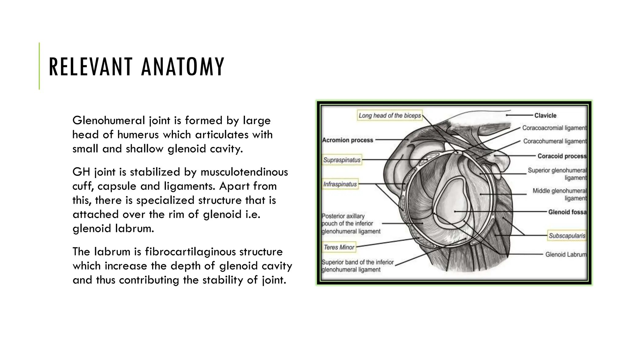
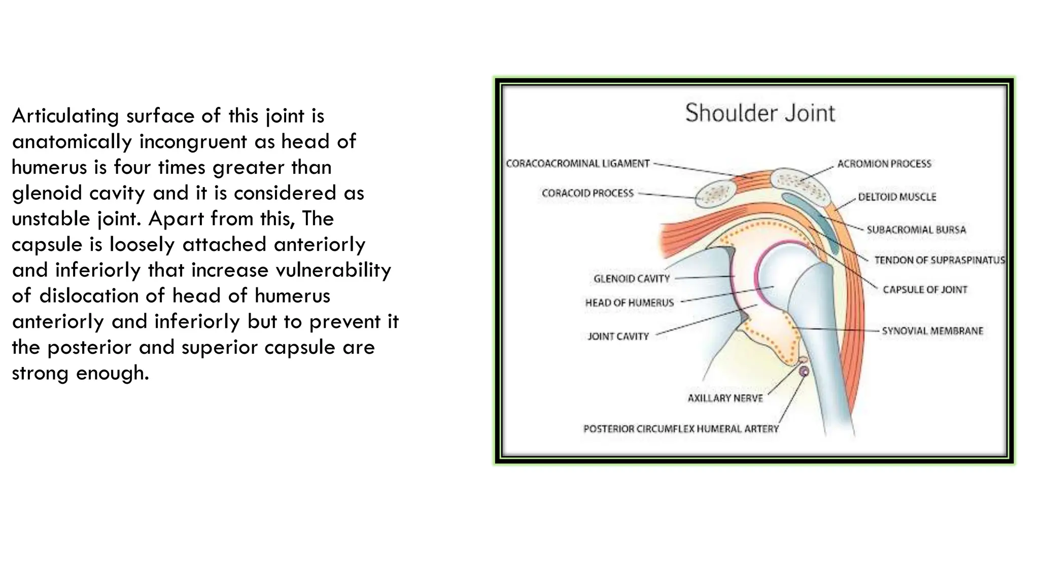
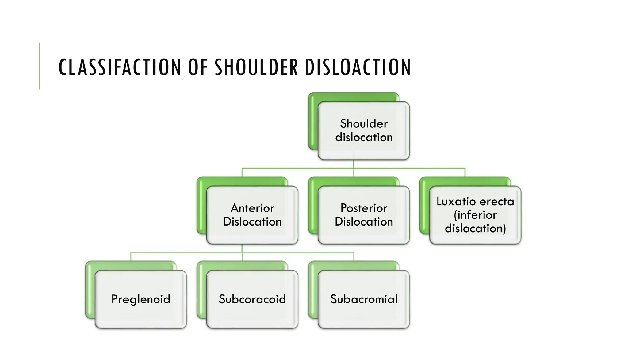
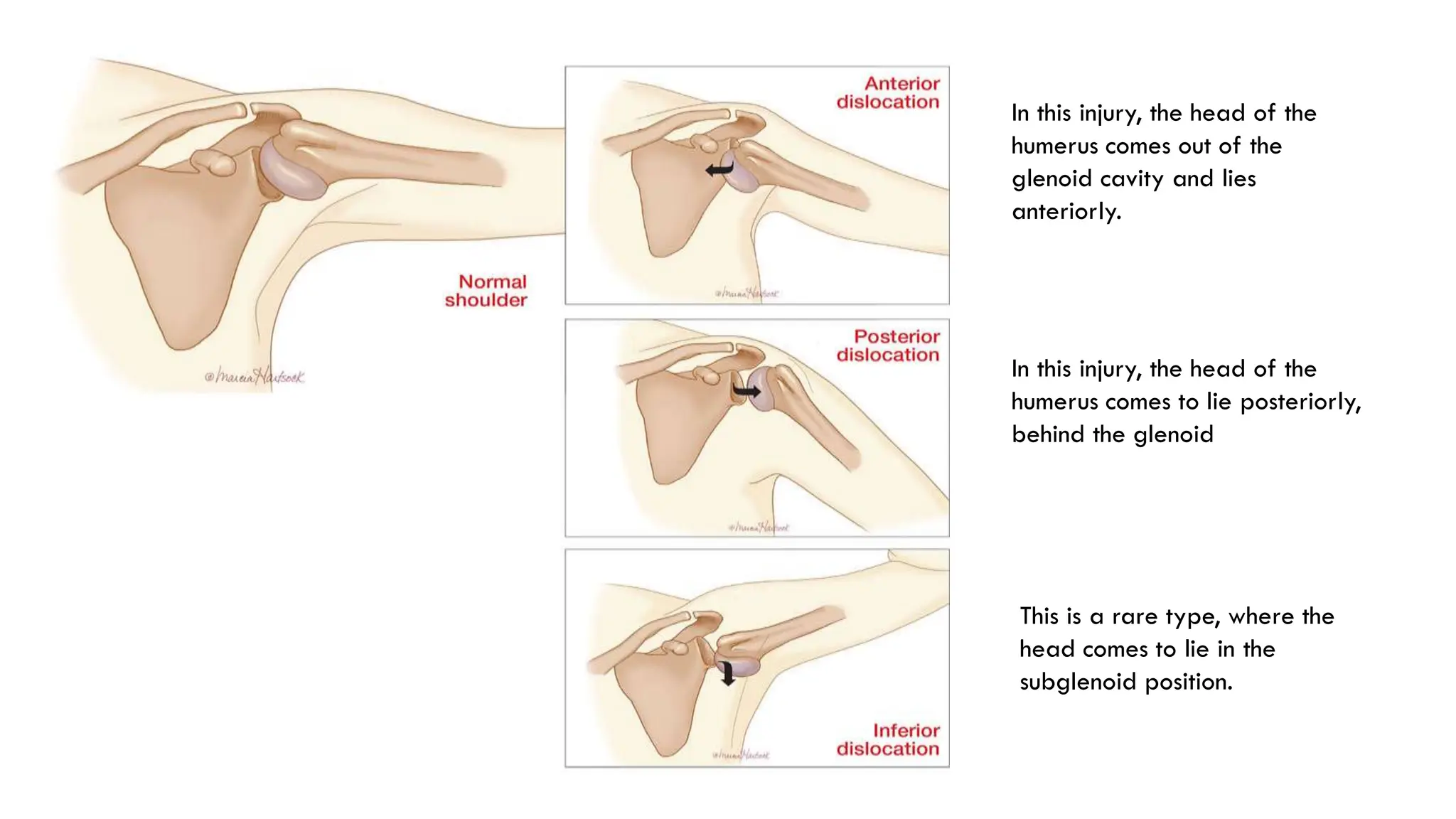
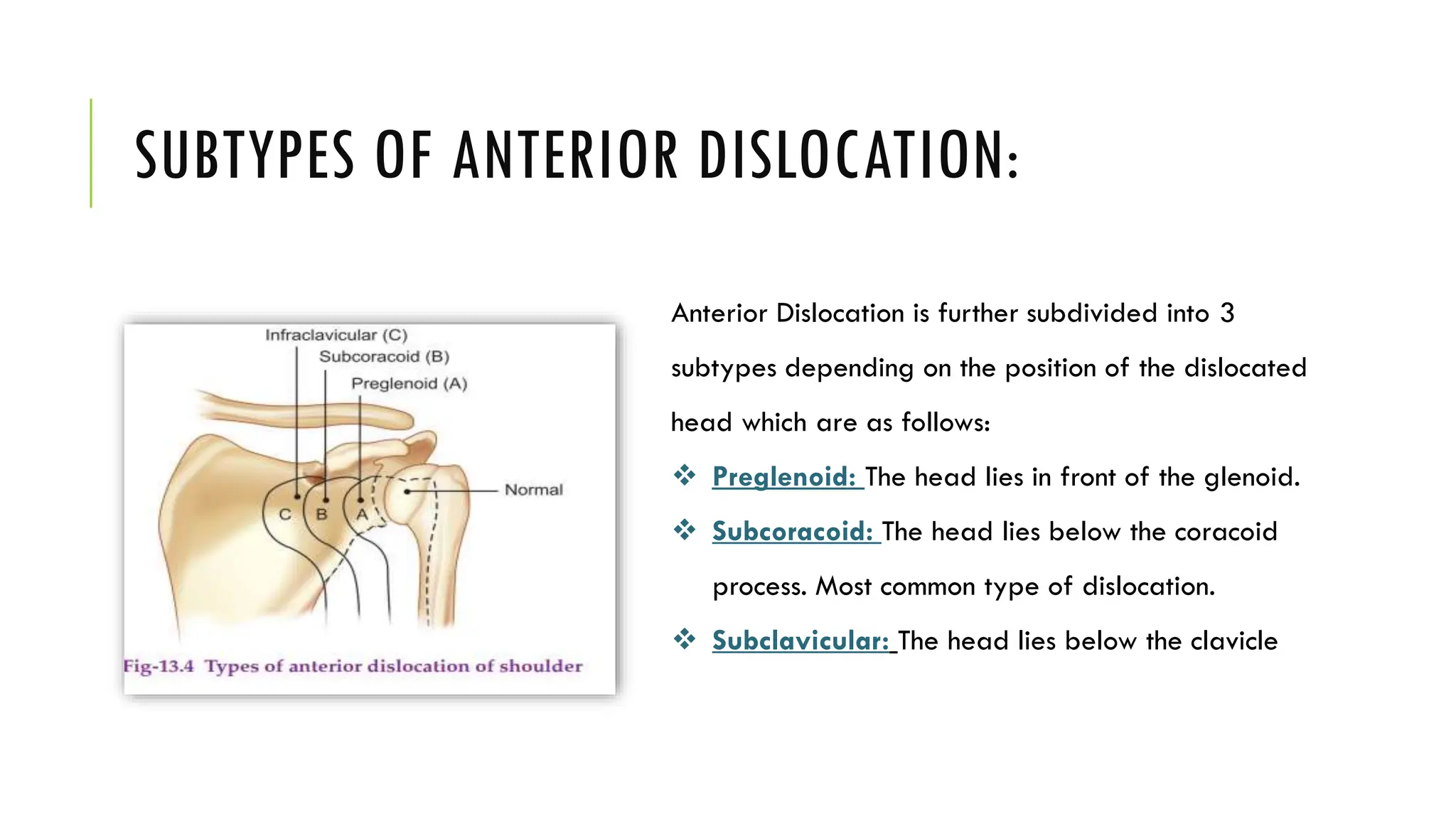
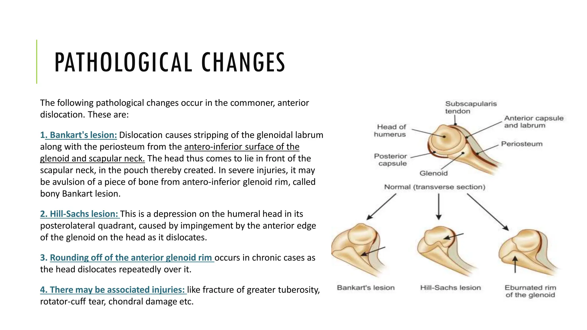
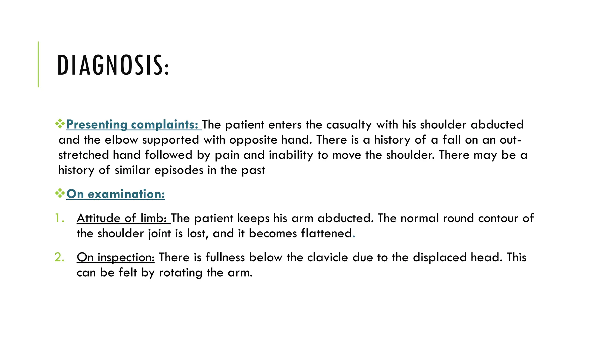
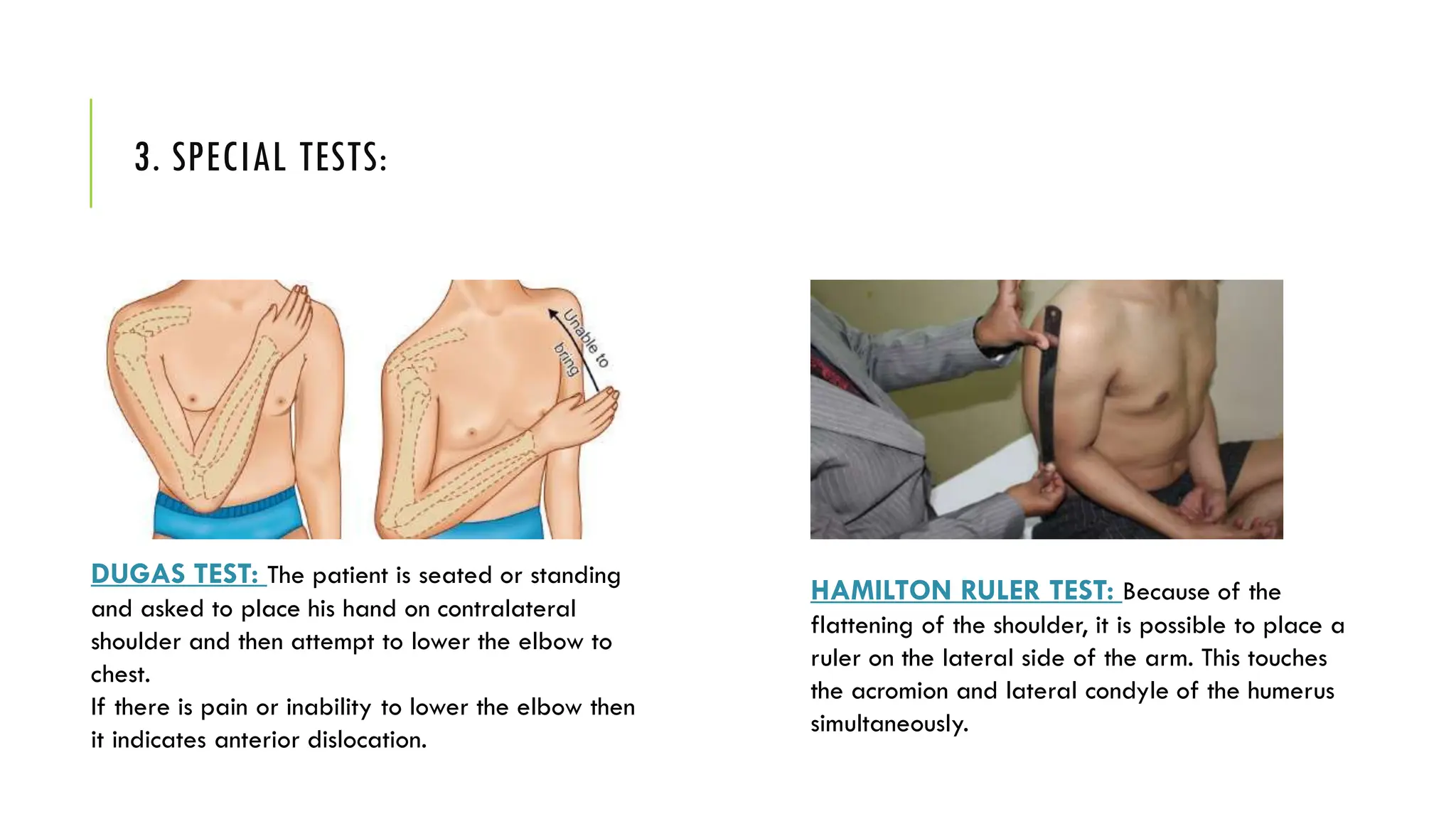
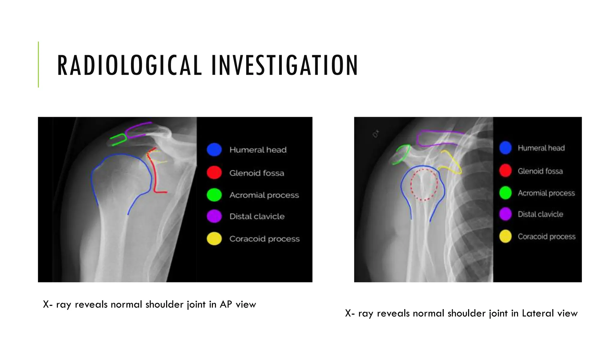
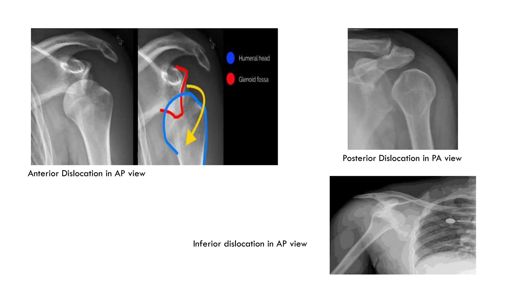
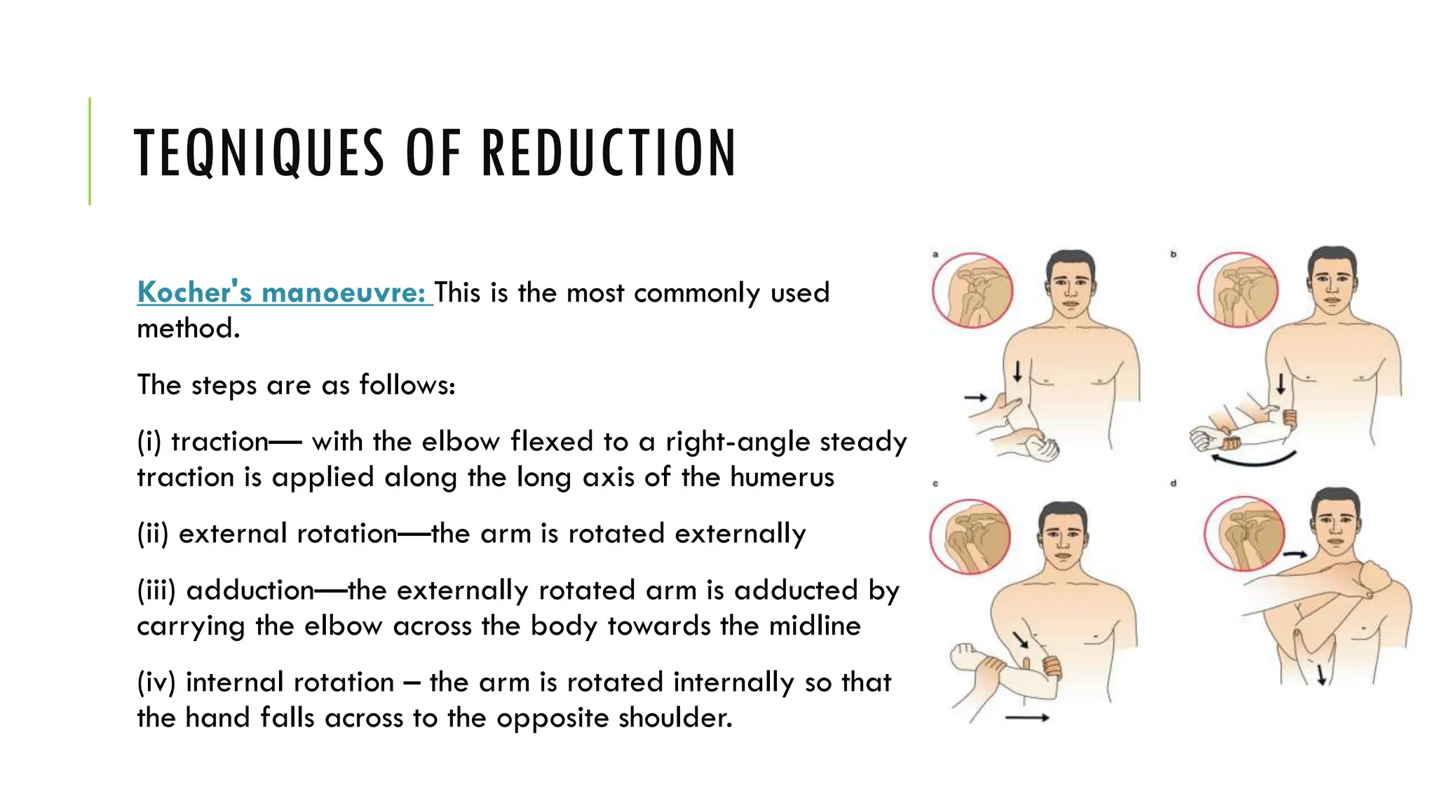
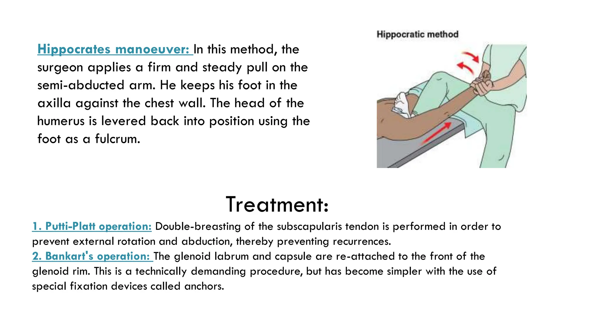
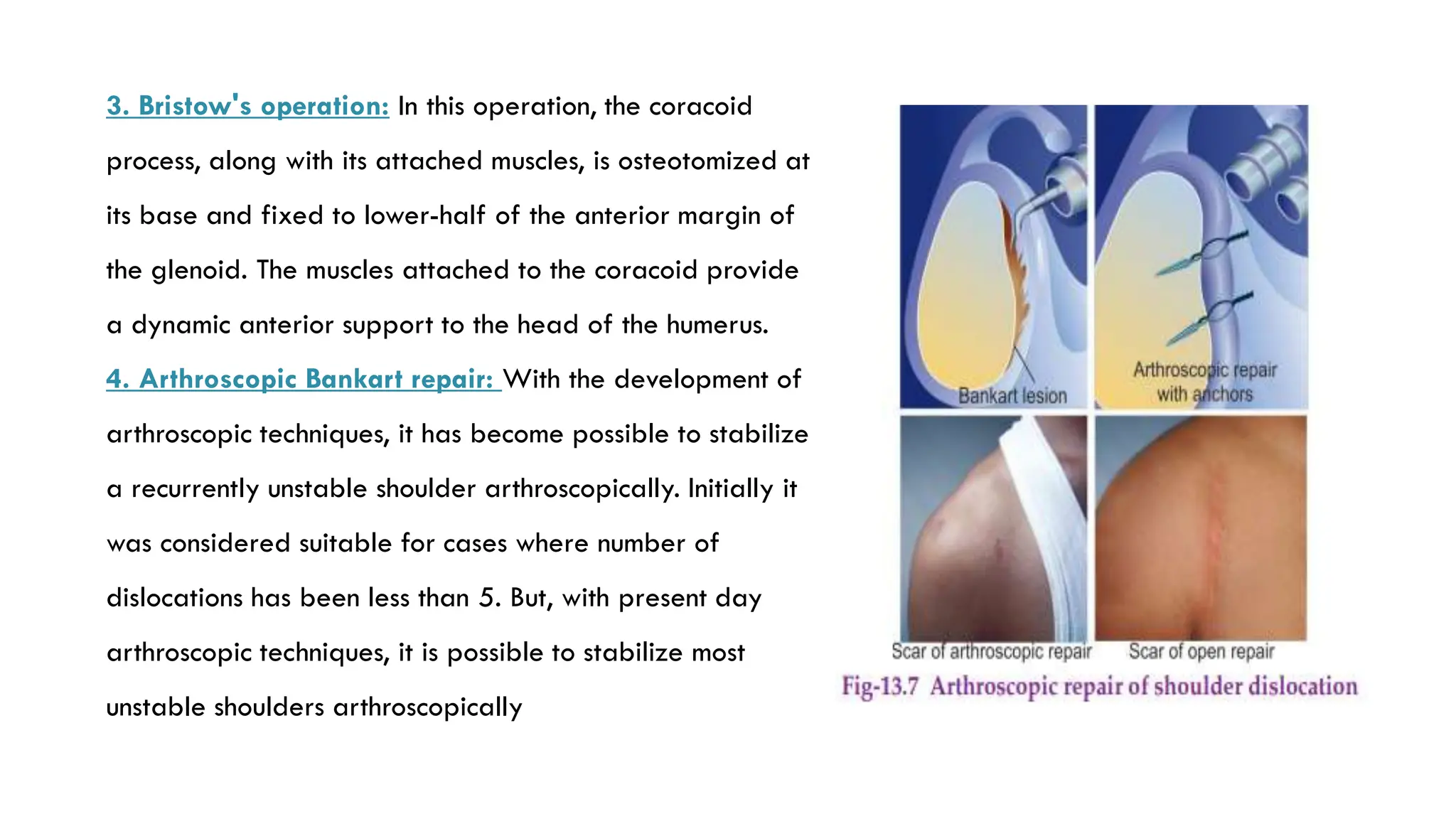
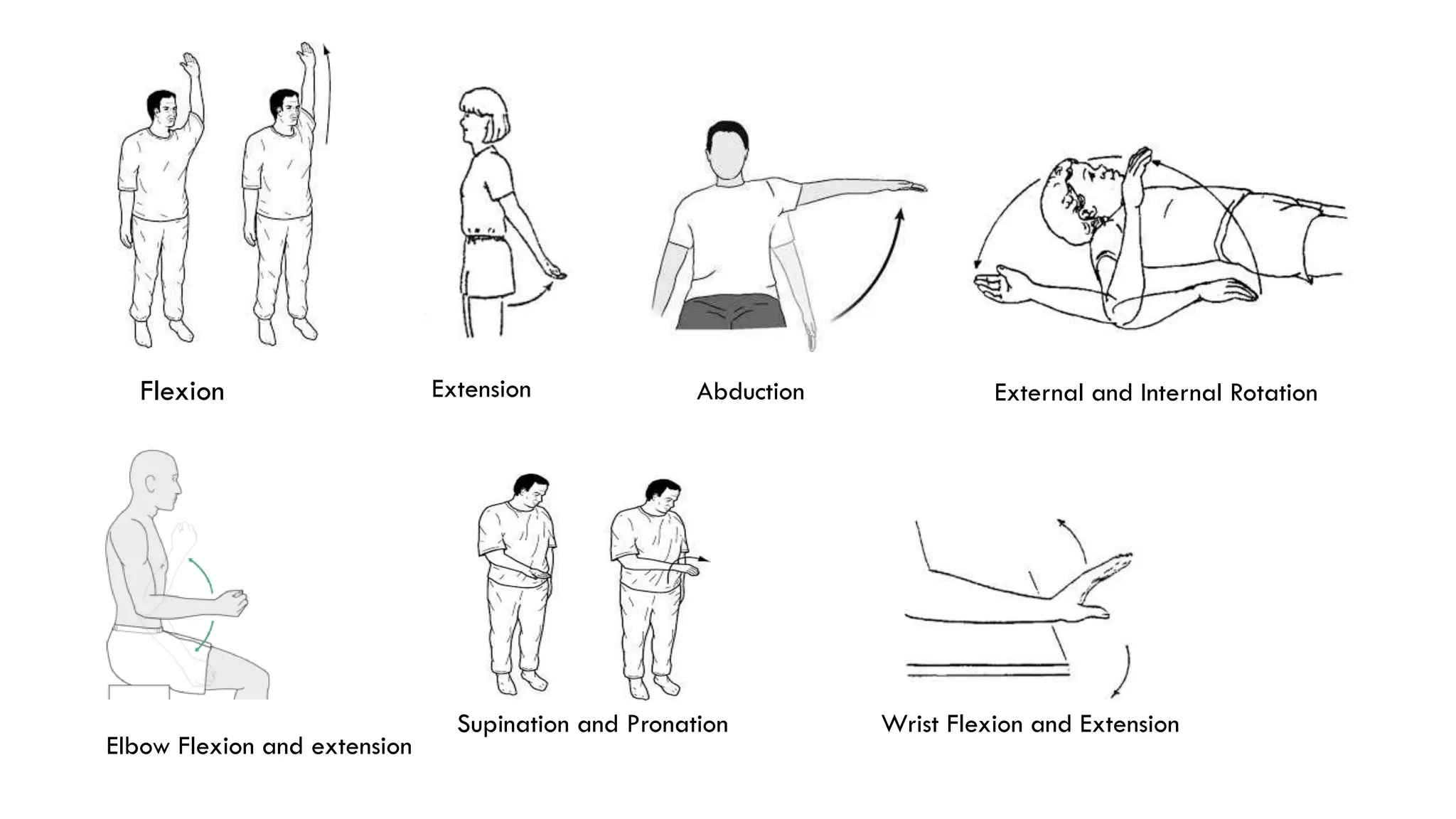
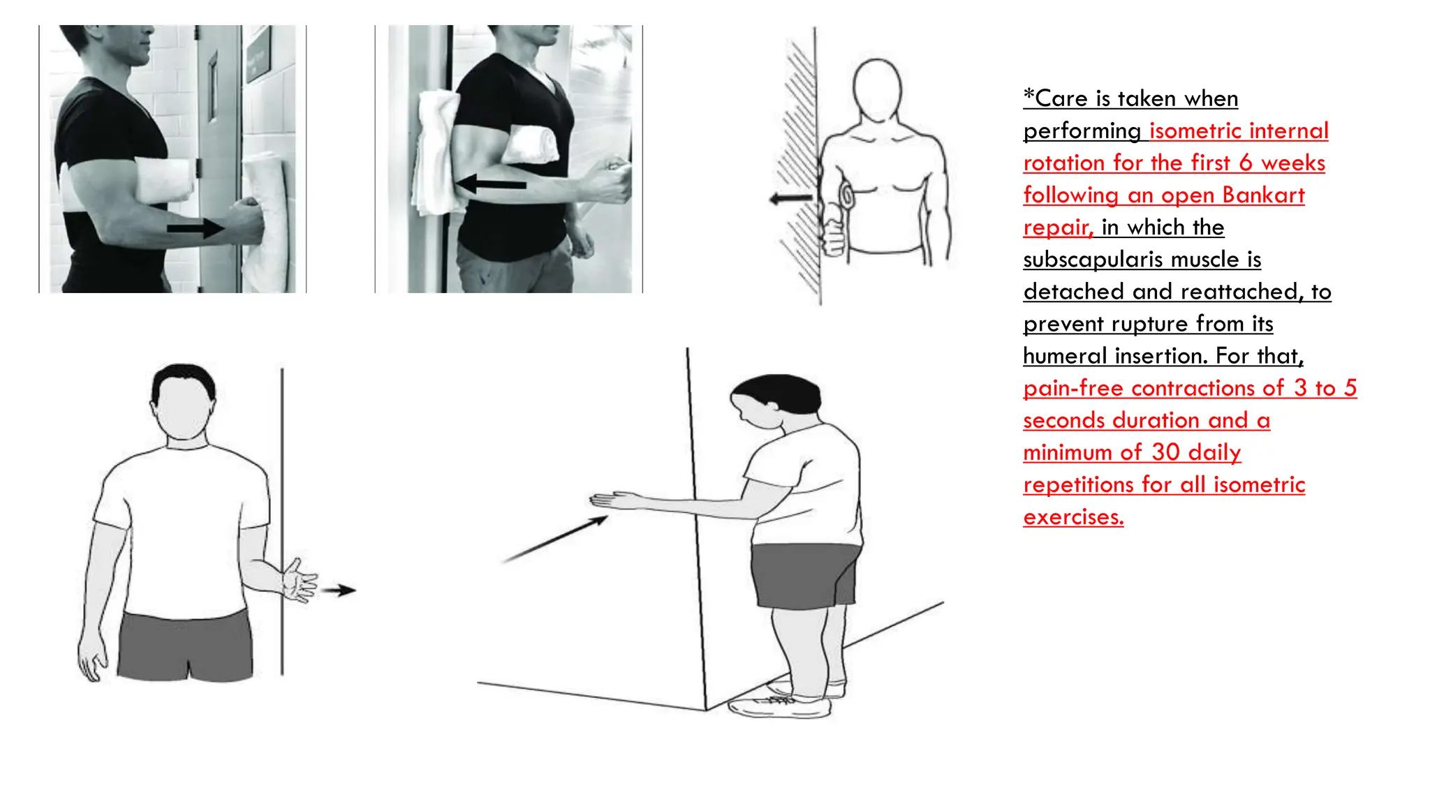
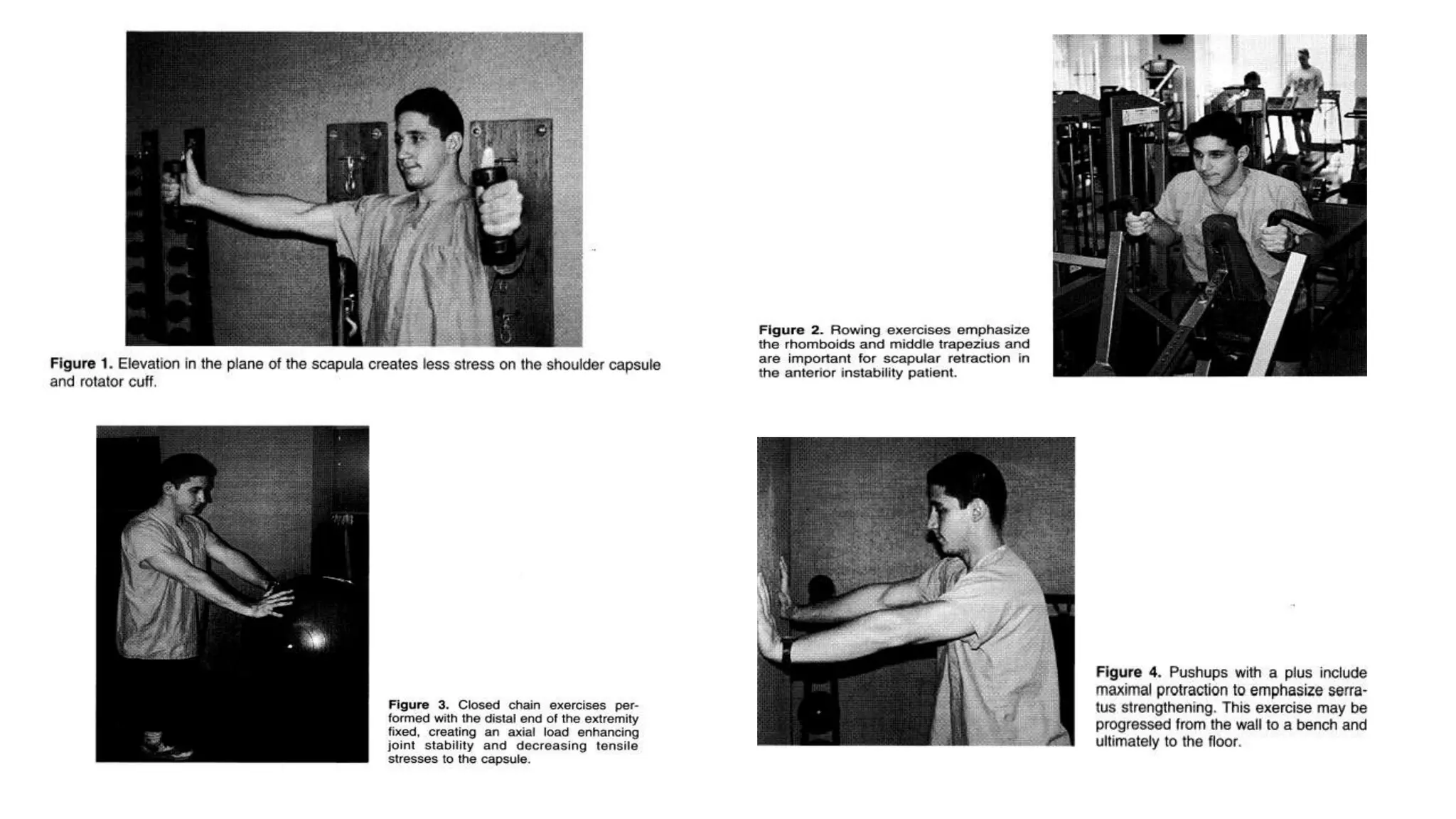
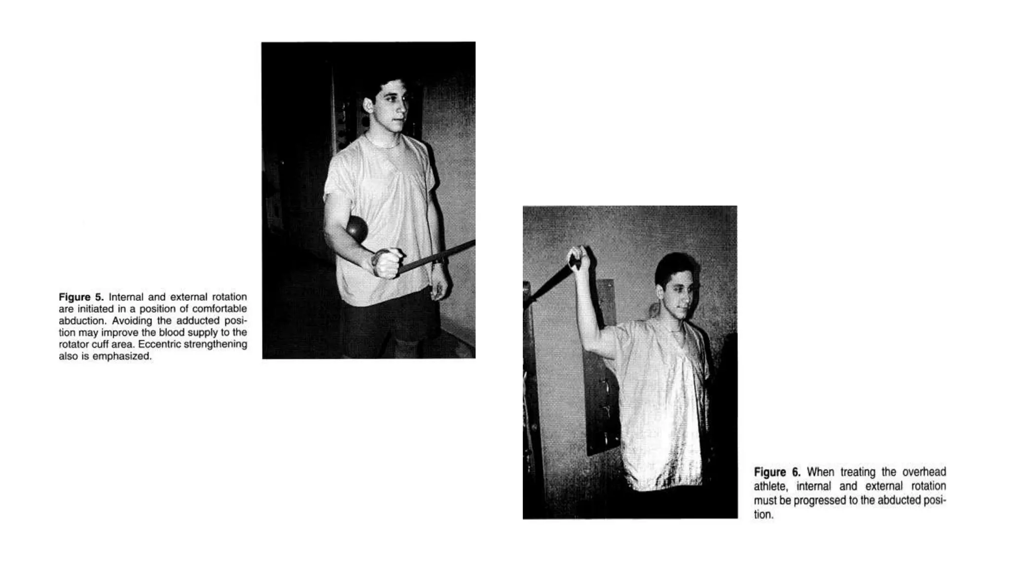
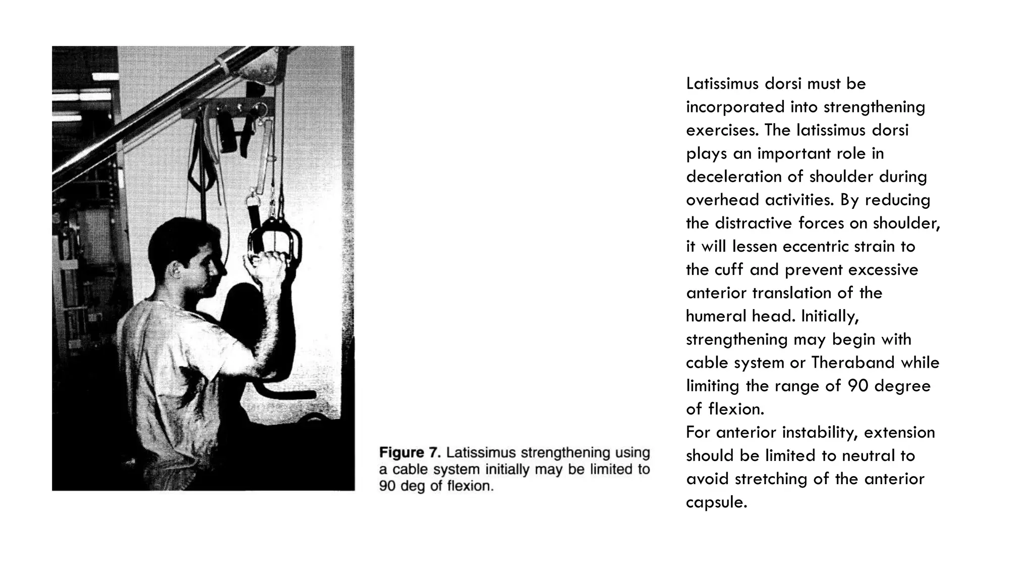
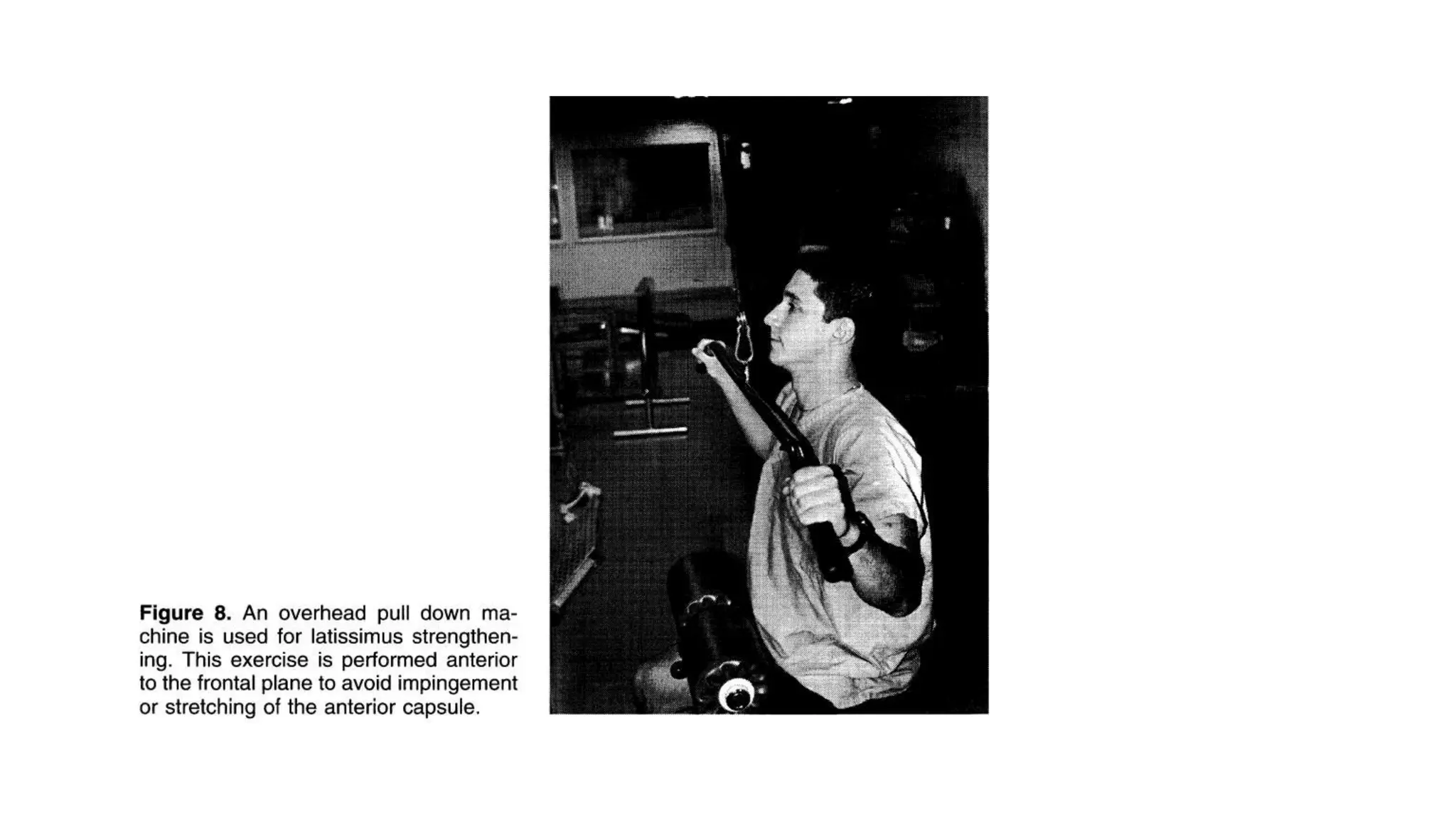
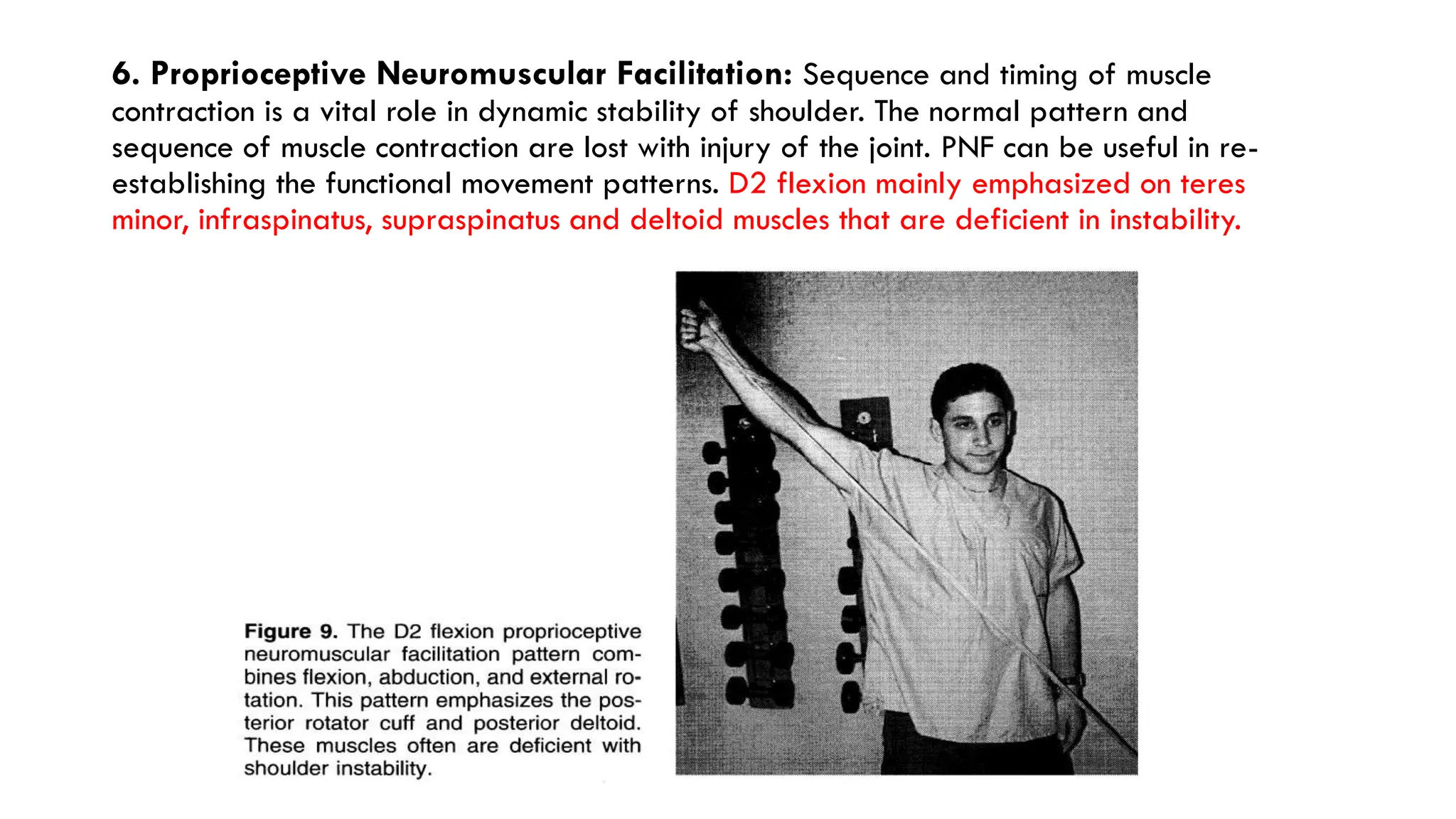
DISLOCATION
PRESENTED BY JASLEEN
KAUR
MASTERS IN ORTHOPAEDICS
2nd PROF.
POINTS TO BE STUDIED:
Relevant Anatomy
Shoulder dislocation definition and mechanism
Classification
Pathological Changes
Diagnosis
Special Tests
Radiological Investigations
Technique of Reduction
Management- Surgical and Post-surgical Physiotherapy Rehabilitation
RELEVANT ANATOMY
Glenohumeral joint is formed by large
head of humerus which articulates with
small and shallow glenoid cavity.
GH joint is stabilized by musculotendinous
cuff, capsule and ligaments. Apart from
this, there is specialized structure that is
attached over the rim of glenoid i.e.
glenoid labrum.
The labrum is fibrocartilaginous structure
which increase the depth of glenoid cavity
and thus contributing the stability of joint.
Articulating surface of this joint is
anatomically incongruent as head of
humerus is four times greater than
glenoid cavity and it is considered as
unstable joint. Apart from this, The
capsule is loosely attached anteriorly
and inferiorly that increase vulnerability
of dislocation of head of humerus
anteriorly and inferiorly but to prevent it
the posterior and superior capsule are
strong enough.
SHOULDER DISLOCATION
This is the commonest joint in the human body to dislocate. It occurs more commonly in adults,
and is rare in children. Anterior dislocation is much more common than posterior dislocation.
MECHANISM:
Fall on an out-stretched hand with the shoulder abducted and externally rotated, is the
common mechanism of injury.
Occasionally, it results from a direct force pushing the humerus head out of the glenoid
cavity Like posterior dislocation may result from a direct blow on the front of the shoulder,
driving the head backwards.
More often, however, posterior dislocation is the consequence of an electric shock or an
epileptiform convulsion
Shoulder
dislocation
Anterior
Dislocation
Preglenoid Subcoracoid Subacromial
Posterior
Dislocation
Luxatio erecta
(inferior
dislocation)
CLASSIFACTION OF SHOULDER DISLOACTION
In this injury, the head of the
humerus comes out of the
glenoid cavity and lies
anteriorly.
In this injury, the head of the
humerus comes to lie posteriorly,
behind the glenoid
This is a rare type, where the
head comes to lie in the
subglenoid position.
Anterior Dislocation is further subdivided into 3
subtypes depending on the position of the dislocated
head which are as follows:
 Preglenoid: The head lies in front of the glenoid.
 Subcoracoid: The head lies below the coracoid
process. Most common type of dislocation.
 Subclavicular: The head lies below the clavicle
SUBTYPES OF ANTERIOR DISLOCATION:
PATHOLOGICAL CHANGES
The following pathological changes occur in the commoner, anterior
dislocation. These are:
1. Bankart's lesion: Dislocation causes stripping of the glenoidal labrum
along with the periosteum from the antero-inferior surface of the
glenoid and scapular neck. The head thus comes to lie in front of the
scapular neck, in the pouch thereby created. In severe injuries, it may
be avulsion of a piece of bone from antero-inferior glenoid rim, called
bony Bankart lesion.
2. Hill-Sachs lesion: This is a depression on the humeral head in its
posterolateral quadrant, caused by impingement by the anterior edge
of the glenoid on the head as it dislocates.
3. Rounding off of the anterior glenoid rim occurs in chronic cases as
the head dislocates repeatedly over it.
4. There may be associated injuries: like fracture of greater tuberosity,
rotator-cuff tear, chondral damage etc.
DIAGNOSIS:
Presenting complaints: The patient enters the casualty with his shoulder abducted
and the elbow supported with opposite hand. There is a history of a fall on an out-
stretched hand followed by pain and inability to move the shoulder. There may be a
history of similar episodes in the past
On examination:
1. Attitude of limb: The patient keeps his arm abducted. The normal round contour of
the shoulder joint is lost, and it becomes flattened.
2. On inspection: There is fullness below the clavicle due to the displaced head. This
can be felt by rotating the arm.
3. SPECIAL TESTS:
DUGAS TEST: The patient is seated or standing
and asked to place his hand on contralateral
shoulder and then attempt to lower the elbow to
chest.
If there is pain or inability to lower the elbow then
it indicates anterior dislocation.
HAMILTON RULER TEST: Because of the
flattening of the shoulder, it is possible to place a
ruler on the lateral side of the arm. This touches
the acromion and lateral condyle of the humerus
simultaneously.
RADIOLOGICAL INVESTIGATION
X- ray reveals normal shoulder joint in AP view
X- ray reveals normal shoulder joint in Lateral view
Anterior Dislocation in AP view
Posterior Dislocation in PA view
Inferior dislocation in AP view
TEQNIQUES OF REDUCTION
Kocher's manoeuvre: This is the most commonly used
method.
The steps are as follows:
(i) traction— with the elbow flexed to a right-angle steady
traction is applied along the long axis of the humerus
(ii) external rotation—the arm is rotated externally
(iii) adduction—the externally rotated arm is adducted by
carrying the elbow across the body towards the midline
(iv) internal rotation – the arm is rotated internally so that
the hand falls across to the opposite shoulder.
Hippocrates manoeuver: In this method, the
surgeon applies a firm and steady pull on the
semi-abducted arm. He keeps his foot in the
axilla against the chest wall. The head of the
humerus is levered back into position using the
foot as a fulcrum.
Treatment:
1. Putti-Platt operation: Double-breasting of the subscapularis tendon is performed in order to
prevent external rotation and abduction, thereby preventing recurrences.
2. Bankart's operation: The glenoid labrum and capsule are re-attached to the front of the
glenoid rim. This is a technically demanding procedure, but has become simpler with the use of
special fixation devices called anchors.
3. Bristow's operation: In this operation, the coracoid
process, along with its attached muscles, is osteotomized at
its base and fixed to lower-half of the anterior margin of
the glenoid. The muscles attached to the coracoid provide
a dynamic anterior support to the head of the humerus.
4. Arthroscopic Bankart repair: With the development of
arthroscopic techniques, it has become possible to stabilize
a recurrently unstable shoulder arthroscopically. Initially it
was considered suitable for cases where number of
dislocations has been less than 5. But, with present day
arthroscopic techniques, it is possible to stabilize most
unstable shoulders arthroscopically
POSTOPERATIVE REHABILITATION
1. Cryotherapy: Cryotherapy in the postoperative shoulder (applied for 15-minute durations
every 1 to 2 waking hours for the first 24 hours, and 4 to 6 times daily for the ensuing 9 days) has
been shown to significantly decrease the frequency and intensity of shoulder pain both at rest and
during rehabilitation.
2. Activity Restriction: The postoperative management of anterior instability has typically
involved a minimum of 6 weeks of activity restriction to minimize stress to healing structures.
During this period of limited upper extremity use, we recommend active exercise of noninvolved
joints (elbow, wrist, and hand).
3. Isometric Exercise: Isometric shoulder muscle exercises, initially performed with the arm
adducted by the side of the body, provide a means for preventing muscular inhibition during the
period of activity restriction.
Isometric exercises for the scapulothoracic muscles are commenced during the first postoperative
week. Isometric exercises for the humeral muscles are commenced during the second postoperative
week.
Flexion Extension Abduction External and Internal Rotation
Wrist Flexion and Extension
Supination and Pronation
Elbow Flexion and extension
*Care is taken when
performing isometric internal
rotation for the first 6 weeks
following an open Bankart
repair, in which the
subscapularis muscle is
detached and reattached, to
prevent rupture from its
humeral insertion. For that,
pain-free contractions of 3 to 5
seconds duration and a
minimum of 30 daily
repetitions for all isometric
exercises.
4. Range of Motion Exercises: Assisted shoulder exercises initially performed within a limited
range of motion are designed to protect the surgical repair and prevent adhesion formation in the early
postoperative period.
These exercises are commenced during the second postoperative week. External rotation range of motion
is limited to 30° (0° abduction) for the first 4 postoperative weeks.
Combined external rotation and abduction range of motion is avoided for the first 6 postoperative
weeks.
Assisted elevation is initially performed in the plane of the scapula to maximize humeral and glenoid
congruency.
Rehabilitation aims to restore full active range of motion by 12 weeks after arthroscopic29 and open
anterior stabilization.
5. Scapulothoracic Muscle Retraining: Upper extremity weight-bearing exercises that
incorporate specific scapular movements at glenohumeral angles of less than 60° elevation are introduced
during the third postoperative week.
Light resistance exercises are commenced during the fourth postoperative week. We emphasize
retraining for scapular protraction and retraction and advocate multiple sets of up to 30 repetitions for
exercises that involve both concentric and eccentric modes of contraction.
Once these goals have been achieved, upper extremity weight-bearing exercises are advanced to
higher angles of elevation and weight-bearing loads are increased (e.g, press-ups, push-ups, and
quadruped exercises).
Latissimus dorsi must be
incorporated into strengthening
exercises. The latissimus dorsi
plays an important role in
deceleration of shoulder during
overhead activities. By reducing
the distractive forces on shoulder,
it will lessen eccentric strain to
the cuff and prevent excessive
anterior translation of the
humeral head. Initially,
strengthening may begin with
cable system or Theraband while
limiting the range of 90 degree
of flexion.
For anterior instability, extension
should be limited to neutral to
avoid stretching of the anterior
capsule.
6. Proprioceptive Neuromuscular Facilitation: Sequence and timing of muscle
contraction is a vital role in dynamic stability of shoulder. The normal pattern and
sequence of muscle contraction are lost with injury of the joint. PNF can be useful in re-
establishing the functional movement patterns. D2 flexion mainly emphasized on teres
minor, infraspinatus, supraspinatus and deltoid muscles that are deficient in instability.
REFERENCES:
BD Chaurasia, Human anatomy Volume 1.
Cynthia Norkin, Joint-structure-and-function-a-comprehensive-analysis.
 Maheshwari and Mhaskar, Essentials of orthopedics
Hayes K, Callanan M, Walton J, Paxinos A, Murrell GA. Shoulder instability:
management and rehabilitation. Journal of Orthopaedics & Sports Physical
Therapy. 2002 Oct;32(10):497-509.
Dines DM, Levinson M. The conservative management of the unstable shoulder
including rehabilitation. Clin Sports Med. 1995;14:797–816.

Shoulder Dislocation slideShare. PDF.pdf

  • 1.
  • 2.
    POINTS TO BESTUDIED: Relevant Anatomy Shoulder dislocation definition and mechanism Classification Pathological Changes Diagnosis Special Tests Radiological Investigations Technique of Reduction Management- Surgical and Post-surgical Physiotherapy Rehabilitation
  • 3.
    RELEVANT ANATOMY Glenohumeral jointis formed by large head of humerus which articulates with small and shallow glenoid cavity. GH joint is stabilized by musculotendinous cuff, capsule and ligaments. Apart from this, there is specialized structure that is attached over the rim of glenoid i.e. glenoid labrum. The labrum is fibrocartilaginous structure which increase the depth of glenoid cavity and thus contributing the stability of joint.
  • 4.
    Articulating surface ofthis joint is anatomically incongruent as head of humerus is four times greater than glenoid cavity and it is considered as unstable joint. Apart from this, The capsule is loosely attached anteriorly and inferiorly that increase vulnerability of dislocation of head of humerus anteriorly and inferiorly but to prevent it the posterior and superior capsule are strong enough.
  • 5.
    SHOULDER DISLOCATION This isthe commonest joint in the human body to dislocate. It occurs more commonly in adults, and is rare in children. Anterior dislocation is much more common than posterior dislocation. MECHANISM: Fall on an out-stretched hand with the shoulder abducted and externally rotated, is the common mechanism of injury. Occasionally, it results from a direct force pushing the humerus head out of the glenoid cavity Like posterior dislocation may result from a direct blow on the front of the shoulder, driving the head backwards. More often, however, posterior dislocation is the consequence of an electric shock or an epileptiform convulsion
  • 6.
  • 7.
    In this injury,the head of the humerus comes out of the glenoid cavity and lies anteriorly. In this injury, the head of the humerus comes to lie posteriorly, behind the glenoid This is a rare type, where the head comes to lie in the subglenoid position.
  • 8.
    Anterior Dislocation isfurther subdivided into 3 subtypes depending on the position of the dislocated head which are as follows:  Preglenoid: The head lies in front of the glenoid.  Subcoracoid: The head lies below the coracoid process. Most common type of dislocation.  Subclavicular: The head lies below the clavicle SUBTYPES OF ANTERIOR DISLOCATION:
  • 9.
    PATHOLOGICAL CHANGES The followingpathological changes occur in the commoner, anterior dislocation. These are: 1. Bankart's lesion: Dislocation causes stripping of the glenoidal labrum along with the periosteum from the antero-inferior surface of the glenoid and scapular neck. The head thus comes to lie in front of the scapular neck, in the pouch thereby created. In severe injuries, it may be avulsion of a piece of bone from antero-inferior glenoid rim, called bony Bankart lesion. 2. Hill-Sachs lesion: This is a depression on the humeral head in its posterolateral quadrant, caused by impingement by the anterior edge of the glenoid on the head as it dislocates. 3. Rounding off of the anterior glenoid rim occurs in chronic cases as the head dislocates repeatedly over it. 4. There may be associated injuries: like fracture of greater tuberosity, rotator-cuff tear, chondral damage etc.
  • 10.
    DIAGNOSIS: Presenting complaints: Thepatient enters the casualty with his shoulder abducted and the elbow supported with opposite hand. There is a history of a fall on an out- stretched hand followed by pain and inability to move the shoulder. There may be a history of similar episodes in the past On examination: 1. Attitude of limb: The patient keeps his arm abducted. The normal round contour of the shoulder joint is lost, and it becomes flattened. 2. On inspection: There is fullness below the clavicle due to the displaced head. This can be felt by rotating the arm.
  • 11.
    3. SPECIAL TESTS: DUGASTEST: The patient is seated or standing and asked to place his hand on contralateral shoulder and then attempt to lower the elbow to chest. If there is pain or inability to lower the elbow then it indicates anterior dislocation. HAMILTON RULER TEST: Because of the flattening of the shoulder, it is possible to place a ruler on the lateral side of the arm. This touches the acromion and lateral condyle of the humerus simultaneously.
  • 12.
    RADIOLOGICAL INVESTIGATION X- rayreveals normal shoulder joint in AP view X- ray reveals normal shoulder joint in Lateral view
  • 13.
    Anterior Dislocation inAP view Posterior Dislocation in PA view Inferior dislocation in AP view
  • 14.
    TEQNIQUES OF REDUCTION Kocher'smanoeuvre: This is the most commonly used method. The steps are as follows: (i) traction— with the elbow flexed to a right-angle steady traction is applied along the long axis of the humerus (ii) external rotation—the arm is rotated externally (iii) adduction—the externally rotated arm is adducted by carrying the elbow across the body towards the midline (iv) internal rotation – the arm is rotated internally so that the hand falls across to the opposite shoulder.
  • 15.
    Hippocrates manoeuver: Inthis method, the surgeon applies a firm and steady pull on the semi-abducted arm. He keeps his foot in the axilla against the chest wall. The head of the humerus is levered back into position using the foot as a fulcrum. Treatment: 1. Putti-Platt operation: Double-breasting of the subscapularis tendon is performed in order to prevent external rotation and abduction, thereby preventing recurrences. 2. Bankart's operation: The glenoid labrum and capsule are re-attached to the front of the glenoid rim. This is a technically demanding procedure, but has become simpler with the use of special fixation devices called anchors.
  • 16.
    3. Bristow's operation:In this operation, the coracoid process, along with its attached muscles, is osteotomized at its base and fixed to lower-half of the anterior margin of the glenoid. The muscles attached to the coracoid provide a dynamic anterior support to the head of the humerus. 4. Arthroscopic Bankart repair: With the development of arthroscopic techniques, it has become possible to stabilize a recurrently unstable shoulder arthroscopically. Initially it was considered suitable for cases where number of dislocations has been less than 5. But, with present day arthroscopic techniques, it is possible to stabilize most unstable shoulders arthroscopically
  • 17.
    POSTOPERATIVE REHABILITATION 1. Cryotherapy:Cryotherapy in the postoperative shoulder (applied for 15-minute durations every 1 to 2 waking hours for the first 24 hours, and 4 to 6 times daily for the ensuing 9 days) has been shown to significantly decrease the frequency and intensity of shoulder pain both at rest and during rehabilitation. 2. Activity Restriction: The postoperative management of anterior instability has typically involved a minimum of 6 weeks of activity restriction to minimize stress to healing structures. During this period of limited upper extremity use, we recommend active exercise of noninvolved joints (elbow, wrist, and hand). 3. Isometric Exercise: Isometric shoulder muscle exercises, initially performed with the arm adducted by the side of the body, provide a means for preventing muscular inhibition during the period of activity restriction. Isometric exercises for the scapulothoracic muscles are commenced during the first postoperative week. Isometric exercises for the humeral muscles are commenced during the second postoperative week.
  • 18.
    Flexion Extension AbductionExternal and Internal Rotation Wrist Flexion and Extension Supination and Pronation Elbow Flexion and extension
  • 19.
    *Care is takenwhen performing isometric internal rotation for the first 6 weeks following an open Bankart repair, in which the subscapularis muscle is detached and reattached, to prevent rupture from its humeral insertion. For that, pain-free contractions of 3 to 5 seconds duration and a minimum of 30 daily repetitions for all isometric exercises.
  • 20.
    4. Range ofMotion Exercises: Assisted shoulder exercises initially performed within a limited range of motion are designed to protect the surgical repair and prevent adhesion formation in the early postoperative period. These exercises are commenced during the second postoperative week. External rotation range of motion is limited to 30° (0° abduction) for the first 4 postoperative weeks. Combined external rotation and abduction range of motion is avoided for the first 6 postoperative weeks. Assisted elevation is initially performed in the plane of the scapula to maximize humeral and glenoid congruency. Rehabilitation aims to restore full active range of motion by 12 weeks after arthroscopic29 and open anterior stabilization. 5. Scapulothoracic Muscle Retraining: Upper extremity weight-bearing exercises that incorporate specific scapular movements at glenohumeral angles of less than 60° elevation are introduced during the third postoperative week. Light resistance exercises are commenced during the fourth postoperative week. We emphasize retraining for scapular protraction and retraction and advocate multiple sets of up to 30 repetitions for exercises that involve both concentric and eccentric modes of contraction. Once these goals have been achieved, upper extremity weight-bearing exercises are advanced to higher angles of elevation and weight-bearing loads are increased (e.g, press-ups, push-ups, and quadruped exercises).
  • 23.
    Latissimus dorsi mustbe incorporated into strengthening exercises. The latissimus dorsi plays an important role in deceleration of shoulder during overhead activities. By reducing the distractive forces on shoulder, it will lessen eccentric strain to the cuff and prevent excessive anterior translation of the humeral head. Initially, strengthening may begin with cable system or Theraband while limiting the range of 90 degree of flexion. For anterior instability, extension should be limited to neutral to avoid stretching of the anterior capsule.
  • 25.
    6. Proprioceptive NeuromuscularFacilitation: Sequence and timing of muscle contraction is a vital role in dynamic stability of shoulder. The normal pattern and sequence of muscle contraction are lost with injury of the joint. PNF can be useful in re- establishing the functional movement patterns. D2 flexion mainly emphasized on teres minor, infraspinatus, supraspinatus and deltoid muscles that are deficient in instability.
  • 26.
    REFERENCES: BD Chaurasia, Humananatomy Volume 1. Cynthia Norkin, Joint-structure-and-function-a-comprehensive-analysis.  Maheshwari and Mhaskar, Essentials of orthopedics Hayes K, Callanan M, Walton J, Paxinos A, Murrell GA. Shoulder instability: management and rehabilitation. Journal of Orthopaedics & Sports Physical Therapy. 2002 Oct;32(10):497-509. Dines DM, Levinson M. The conservative management of the unstable shoulder including rehabilitation. Clin Sports Med. 1995;14:797–816.