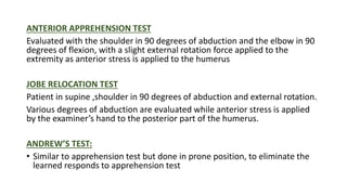
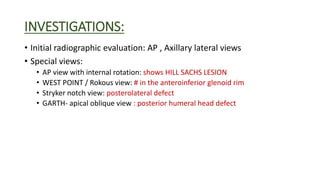
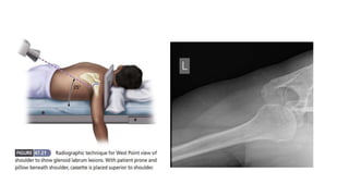
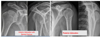
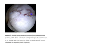
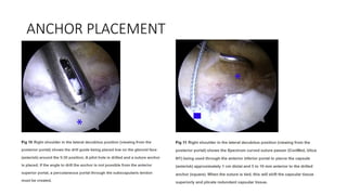
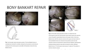
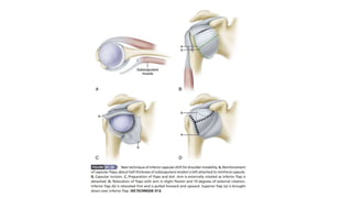
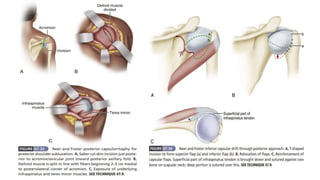
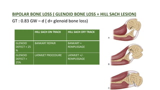
SHOULDER INSTABILITY
BY DR K GOUTHAM
JUNIOR RESIDENT
DEPT OF ORTHOPAEDICS
HISTORY:
• Onset of the pathology : traumatic or chronic
• Mechanism and severity of initial trauma
High- energy trauma: sports collision and RTA increases the risk of
bony defects
• Patients age at initial presentation
Age <20 yrs : 80% of RDS
Age > 30 yrs : 20% of RDS
• h/o recurrent subluxation( shoulder sliding in and out), movement
which cause the same
• Frequency of dislocation
• Ease with which relocation occurs
• Precipitating factors [like seizure]
• Generalized ligament laxity: dislocation reduces by the patient himself
• h/o Dislocations during sleep or with the arm in an overhead position
• h/o having a dead arm
Physical Examination:
• General examination :look for ligament laxity
• Asymmetry/ Atrophy of shoulder
• Tenderness in anterior and posterior capsule
• Rotator cuff and AC joint tenderness
• Active and passive range of movement
• Strength of deltoid and rotator cuff muscles
• Test for scapular winging
SPECIAL TESTS:
• SHIFT AND LOAD TEST
• DRAWER TEST
• SULCUS TEST
• ANTERIOR APPREHENSIVE TEST
• JOBE RELOCATION TEST
• SHOULDER LACHMAN TEST
• POSTERIOR CLUNK TEST
• ANDREWS TEST
• FULCRUM TEST
LOAD SHIFT TEST:
Patient in sitting position with arm hanging by the side and forearm in his lap.
Stabilize the scapula with one hand and grasp the humerus head with the other
hand , thumb in posterior and index , middle finger anterior
Apply compression and shift anteromedially towards coracoid and posterolaterally
• Sulcus test (NEER and FOSTER)
oLimb is pulled distally in neutral rotation and observing for the sulcus
oPositive test : multidirectional instability
oDone at arm 0 degree
and 45 degree abduction
Grading :
1+ : subluxation < 1cm
2+ : subluxation < 2 cm
3+ : subluxation > 2 cm
( subluxation at 0 degree of
Abduction – laxity at the rotator interval, at 45 degree – laxity of
inferior glenohumeral ligament complex)
Shoulder Lachman test:
• Left hand grasps the proximal humerus and right hand holds the
elbow, forward pressure is given from posterior aspect of the
shoulder , translation is graded
Posterior clunk test:
• Shoulder at 90degree abduction, forward
Flexion and internal rotated
• posterior stess is given
ANTERIOR APPREHENSION TEST
Evaluated with the shoulder in 90 degrees of abduction and the elbow in 90
degrees of flexion, with a slight external rotation force applied to the
extremity as anterior stress is applied to the humerus
JOBE RELOCATION TEST
Patient in supine ,shoulder in 90 degrees of abduction and external rotation.
Various degrees of abduction are evaluated while anterior stress is applied
by the examiner’s hand to the posterior part of the humerus.
ANDREW’S TEST:
• Similar to apprehension test but done in prone position, to eliminate the
learned responds to apprehension test
INVESTIGATIONS:
• Initial radiographic evaluation: AP , Axillary lateral views
• Special views:
• AP view with internal rotation: shows HILL SACHS LESION
• WEST POINT / Rokous view: # in the anteroinferior glenoid rim
• Stryker notch view: posterolateral defect
• GARTH- apical oblique view : posterior humeral head defect
The Stryker notch view is obtained with the
patient supine and the elbow elevated over the
head. The x-ray beam is directed 10 degrees
cephalad
Anterior dislocation with
Bankart fracture
Posterior dislocation
Posterior dislocation-fracture
• MRI :
ofor evaluating soft tissue lesion associated with instability
oEvaluation of HAGL- humeral avulsion glenohumeral lesion
oDetermining appropriate surgical intervention: on-track, off-track
lesions
oFindings :
• patulous inferior capsule (IGHL anterior and posterior bands)
• Bankart lesion - may occur in conjunction with traumatic anterior instability
• Kim lesion - may occur in conjunction with traumatic posterior instability
• bony erosion of glenoid - following chronic anterior instability
OSSEUS
BANKART
CT/ 3D CT:
Most sensitive test for detecting and measuring
bone deficiency
Retroversion of glenoid or humerus is also
evaluated
Reverse bankart lesion
EXAMINATION USING ANAESTHETIC AND ARTHROSCOPY:
• Multidirectional instability : shows unsuspected plane of instability
• For anterior instability arm is abducted
• Significant findings: at 40 & 80 degrees of external rotation
• Translation of grade 2: 93% sensitivity
100% specificity for instability
• For posterior instability : arm is pushed posteriorly
Physiotherapy : help strengthen the joint, helping to prevent further
dislocations, and provide a solid platform for post surgical rehab
Treatment options available include:
• Cryotherapy
• Pain management Acute management of symptoms
• Protective taping and bracing
• Muscular strengthening programme
• Proprioceptive training
• Manual therapy
• Advice and post surgical management
ANTERIOR INSTABILITY OF SHOULDER:
• Procedures should include:
• Low recurrence rate
• Low complication rate
• Low reoperation rate
• Maintain motion
• Allows observation of the joint
• Correct the pathologic conditions
• Instability severity index score :
Arthroscopic surgery:
• Bankarts repair
• Capsular plication
Open surgery includes:
• Jobe capsulolabral reconstruction
• Neer capsular shift for anterior instability
• Glenoid-based shift for posterior instability
• Reconstruction of anterior Glenoid using iliac crest bone Autograft
OPEN BANKART REPAIR:
INDICATION
• when the labrum and the capsule are separated from the glenoid rim
• if the capsule is thin
ADVANTAGE:
• it corrects the labral defect and imbricates the capsule without
requiring any metallic internal fixation devices.
DISADVANTAGE : Technical difficulties
KEYS TO SUCCESS OF THIS PROCEDURE :
• Maximizing the healing potential by abrading the scapular neck
• Restoring glenoid concavity
• Securing anatomic capsular fixation at the edge of the glenoid
articular surface
• Re-creating physiologic capsular tension by superior and inferior
capsular advancement and imbrication
• Performing supervised goal-oriented rehabilitation.
ARTHROSCOPIC BANKART REPAIR
TECHNIQUE
• ANAESTHESIA : General anaesthesia
• POSITION: Lateral decubitus position
• POSITION OF ARM:
45 to 60 degrees of abduction and 20 degrees of forward flexion using 12 to
14 lb of traction.
• POSTERIOR PORTAL:
 2 cm inferior and just medial to the posterolateral edge of the acromion.
 After identifying the quadrant or quadrants of injury to the labrum, next
portal is created.
ANTEROSUPERIOR PORTAL
posterior to the biceps tendon and anterior to the leading edge of the
supraspinatus tendon.
GLENOID PREPARATION
ANCHOR PLACEMENT
BONY BANKART REPAIR
MODIFIED BANKART REPAIR
• By MONTGOMERY AND JOBE
ANTERIOR STABILIZATION WITH ASSOCIATED
GLENOID DEFICIENCY (LATERJET PROCEDURE)
INDICATION:
• instability with glenoid bone loss
• Combinations of glenoid and humeral bone loss
• Complex soft-tissue injury
• Revision of a Bankart repair
• Patients engaged in high-risk sports (climbing, rugby) or occupations
(carpentry), or who have a high risk of recurrence due to the intensity
and action of their activity (throwers), are ideal candidates for the
Latarjet procedure
BIOMECHANICS OF LATARJET
• The conjoint tendon acts as a sling to the inferior subscapularis and
anteroinferior capsule when the arm is abducted and externally
rotate.
• The addition of bone to the glenoid rim increases the anteroposterior
(AP) osseous diameter.
• The inferior capsule is reinforced with a portion of the coracoacromial
ligament.
• Position: beach chair position
• Incision: Make a 4 to 7-cm skin incision beginning under the tip of the
coracoid process
• Steps:
• Division of the subscapularis, capsulotomy, and exposure
• Fixation of the bone block
MULTIDIRECTIONAL INSTABILITY OF THE
SHOULDER
• Primary abnormality in multidirectional instability is a loose,
redundant inferior pouch.
• Principle of the procedure : to detach the capsule from the neck of
the humerus and shift it to the opposite side of the calcar (inferior
portion of the neck of the humerus)
Procedure: CAPSULAR SHIFT(Neer and Foster)
• Place the patient in a tilted position with the front and the back of the
shoulder exposed.
Incision : 9 cm in the skin creases from the anterior border of the axilla
to the coracoid process.
Disection:
• Develop the deltopectoral interval medial to the cephalic vein, and
retract the deltoid laterally.
• Divide the clavipectoral fascia, and retract the muscles attached to
the coracoid process medially
POSTERIOR INSTABILITY OF THE
SHOULDER
Atraumatic type of posterior instability
• Conservative treatment unless they have frequent and significant
disability and conservative treatment has failed.
Procedure:
• NEER INFERIOR CAPSULAR SHIFT PROCEDURE THROUGH A
POSTERIOR APPROACH
Position: lateral decubitus position
Incision: 10-cm incision vertically over the posterior aspect of the
acromion and the spine of the scapula
TIBONE AND BRADLEY TECHNIQUE
CAPSULAR SHIFT RECONSTRUCTION WITH
POSTERIOR GLENOID OSTEOTOMY
MCLAUGHLIN PROCEDURE
REMPLISSAGE
• French term : “to fill”
• Recent arthroscopic procedure’
Indication :
• Anterior shoulder instability with HILL SACH’s lesion
• Used along with Bankart repair
Principle :
Bony defect is filled by infraspinatus tenodesis and posterior capsule
capsulodesis
Calculations for identifying glenoid track:
• NCCT shoulder with 3D reconstruction
• Calculate the glenoid width -GW
• Glenoid track is 83% of glenoid width: 0.83 GW
• Calculate hill sach width HSW
• HILL SACH interval: HSI = HSW + BB
• BB : Bony bridge
• GT> HSI: ‘on track’  Bankart repair
• GT< HSI: ‘off track’  Remplissage
BIPOLAR BONE LOSS ( GLENOID BONE LOSS + HILL SACH LESION)
GT : 0.83 GW – d ( d= glenoid bone loss)
HILL SACH ON TRACK HILL SACH OFF TRACK
GLENOID
DEFECT < 25
%
BANKART REPAIR BANKART +
REMPLISSAGE
GLENOID
DEFECT >
25%
LATARJET PROCEDURE LATARJET +/-
REMPLISSAGE

SHOULDER INSTABILITY.pptx

  • 1.
    SHOULDER INSTABILITY BY DRK GOUTHAM JUNIOR RESIDENT DEPT OF ORTHOPAEDICS
  • 2.
    HISTORY: • Onset ofthe pathology : traumatic or chronic • Mechanism and severity of initial trauma High- energy trauma: sports collision and RTA increases the risk of bony defects • Patients age at initial presentation Age <20 yrs : 80% of RDS Age > 30 yrs : 20% of RDS • h/o recurrent subluxation( shoulder sliding in and out), movement which cause the same
  • 3.
    • Frequency ofdislocation • Ease with which relocation occurs • Precipitating factors [like seizure] • Generalized ligament laxity: dislocation reduces by the patient himself • h/o Dislocations during sleep or with the arm in an overhead position • h/o having a dead arm
  • 4.
    Physical Examination: • Generalexamination :look for ligament laxity • Asymmetry/ Atrophy of shoulder
  • 5.
    • Tenderness inanterior and posterior capsule • Rotator cuff and AC joint tenderness • Active and passive range of movement • Strength of deltoid and rotator cuff muscles • Test for scapular winging
  • 6.
    SPECIAL TESTS: • SHIFTAND LOAD TEST • DRAWER TEST • SULCUS TEST • ANTERIOR APPREHENSIVE TEST • JOBE RELOCATION TEST • SHOULDER LACHMAN TEST • POSTERIOR CLUNK TEST • ANDREWS TEST • FULCRUM TEST
  • 7.
    LOAD SHIFT TEST: Patientin sitting position with arm hanging by the side and forearm in his lap. Stabilize the scapula with one hand and grasp the humerus head with the other hand , thumb in posterior and index , middle finger anterior Apply compression and shift anteromedially towards coracoid and posterolaterally
  • 8.
    • Sulcus test(NEER and FOSTER) oLimb is pulled distally in neutral rotation and observing for the sulcus oPositive test : multidirectional instability oDone at arm 0 degree and 45 degree abduction Grading : 1+ : subluxation < 1cm 2+ : subluxation < 2 cm 3+ : subluxation > 2 cm ( subluxation at 0 degree of Abduction – laxity at the rotator interval, at 45 degree – laxity of inferior glenohumeral ligament complex)
  • 9.
    Shoulder Lachman test: •Left hand grasps the proximal humerus and right hand holds the elbow, forward pressure is given from posterior aspect of the shoulder , translation is graded Posterior clunk test: • Shoulder at 90degree abduction, forward Flexion and internal rotated • posterior stess is given
  • 10.
    ANTERIOR APPREHENSION TEST Evaluatedwith the shoulder in 90 degrees of abduction and the elbow in 90 degrees of flexion, with a slight external rotation force applied to the extremity as anterior stress is applied to the humerus JOBE RELOCATION TEST Patient in supine ,shoulder in 90 degrees of abduction and external rotation. Various degrees of abduction are evaluated while anterior stress is applied by the examiner’s hand to the posterior part of the humerus. ANDREW’S TEST: • Similar to apprehension test but done in prone position, to eliminate the learned responds to apprehension test
  • 11.
    INVESTIGATIONS: • Initial radiographicevaluation: AP , Axillary lateral views • Special views: • AP view with internal rotation: shows HILL SACHS LESION • WEST POINT / Rokous view: # in the anteroinferior glenoid rim • Stryker notch view: posterolateral defect • GARTH- apical oblique view : posterior humeral head defect
  • 14.
    The Stryker notchview is obtained with the patient supine and the elbow elevated over the head. The x-ray beam is directed 10 degrees cephalad
  • 15.
    Anterior dislocation with Bankartfracture Posterior dislocation
  • 16.
  • 17.
    • MRI : oforevaluating soft tissue lesion associated with instability oEvaluation of HAGL- humeral avulsion glenohumeral lesion oDetermining appropriate surgical intervention: on-track, off-track lesions oFindings : • patulous inferior capsule (IGHL anterior and posterior bands) • Bankart lesion - may occur in conjunction with traumatic anterior instability • Kim lesion - may occur in conjunction with traumatic posterior instability • bony erosion of glenoid - following chronic anterior instability
  • 18.
  • 20.
    CT/ 3D CT: Mostsensitive test for detecting and measuring bone deficiency Retroversion of glenoid or humerus is also evaluated
  • 21.
  • 22.
    EXAMINATION USING ANAESTHETICAND ARTHROSCOPY: • Multidirectional instability : shows unsuspected plane of instability • For anterior instability arm is abducted • Significant findings: at 40 & 80 degrees of external rotation • Translation of grade 2: 93% sensitivity 100% specificity for instability • For posterior instability : arm is pushed posteriorly
  • 23.
    Physiotherapy : helpstrengthen the joint, helping to prevent further dislocations, and provide a solid platform for post surgical rehab Treatment options available include: • Cryotherapy • Pain management Acute management of symptoms • Protective taping and bracing • Muscular strengthening programme • Proprioceptive training • Manual therapy • Advice and post surgical management
  • 24.
    ANTERIOR INSTABILITY OFSHOULDER: • Procedures should include: • Low recurrence rate • Low complication rate • Low reoperation rate • Maintain motion • Allows observation of the joint • Correct the pathologic conditions • Instability severity index score :
  • 25.
    Arthroscopic surgery: • Bankartsrepair • Capsular plication Open surgery includes: • Jobe capsulolabral reconstruction • Neer capsular shift for anterior instability • Glenoid-based shift for posterior instability • Reconstruction of anterior Glenoid using iliac crest bone Autograft
  • 26.
    OPEN BANKART REPAIR: INDICATION •when the labrum and the capsule are separated from the glenoid rim • if the capsule is thin ADVANTAGE: • it corrects the labral defect and imbricates the capsule without requiring any metallic internal fixation devices. DISADVANTAGE : Technical difficulties
  • 28.
    KEYS TO SUCCESSOF THIS PROCEDURE : • Maximizing the healing potential by abrading the scapular neck • Restoring glenoid concavity • Securing anatomic capsular fixation at the edge of the glenoid articular surface • Re-creating physiologic capsular tension by superior and inferior capsular advancement and imbrication • Performing supervised goal-oriented rehabilitation.
  • 29.
    ARTHROSCOPIC BANKART REPAIR TECHNIQUE •ANAESTHESIA : General anaesthesia • POSITION: Lateral decubitus position • POSITION OF ARM: 45 to 60 degrees of abduction and 20 degrees of forward flexion using 12 to 14 lb of traction. • POSTERIOR PORTAL:  2 cm inferior and just medial to the posterolateral edge of the acromion.  After identifying the quadrant or quadrants of injury to the labrum, next portal is created. ANTEROSUPERIOR PORTAL posterior to the biceps tendon and anterior to the leading edge of the supraspinatus tendon.
  • 33.
  • 34.
  • 36.
  • 39.
    MODIFIED BANKART REPAIR •By MONTGOMERY AND JOBE
  • 41.
    ANTERIOR STABILIZATION WITHASSOCIATED GLENOID DEFICIENCY (LATERJET PROCEDURE) INDICATION: • instability with glenoid bone loss • Combinations of glenoid and humeral bone loss • Complex soft-tissue injury • Revision of a Bankart repair • Patients engaged in high-risk sports (climbing, rugby) or occupations (carpentry), or who have a high risk of recurrence due to the intensity and action of their activity (throwers), are ideal candidates for the Latarjet procedure
  • 42.
    BIOMECHANICS OF LATARJET •The conjoint tendon acts as a sling to the inferior subscapularis and anteroinferior capsule when the arm is abducted and externally rotate. • The addition of bone to the glenoid rim increases the anteroposterior (AP) osseous diameter. • The inferior capsule is reinforced with a portion of the coracoacromial ligament.
  • 43.
    • Position: beachchair position • Incision: Make a 4 to 7-cm skin incision beginning under the tip of the coracoid process • Steps: • Division of the subscapularis, capsulotomy, and exposure • Fixation of the bone block
  • 45.
    MULTIDIRECTIONAL INSTABILITY OFTHE SHOULDER • Primary abnormality in multidirectional instability is a loose, redundant inferior pouch. • Principle of the procedure : to detach the capsule from the neck of the humerus and shift it to the opposite side of the calcar (inferior portion of the neck of the humerus) Procedure: CAPSULAR SHIFT(Neer and Foster) • Place the patient in a tilted position with the front and the back of the shoulder exposed.
  • 46.
    Incision : 9cm in the skin creases from the anterior border of the axilla to the coracoid process. Disection: • Develop the deltopectoral interval medial to the cephalic vein, and retract the deltoid laterally. • Divide the clavipectoral fascia, and retract the muscles attached to the coracoid process medially
  • 48.
    POSTERIOR INSTABILITY OFTHE SHOULDER Atraumatic type of posterior instability • Conservative treatment unless they have frequent and significant disability and conservative treatment has failed. Procedure: • NEER INFERIOR CAPSULAR SHIFT PROCEDURE THROUGH A POSTERIOR APPROACH Position: lateral decubitus position Incision: 10-cm incision vertically over the posterior aspect of the acromion and the spine of the scapula
  • 50.
  • 51.
    CAPSULAR SHIFT RECONSTRUCTIONWITH POSTERIOR GLENOID OSTEOTOMY
  • 52.
  • 53.
    REMPLISSAGE • French term: “to fill” • Recent arthroscopic procedure’ Indication : • Anterior shoulder instability with HILL SACH’s lesion • Used along with Bankart repair Principle : Bony defect is filled by infraspinatus tenodesis and posterior capsule capsulodesis
  • 54.
    Calculations for identifyingglenoid track: • NCCT shoulder with 3D reconstruction • Calculate the glenoid width -GW • Glenoid track is 83% of glenoid width: 0.83 GW • Calculate hill sach width HSW • HILL SACH interval: HSI = HSW + BB • BB : Bony bridge • GT> HSI: ‘on track’  Bankart repair • GT< HSI: ‘off track’  Remplissage
  • 55.
    BIPOLAR BONE LOSS( GLENOID BONE LOSS + HILL SACH LESION) GT : 0.83 GW – d ( d= glenoid bone loss) HILL SACH ON TRACK HILL SACH OFF TRACK GLENOID DEFECT < 25 % BANKART REPAIR BANKART + REMPLISSAGE GLENOID DEFECT > 25% LATARJET PROCEDURE LATARJET +/- REMPLISSAGE