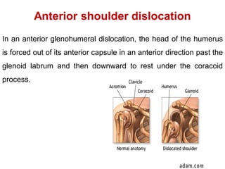
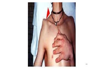
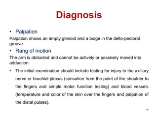
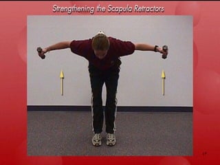
Shoulder Dislocation
1
Shoulder dislocation
Definition:
Dislocation of the glenohumeral joint is a displacement of the
humeral head from its normal position in the glenoid labral
fossa(complete separation of the articular surfaces of the
glenohumeral joint).
Prevalence:
• Shoulder dislocation is a common reason for emergency
room visits and accounts for about 45% of all dislocations
• Traumatic shoulder dislocations are far more common than
non-traumatic forms
Types of shoulder dislocation
According to direction of dislocation
1- Anterior shoulder dislocation ( very common)
2- Posterior shoulder dislocation ( un common)
3- Inferior shoulder dislocation( extremely rare)
According to mechanism of injury
1- Traumatic
2- A traumatic
3
Posterior dislocation
Definition
In a posterior glenohumeral dislocation, the head of the
humerus is forced out of its posterior capsule in an posterior
direction past the glenoid labrm
Types
• Subacromial
• Subglenoid
• Subspinous
5
Prevalence and mechanism of injury
• posterior dislocation represents 3 to 4 % of shoulder
dislocations
• It is caused by posterior driving force of the humerus
while it is adducted, flexed, and internally rotated
Risk factors for posterior shoulder dislocations include:
• seizure
• electric shock
• follow-through from throwing an object
• underlying lesions of the shoulder, such as reverse
Bankart and reverse Hill-Sachs defects
6
Anterior shoulder dislocation
In an anterior glenohumeral dislocation, the head of the humerus
is forced out of its anterior capsule in an anterior direction past the
glenoid labrum and then downward to rest under the coracoid
process.
prevalence
• Anterior shoulder dislocations contribute 96% to 98% of all
shoulder dislocations
• In 90% of cases, anterior shoulder dislocation affects
young individuals(9 out of 10 patients are 21 to 30 years of
age), many of whom are athletes
• The frequency of anterior dislocation exhibits two peaks,
during the second and sixth decades, respectively.
• Men are affected 3 times more often than women
Mechanism of injury
The mechanism of anterior type may be:
• Direct a forward impulse of the elevated,
abducted, and externally rotated arm (e.g.,
during a basketball smash) or
• A fall on the palm of the hand with the arm
outstretched.
9
Diagnosis
• History
Personal data
Chief complaint:
• pain and muscle guarding due to bleeding and
inflammation
• Inability to move the affected UL
10
11
Inspection
The patient supports the injured arm with
the hand of the uninjured side. Signs that
suggest anterior dislocation include a
squared-off appearance of the shoulder
with loss of the normal rounded contour,
bulging of the acromion, and filling of the
delto-pectoral groove
12
Diagnosis
• Palpation
Palpation shows an empty glenoid and a bulge in the delto-pectoral
groove
• Rang of motion
The arm is abducted and cannot be actively or passively moved into
adduction.
• The initial examination should include testing for injury to the axillary
nerve or brachial plexus (sensation from the point of the shoulder to
the fingers and simple motor function testing) and blood vessels
(temperature and color of the skin over the fingers and palpation of
the distal pulses).
13
Diagnosis
Radiological assessment
• Both an antero-posterior and a lateral radiographic view
should be obtained
• Computed tomography (CT) offers the best accuracy
and sensitivity for detecting and evaluating a fracture
andfor assessing the extent of impaction damage.
14
Treatment
1-Reduction
The traditional treatment for anterior shoulder dislocation is reduction
followed by immobilization with the arm in internal rotation for 3 to 6
weeks followed by rehabilitation therapy.
The efficacy of this treatment remains unclear. The recurrence rate can
reach 95% depending on the risk factors, particularly patient age at the
first episode.
15
treatment
Immobilization with the arm in external rotation (ER) after
the first episode has been suggested based on magnetic
resonance imaging (MRI) studies showing that external
rotation increases the amount of tension on the sub-
scapularis muscle and maintains the labrum and capsule in
close contact with the glenoid
16
17
However the ER immobilization could not
reduce the rates of recurrence after primary
anterior shoulder dislocation or improve the
quality of life compared with the IR
immobilization.
treatment
Reduction maneuvers may be :
1-Reduction methods without counter support
e.g Hippo-cratic method (simple traction along
the axis of the arm )
2-Reduction with countersupport on the axilla
18
treatment
2- Post-reduction management
• An antero-posterior radiograph should be obtained to
confirm that complete reduction has been achieved and
to look for concomitant lesions
• After reduction, the patient should be re-evaluated for
nerve and vessel injuries
19
Rehabilitation program
Goals of rehabilitation program
• restoring the normal axis of rotation for the
glenohumeral joint
• optimizing the stabilizing muscles length –
tension relationship
• restoring proper neuromuscular control to the
shoulder complex.
20
I. Protection Phase
A) Protect Tissue Healing
• After reduction shoulder is protected in a shoulder sling
for an average of 6-8 weeks (this have been found to be
the most recommended period to minimize possibility of
recurrent dislocation specially in young patients.
• During the post traumatic period (first 1week), the patient
arm may need to be continuous immobilized to reduce
pain & spasm.
• Afterwards, the arm is only removed from sling during
exercises.
21
B) Promote Tissue Healing
According to patient tolerance, the Following exercises
are gradually administrated:
1- Protected ROM
2- Intermittent muscle setting of the rotator cuff, deltoid and
Biceps Brachii.
3- Grade II are initiated as soon the patient tolerate them.
(Arm is positioned beside body or in resting position).
22
4- Initiate a scapulothoracic exercise program, avoiding
elevated positions of the upper extremity that put stability
at risk.
6- Patients should begin an aerobic training regime with the
lower extremity like stationary biking
23
Precautions
• To avoid disruption the healing of the capsule and other damaged
tissues, ROM into external rotation is performed with the elbow at the
patient’s side, with the shoulder flexed in the sagittal plane, and with
the shoulder in the resting position (in the plane of the scapula,
abducted 55 and 30 to 45 anterior to the frontal plane)
• forearm is moved from in front of the trunk (maximal internal
rotation) to 0 or possibly 10 to 15 external rotation. 24
Contraindications at this stage
• External rotation at
90 degrees
abduction
• Shoulder
extension beyond
0 degree
. 25
26
27
II. Controlled Motion Phase
A) Provide Protection
• Avoid full return to unrestricted activity
• Use sling only when the shoulder is tired or
during activities where protection is needed.
28
B) Increase Shoulder Mobility
• The program begins with the use of an active assistive
ROM exercises
• Mobilization techniques are initiated using all appropriate
glides except the anterior glide
• The posterior joint structures are passively stretched with
horizontal adduction self-stretching techniques.
29
30
31
C) Increase stability and strength
The following muscle groups should be strengthened
1- Shoulder Internal rotators & Adductors: to increase
anterior stability, and support anterior capsule.
2- Shoulder external rotators: To reduce anterior translation
forces, and participate in the deltoid-rotator cuff force
couple when abducting and laterally rotating the humerus
3- Scapular stability is essential for normal shoulder
function.
4- PNF techniques to help reestablish neuromuscular
control.
32
Strengthening exercises are graduated as follows:
1- Isometric exercises with joint position at the side of the
trunk and progressed to various pain-free positions
2- Partial weight bearing and stabilization exercises
3- Dynamic resistance limiting external rotation to 50
degree and avoiding the position of dislocation
33
4- At 3 weeks, supervised isokinetic resistance for internal
rotation and adduction are initiated.
5- At 5 weeks, all shoulder motions are incorporated into
isokinetic program except for external rotation in 90
degrees abduction
34
35
36
Stabilization Exercises
Closed chain exercises (CKC)
• Start with the hands on the ground or table for
strengthening the scapular stabilizers more aggressively.
These exercises should begin on a stable surface like a
table, progressing the amount of weight bearing by
advancing from the table to the ground.
• Advancing to a less stable surface like a BAPS board or
Swiss ball to reestablish neuromuscular control
"proprioceptive training". 37
38
39
III. Return to function phase
A) Restore functional control
• Coordination training to synchronize action of shoulder
and scapular muscles.
• Endurance training for all shoulder and scapular
muscles.
• Prophylactic stretching to gain full ROM.
• Scapular and rotator cuff strengthening ex.
• As stability improves, progress to:
1-Eccentric training to maximum load
2-Increasing speed and control
3-Simulating desired functional patterns for activity
40
• CKC become more challenging by adding motion to the
demands of the stabilization e.g. weight shifting on a
Fitter and closed kinetic strengthening on a stair climber
for endurance are started.
• Plyometric exercises can be added to improve dynamic
control.
41
42
43
B) Return to activity
• It is important that the patient learns to recognize signs of fatigue
and impingement and stays within the tolerance of the tissues.
• Return to full activity when there is
1- No muscle imbalance
2- Good coordination
3- Apprehension test is normal
• Total rehabilitation time usually lasts from 2.5-4 months. But it may It
last as long as 20 weeks depending on the patient's shoulder
strength, lack of pain, and the ability to protect the involved
shoulder.
44
• Between 20 and 26 weeks will be required for
rehabilitation of athletes.
• Some therapist and physicians recommend use
of a protective shoulders harness while
participating in athletic activities.
45
Posterior dislocation
• the management approach is the same as anterior
dislocation with one exception
• Avoid the position of flexion/ adduction / internal rotation
in the acute (protection 0 phase.
• Immobilization with hand hanging freely by the side of
the body is used if it is more comfortable than the sling
used
46

shoulder dislocation.pdf

  • 1.
  • 2.
    Shoulder dislocation Definition: Dislocation ofthe glenohumeral joint is a displacement of the humeral head from its normal position in the glenoid labral fossa(complete separation of the articular surfaces of the glenohumeral joint). Prevalence: • Shoulder dislocation is a common reason for emergency room visits and accounts for about 45% of all dislocations • Traumatic shoulder dislocations are far more common than non-traumatic forms
  • 3.
    Types of shoulderdislocation According to direction of dislocation 1- Anterior shoulder dislocation ( very common) 2- Posterior shoulder dislocation ( un common) 3- Inferior shoulder dislocation( extremely rare) According to mechanism of injury 1- Traumatic 2- A traumatic 3
  • 4.
    Posterior dislocation Definition In aposterior glenohumeral dislocation, the head of the humerus is forced out of its posterior capsule in an posterior direction past the glenoid labrm Types • Subacromial • Subglenoid • Subspinous
  • 5.
  • 6.
    Prevalence and mechanismof injury • posterior dislocation represents 3 to 4 % of shoulder dislocations • It is caused by posterior driving force of the humerus while it is adducted, flexed, and internally rotated Risk factors for posterior shoulder dislocations include: • seizure • electric shock • follow-through from throwing an object • underlying lesions of the shoulder, such as reverse Bankart and reverse Hill-Sachs defects 6
  • 7.
    Anterior shoulder dislocation Inan anterior glenohumeral dislocation, the head of the humerus is forced out of its anterior capsule in an anterior direction past the glenoid labrum and then downward to rest under the coracoid process.
  • 8.
    prevalence • Anterior shoulderdislocations contribute 96% to 98% of all shoulder dislocations • In 90% of cases, anterior shoulder dislocation affects young individuals(9 out of 10 patients are 21 to 30 years of age), many of whom are athletes • The frequency of anterior dislocation exhibits two peaks, during the second and sixth decades, respectively. • Men are affected 3 times more often than women
  • 9.
    Mechanism of injury Themechanism of anterior type may be: • Direct a forward impulse of the elevated, abducted, and externally rotated arm (e.g., during a basketball smash) or • A fall on the palm of the hand with the arm outstretched. 9
  • 10.
    Diagnosis • History Personal data Chiefcomplaint: • pain and muscle guarding due to bleeding and inflammation • Inability to move the affected UL 10
  • 11.
    11 Inspection The patient supportsthe injured arm with the hand of the uninjured side. Signs that suggest anterior dislocation include a squared-off appearance of the shoulder with loss of the normal rounded contour, bulging of the acromion, and filling of the delto-pectoral groove
  • 12.
  • 13.
    Diagnosis • Palpation Palpation showsan empty glenoid and a bulge in the delto-pectoral groove • Rang of motion The arm is abducted and cannot be actively or passively moved into adduction. • The initial examination should include testing for injury to the axillary nerve or brachial plexus (sensation from the point of the shoulder to the fingers and simple motor function testing) and blood vessels (temperature and color of the skin over the fingers and palpation of the distal pulses). 13
  • 14.
    Diagnosis Radiological assessment • Bothan antero-posterior and a lateral radiographic view should be obtained • Computed tomography (CT) offers the best accuracy and sensitivity for detecting and evaluating a fracture andfor assessing the extent of impaction damage. 14
  • 15.
    Treatment 1-Reduction The traditional treatmentfor anterior shoulder dislocation is reduction followed by immobilization with the arm in internal rotation for 3 to 6 weeks followed by rehabilitation therapy. The efficacy of this treatment remains unclear. The recurrence rate can reach 95% depending on the risk factors, particularly patient age at the first episode. 15
  • 16.
    treatment Immobilization with thearm in external rotation (ER) after the first episode has been suggested based on magnetic resonance imaging (MRI) studies showing that external rotation increases the amount of tension on the sub- scapularis muscle and maintains the labrum and capsule in close contact with the glenoid 16
  • 17.
    17 However the ERimmobilization could not reduce the rates of recurrence after primary anterior shoulder dislocation or improve the quality of life compared with the IR immobilization.
  • 18.
    treatment Reduction maneuvers maybe : 1-Reduction methods without counter support e.g Hippo-cratic method (simple traction along the axis of the arm ) 2-Reduction with countersupport on the axilla 18
  • 19.
    treatment 2- Post-reduction management •An antero-posterior radiograph should be obtained to confirm that complete reduction has been achieved and to look for concomitant lesions • After reduction, the patient should be re-evaluated for nerve and vessel injuries 19
  • 20.
    Rehabilitation program Goals ofrehabilitation program • restoring the normal axis of rotation for the glenohumeral joint • optimizing the stabilizing muscles length – tension relationship • restoring proper neuromuscular control to the shoulder complex. 20
  • 21.
    I. Protection Phase A)Protect Tissue Healing • After reduction shoulder is protected in a shoulder sling for an average of 6-8 weeks (this have been found to be the most recommended period to minimize possibility of recurrent dislocation specially in young patients. • During the post traumatic period (first 1week), the patient arm may need to be continuous immobilized to reduce pain & spasm. • Afterwards, the arm is only removed from sling during exercises. 21
  • 22.
    B) Promote TissueHealing According to patient tolerance, the Following exercises are gradually administrated: 1- Protected ROM 2- Intermittent muscle setting of the rotator cuff, deltoid and Biceps Brachii. 3- Grade II are initiated as soon the patient tolerate them. (Arm is positioned beside body or in resting position). 22
  • 23.
    4- Initiate ascapulothoracic exercise program, avoiding elevated positions of the upper extremity that put stability at risk. 6- Patients should begin an aerobic training regime with the lower extremity like stationary biking 23
  • 24.
    Precautions • To avoiddisruption the healing of the capsule and other damaged tissues, ROM into external rotation is performed with the elbow at the patient’s side, with the shoulder flexed in the sagittal plane, and with the shoulder in the resting position (in the plane of the scapula, abducted 55 and 30 to 45 anterior to the frontal plane) • forearm is moved from in front of the trunk (maximal internal rotation) to 0 or possibly 10 to 15 external rotation. 24
  • 25.
    Contraindications at thisstage • External rotation at 90 degrees abduction • Shoulder extension beyond 0 degree . 25
  • 26.
  • 27.
  • 28.
    II. Controlled MotionPhase A) Provide Protection • Avoid full return to unrestricted activity • Use sling only when the shoulder is tired or during activities where protection is needed. 28
  • 29.
    B) Increase ShoulderMobility • The program begins with the use of an active assistive ROM exercises • Mobilization techniques are initiated using all appropriate glides except the anterior glide • The posterior joint structures are passively stretched with horizontal adduction self-stretching techniques. 29
  • 30.
  • 31.
  • 32.
    C) Increase stabilityand strength The following muscle groups should be strengthened 1- Shoulder Internal rotators & Adductors: to increase anterior stability, and support anterior capsule. 2- Shoulder external rotators: To reduce anterior translation forces, and participate in the deltoid-rotator cuff force couple when abducting and laterally rotating the humerus 3- Scapular stability is essential for normal shoulder function. 4- PNF techniques to help reestablish neuromuscular control. 32
  • 33.
    Strengthening exercises aregraduated as follows: 1- Isometric exercises with joint position at the side of the trunk and progressed to various pain-free positions 2- Partial weight bearing and stabilization exercises 3- Dynamic resistance limiting external rotation to 50 degree and avoiding the position of dislocation 33
  • 34.
    4- At 3weeks, supervised isokinetic resistance for internal rotation and adduction are initiated. 5- At 5 weeks, all shoulder motions are incorporated into isokinetic program except for external rotation in 90 degrees abduction 34
  • 35.
  • 36.
  • 37.
    Stabilization Exercises Closed chainexercises (CKC) • Start with the hands on the ground or table for strengthening the scapular stabilizers more aggressively. These exercises should begin on a stable surface like a table, progressing the amount of weight bearing by advancing from the table to the ground. • Advancing to a less stable surface like a BAPS board or Swiss ball to reestablish neuromuscular control "proprioceptive training". 37
  • 38.
  • 39.
  • 40.
    III. Return tofunction phase A) Restore functional control • Coordination training to synchronize action of shoulder and scapular muscles. • Endurance training for all shoulder and scapular muscles. • Prophylactic stretching to gain full ROM. • Scapular and rotator cuff strengthening ex. • As stability improves, progress to: 1-Eccentric training to maximum load 2-Increasing speed and control 3-Simulating desired functional patterns for activity 40
  • 41.
    • CKC becomemore challenging by adding motion to the demands of the stabilization e.g. weight shifting on a Fitter and closed kinetic strengthening on a stair climber for endurance are started. • Plyometric exercises can be added to improve dynamic control. 41
  • 42.
  • 43.
  • 44.
    B) Return toactivity • It is important that the patient learns to recognize signs of fatigue and impingement and stays within the tolerance of the tissues. • Return to full activity when there is 1- No muscle imbalance 2- Good coordination 3- Apprehension test is normal • Total rehabilitation time usually lasts from 2.5-4 months. But it may It last as long as 20 weeks depending on the patient's shoulder strength, lack of pain, and the ability to protect the involved shoulder. 44
  • 45.
    • Between 20and 26 weeks will be required for rehabilitation of athletes. • Some therapist and physicians recommend use of a protective shoulders harness while participating in athletic activities. 45
  • 46.
    Posterior dislocation • themanagement approach is the same as anterior dislocation with one exception • Avoid the position of flexion/ adduction / internal rotation in the acute (protection 0 phase. • Immobilization with hand hanging freely by the side of the body is used if it is more comfortable than the sling used 46