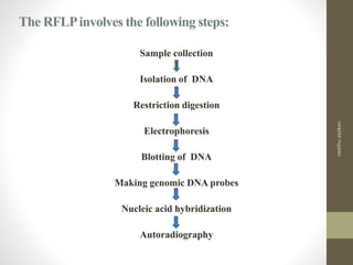
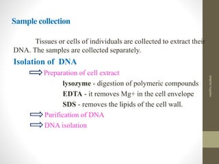
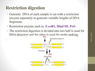
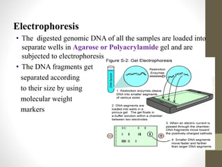
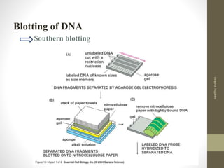
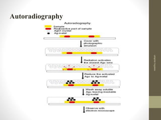
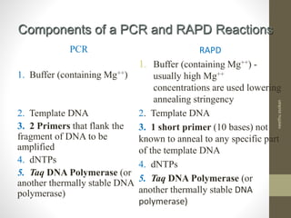
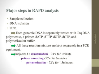
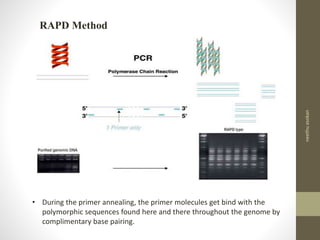
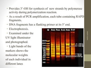
neethuasokan
RFLP
Definition
• The variation in the restriction DNA fragment lengths
between individuals of a species is called restriction
fragment length polymorphism (RFLP).
• Laboratory technique to analyze and compare DNAs of
two or more individuals of a species or of different
species.
• Genetic structure of all the individuals of a species is
same, but at DNA level there are so many single base
variations between the individuals due to point mutation.
• If a single base is altered due to mutation restriction
enzymes never cut at the target sites.
neethuasokan
HISTORY
• Shortly after Kary Mullis invented the Polymerase Chain
Reaction (PCR) it was realized that short primers would
bind to several locations in a genome and thus could
produce multiple fragments.
• Williams et al. (1990) developed Random Amplified
Polymorphic DNA (RAPD) a technique using very short
10 base primers to generate random fragments from
template DNAs.
• RAPD fragments can be separated and used as genetic
markers or a kind of DNA fingerprint.
neethuasokan
RFLPTECHNIQUES
neethuasokan
The RFLPinvolves the following steps:
Sample collection
Isolation of DNA
Restriction digestion
Electrophoresis
Blotting of DNA
Making genomic DNA probes
Nucleic acid hybridization
Autoradiography
neethuasokan
Sample collection
Tissues or cells of individuals are collected to extract their
DNA. The samples are collected separately.
Isolation of DNA
Preparation of cell extract
lysozyme - digestion of polymeric compounds
EDTA - it removes Mg+ in the cell envelope
SDS - removes the lipids of the cell wall.
Purification of DNA
DNA isolation
neethuasokan
Restriction digestion
• Genomic DNA of each sample is cut with a restriction
enzyme separately to generate variable lengths of DNA
fragments.
• Restriction enzyme such as E.coR1, Hind ІІІ, Pst1.
• The restriction digestion is divided into two half is used for
DNA detection and the other is used for probe making.
.
neethuasokan
Electrophoresis
• The digested genomic DNA of all the samples are loaded into
separate wells in Agarose or Polyacrylamide gel and are
subjected to electrophoresis.
• The DNA fragments get
separated according
to their size by using
molecular weight
markers
neethuasokan
Blotting of DNA
Southern blotting
neethuasokan
• Each sample is electrophoresed to separate the DNA
fragments.
• Fragments of 0.5-2 kb are extracted from the gel and cloned
in pUC21 vector to construct rDNAs.
• These rDNAs are amplified by introducing them into
bacterial host and the amplified rDNAs are reisolated.
• Restriction digestion & electrophoresis.
• Radiolabelling by Nick translation or End labelling
• Genomic probes are formed.
Making genomic DNA probes
neethuasokan
Nucleic acid hybridization
neethuasokan
Autoradiography
neethuasokan
RFLPs are considered as the first class genetic markers to
construct high resolution linkage maps of chromosomes.
Used to construct chromosome map of human being, rice,
wheat, maize, and microbes.
Single gene diseases in man, plants and animals can be
identified with RFLP markers.
Monitoring inheritance of agronomic traits
Diagnostic in genetically inherited disease
Pedigree analysis,
Forensic typing - Parentage analysis
Identifying hybrids
Species level relationship
 Also in some case at higher level relationship
Applications of RFLP
neethuasokan
Advantages
Fingerprinting technique replacing RFLP
Highly polymorphic
High reproducibility
Identify through absent or present of fragment
Characters can be increased by changing the RE and
nucleotide at selective primers
Codominantly inherited.
neethuasokan
Disadvantages
Dominant – lose the codominant character
Homology – ability to differentiate different fragment with similar
size
 Mutation rate – high homoplasy
– High levels of variation - similarity between two taxa are low, so
both character and distance measures and tree reconstruction
programmes are increasingly inaccurate
– if levels of variation are high - Homoplasy
 Scoring - bias
neethuasokan
Definition
• The RAPD is a PCR based method to detect variations
between individuals of a species by selective amplification of
some polymorphic sequences in their genomes.
• Developed by J.G.K.Williams et.al. in 1991.
• Only least number of DNA fragments are considered for
RAPD analysis.
• The set of DNAs generated by the random PCR is called
RAPD.
• The RAPDs of one individual is differ from the RAPDs of
other individuals both in number and size of DNA fragments.
RAPD
neethuasokan
Components of a PCR and RAPD Reactions
PCR
1. Buffer (containing Mg++)
2. Template DNA
3. 2 Primers that flank the
fragment of DNA to be
amplified
4. dNTPs
5. Taq DNA Polymerase (or
another thermally stable DNA
polymerase)
RAPD
1. Buffer (containing Mg++) -
usually high Mg++
concentrations are used lowering
annealing stringency
2. Template DNA
3. 1 short primer (10 bases) not
known to anneal to any specific part
of the template DNA
4. dNTPs
5. Taq DNA Polymerase (or
another thermally stable DNA
polymerase)
neethuasokan
Major steps in RAPD analysis
• Sample collection
• DNA isolation
• PCR
Each genomic DNA is separately treated with Taq DNA
polymerase, a primer, dATP ,dTTP, dGTP, dCTP, and
polymerization buffer.
All these reaction mixture are kept separately in a PCR
equipment.
subjected t o denaturation – 94°c for 1minute
primer annealing -36°c for 2minutes
polymerization – 72°c for 1.5minutes.
neethuasokan
• During the primer annealing, the primer molecules get bind with the
polymorphic sequences found here and there throughout the genome by
complimentary base pairing.
RAPD Method
neethuasokan
• Provides 3’-OH for synthesis of new strands by polymerase
activity during polymerization reaction.
• As a result of PCR amplification, each tube containing RAPD
fragments.
• DNA fragments has a flanking primer at its 5’ end.
• Electrophoresis.
• Examined under the
UV light illuminator
and photographed.
• Light bands of the
markers shows the
molecular weights
of each individual in
different lanes
neethuasokan
Uses of RAPD
• RAPDs are used as genetic markers for constructing genetic maps of
higher organisms.
• Helps to identify genes of high economic value through comparison
of RAPD fragments.
• Distinguishes one individuals from others of a species as
fingerprinting analysis
• Determine specific genes in chromosomes
It has become widely used in the study of
• Genetic diversity/polymorphism,
• Germplasm characterization,
• Genetic structure of populations,
• Domestication,
neethuasokan
• Detection of somoclonal variation,
• Cultivar identification,
• Hybrid purity,
• Genome mapping,
• Developing genetic markers linked to a trait in question,
• Population and evolutionary genetics,
• Plant and animal breeding,
• Animal-plant-microbe interactions,
• Pesticide/Herbicide resistance,
neethuasokan
Advantages of RAPD
• No need for species specific probes in RAPD
• Probes prepared for one species may also used for other
species
• Quick method
• Can be performed in crude DNA samples also
• Requires only a small amount of samples
• Doesn’t require radioactive probes and hybridization.
• It detects dominant variations in the genome
neethuasokan
Disadvantages
• Profiling is dependent on the reaction conditions
• Profiles are not able to distinguish heterozygous from
homozygous individuals- dominant marker
• Lack of a prior knowledge on the identity of the amplification
products.
• Problems with reproducibility (sensitive to changes in the
quality of DNA, PCR components and PCR conditions).
• Problems of co-migration (do equal-sized bands correspond to
the same homologous DNA fragment). Gel electrophoresis can
separate DNA quantitatively, cannot separate equal-sized
fragments qualitatively (i.e. according to base sequence).
neethuasokan
References
• N.Senthikumar & G.Gurusubramanian, Random amplified
polymorphic DNA (RAPD) markers and its applications 2011,
www.sciencevision.in
• Timothy G. Standish, Ph. D. , Random Amplified Polymorphic
DNA , (ppt).
• M.O. Dayhoff and R.V. ECK, Molecular techniques,(ppt).
• Biotechnology, V.Kumaresan, Saras publications.
• Biotechnology, U.Sathyanarayana.
• Gene Biotechnology, S.Jogdand.
neethuasokan

RAPD, RFLP

  • 1.
  • 2.
    RFLP Definition • The variationin the restriction DNA fragment lengths between individuals of a species is called restriction fragment length polymorphism (RFLP). • Laboratory technique to analyze and compare DNAs of two or more individuals of a species or of different species. • Genetic structure of all the individuals of a species is same, but at DNA level there are so many single base variations between the individuals due to point mutation. • If a single base is altered due to mutation restriction enzymes never cut at the target sites. neethuasokan
  • 3.
    HISTORY • Shortly afterKary Mullis invented the Polymerase Chain Reaction (PCR) it was realized that short primers would bind to several locations in a genome and thus could produce multiple fragments. • Williams et al. (1990) developed Random Amplified Polymorphic DNA (RAPD) a technique using very short 10 base primers to generate random fragments from template DNAs. • RAPD fragments can be separated and used as genetic markers or a kind of DNA fingerprint. neethuasokan
  • 4.
  • 5.
    The RFLPinvolves thefollowing steps: Sample collection Isolation of DNA Restriction digestion Electrophoresis Blotting of DNA Making genomic DNA probes Nucleic acid hybridization Autoradiography neethuasokan
  • 6.
    Sample collection Tissues orcells of individuals are collected to extract their DNA. The samples are collected separately. Isolation of DNA Preparation of cell extract lysozyme - digestion of polymeric compounds EDTA - it removes Mg+ in the cell envelope SDS - removes the lipids of the cell wall. Purification of DNA DNA isolation neethuasokan
  • 7.
    Restriction digestion • GenomicDNA of each sample is cut with a restriction enzyme separately to generate variable lengths of DNA fragments. • Restriction enzyme such as E.coR1, Hind ІІІ, Pst1. • The restriction digestion is divided into two half is used for DNA detection and the other is used for probe making. . neethuasokan
  • 8.
    Electrophoresis • The digestedgenomic DNA of all the samples are loaded into separate wells in Agarose or Polyacrylamide gel and are subjected to electrophoresis. • The DNA fragments get separated according to their size by using molecular weight markers neethuasokan
  • 9.
    Blotting of DNA Southernblotting neethuasokan
  • 10.
    • Each sampleis electrophoresed to separate the DNA fragments. • Fragments of 0.5-2 kb are extracted from the gel and cloned in pUC21 vector to construct rDNAs. • These rDNAs are amplified by introducing them into bacterial host and the amplified rDNAs are reisolated. • Restriction digestion & electrophoresis. • Radiolabelling by Nick translation or End labelling • Genomic probes are formed. Making genomic DNA probes neethuasokan
  • 11.
  • 12.
  • 13.
    RFLPs are consideredas the first class genetic markers to construct high resolution linkage maps of chromosomes. Used to construct chromosome map of human being, rice, wheat, maize, and microbes. Single gene diseases in man, plants and animals can be identified with RFLP markers. Monitoring inheritance of agronomic traits Diagnostic in genetically inherited disease Pedigree analysis, Forensic typing - Parentage analysis Identifying hybrids Species level relationship  Also in some case at higher level relationship Applications of RFLP neethuasokan
  • 14.
    Advantages Fingerprinting technique replacingRFLP Highly polymorphic High reproducibility Identify through absent or present of fragment Characters can be increased by changing the RE and nucleotide at selective primers Codominantly inherited. neethuasokan
  • 15.
    Disadvantages Dominant – losethe codominant character Homology – ability to differentiate different fragment with similar size  Mutation rate – high homoplasy – High levels of variation - similarity between two taxa are low, so both character and distance measures and tree reconstruction programmes are increasingly inaccurate – if levels of variation are high - Homoplasy  Scoring - bias neethuasokan
  • 16.
    Definition • The RAPDis a PCR based method to detect variations between individuals of a species by selective amplification of some polymorphic sequences in their genomes. • Developed by J.G.K.Williams et.al. in 1991. • Only least number of DNA fragments are considered for RAPD analysis. • The set of DNAs generated by the random PCR is called RAPD. • The RAPDs of one individual is differ from the RAPDs of other individuals both in number and size of DNA fragments. RAPD neethuasokan
  • 17.
    Components of aPCR and RAPD Reactions PCR 1. Buffer (containing Mg++) 2. Template DNA 3. 2 Primers that flank the fragment of DNA to be amplified 4. dNTPs 5. Taq DNA Polymerase (or another thermally stable DNA polymerase) RAPD 1. Buffer (containing Mg++) - usually high Mg++ concentrations are used lowering annealing stringency 2. Template DNA 3. 1 short primer (10 bases) not known to anneal to any specific part of the template DNA 4. dNTPs 5. Taq DNA Polymerase (or another thermally stable DNA polymerase) neethuasokan
  • 18.
    Major steps inRAPD analysis • Sample collection • DNA isolation • PCR Each genomic DNA is separately treated with Taq DNA polymerase, a primer, dATP ,dTTP, dGTP, dCTP, and polymerization buffer. All these reaction mixture are kept separately in a PCR equipment. subjected t o denaturation – 94°c for 1minute primer annealing -36°c for 2minutes polymerization – 72°c for 1.5minutes. neethuasokan
  • 19.
    • During theprimer annealing, the primer molecules get bind with the polymorphic sequences found here and there throughout the genome by complimentary base pairing. RAPD Method neethuasokan
  • 20.
    • Provides 3’-OHfor synthesis of new strands by polymerase activity during polymerization reaction. • As a result of PCR amplification, each tube containing RAPD fragments. • DNA fragments has a flanking primer at its 5’ end. • Electrophoresis. • Examined under the UV light illuminator and photographed. • Light bands of the markers shows the molecular weights of each individual in different lanes neethuasokan
  • 21.
    Uses of RAPD •RAPDs are used as genetic markers for constructing genetic maps of higher organisms. • Helps to identify genes of high economic value through comparison of RAPD fragments. • Distinguishes one individuals from others of a species as fingerprinting analysis • Determine specific genes in chromosomes It has become widely used in the study of • Genetic diversity/polymorphism, • Germplasm characterization, • Genetic structure of populations, • Domestication, neethuasokan
  • 22.
    • Detection ofsomoclonal variation, • Cultivar identification, • Hybrid purity, • Genome mapping, • Developing genetic markers linked to a trait in question, • Population and evolutionary genetics, • Plant and animal breeding, • Animal-plant-microbe interactions, • Pesticide/Herbicide resistance, neethuasokan
  • 23.
    Advantages of RAPD •No need for species specific probes in RAPD • Probes prepared for one species may also used for other species • Quick method • Can be performed in crude DNA samples also • Requires only a small amount of samples • Doesn’t require radioactive probes and hybridization. • It detects dominant variations in the genome neethuasokan
  • 24.
    Disadvantages • Profiling isdependent on the reaction conditions • Profiles are not able to distinguish heterozygous from homozygous individuals- dominant marker • Lack of a prior knowledge on the identity of the amplification products. • Problems with reproducibility (sensitive to changes in the quality of DNA, PCR components and PCR conditions). • Problems of co-migration (do equal-sized bands correspond to the same homologous DNA fragment). Gel electrophoresis can separate DNA quantitatively, cannot separate equal-sized fragments qualitatively (i.e. according to base sequence). neethuasokan
  • 25.
    References • N.Senthikumar &G.Gurusubramanian, Random amplified polymorphic DNA (RAPD) markers and its applications 2011, www.sciencevision.in • Timothy G. Standish, Ph. D. , Random Amplified Polymorphic DNA , (ppt). • M.O. Dayhoff and R.V. ECK, Molecular techniques,(ppt). • Biotechnology, V.Kumaresan, Saras publications. • Biotechnology, U.Sathyanarayana. • Gene Biotechnology, S.Jogdand. neethuasokan