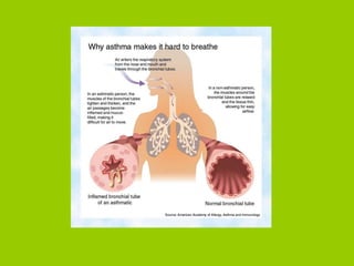
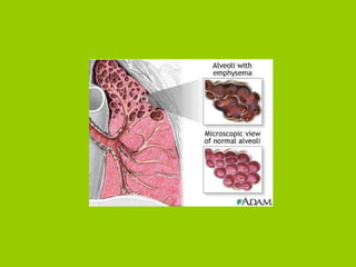
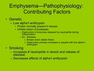
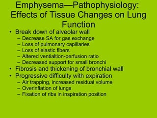
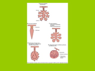
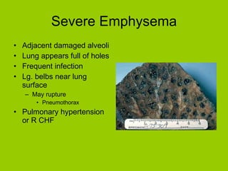
Respiratory Disorders Chapter 19 Pgs 310-360
Homework Assignment Due Tuesday Oct 18  It does NOT have to be typed! You may work in groups Answers MUST be in your own words Case Study A a, b,c, d, f, g, h, o Case Study B a, c, g, h, i, j Case Study C a, b, g, h, i
Overview Diagnostic Tests General Manifestations of Respiratory Disease Infectious Diseases Upper respiratory tract infections Common cold Sinusitis Lower respiratory tract infections RSV Pneumonia Obstructive Lung Diseases Lung Cancer Asthma Chronic Obstructive Pulmonary Disease (COPD) Emphysema Chronic Bronchitis
Diagnostic Tests Spirometry Arterial blood gas determination Oximeters Exercise tolerance Radiography Bronchoscopy Culture, sensitivity tests
General Manifestations of Respiratory Disease Sneezing Coughing Irritation Controlled by medulla Constant, dry unproductive vs. productive cough Sputum Mucus discharge Yellowish-green Rusty, dark-colored Thick, sticky Hemoptysis
Manifestations Breathing patterns and characteristics Kussmaul respiration Labored respiration, prolonged inspiration/expiration times Wheezing Stridors Breath sounds Rales Rhonchi Absence
Manifestations Dyspnea Severe Orthopnea Paroxysmal nocturnal dyspnea Cyanosis Pleural pain Friction rub Clubbed fingers Changes in ABG (arterial blood gases Hypoxemia    inadequate oxygen in blood Hypoxia    inadequate oxygen supply to cells
Causes of Hypoxia Low RBC, Hb Circulation impairment Excessive release of oxygen from RBC Impaired respiratory function CO poisoning
Upper Respiratory Tract Infections:  Common Cold (Infectious Rhinitis) Viral (rhinovirus) Spread thru respiratory droplets Highly contagious Initially mucous membranes of nose, pharynx swollen, increased secretions Signs Nasal congestion and watery discharge Mouth breathing Change in tone of voice Sore throat, headache, slight fever Cough
Common Cold Infection, inflammation can spread Laryngitis Bronchitis Treatment is symptomatic Acetaminophen Decongestant Antihistamine Humidifiers Are antibiotics prescribed?
Secondary Bacterial Infections
Sinusitis Secondary bacterial infection Obstruct drainage in 1 or more paranasal sinuses Common causative organisms Pneumococci Streptococci Haemophilus influenzae Exudate accumulates Signs Nasal congestion, fever, sore throat Diagnosis confirmed by radiograph, transillumination Decongestants, analgesics Antibiotics
 
Lower Respiratory Tract Infections:  Bronchiolitis (RSV Infection) 2-12 month Caused by syncytial virus Transmitted by oral droplet Predisposing factors (asthma, smoking) Causes necrosis and inflammation of small bronchi and bronchioles Signs Wheezing and dyspnea Rapid, shallow respirations Cough Rales Chest retractions Fever Treatment Supportive and symptomatic
Pneumonia Primary acute or secondary Risk following aspiration, inflammation in lung Transmission Inhaling virus Resident bacteria spreading along mucosa Aspiration in secretions
Classification of the Pneumonias Causative agent Virus, bacteria, fungus Lobar is typically bacterial Pneumococcus Anatomical distribution of lesion Both lungs or lobar Pathophysiologic changes Viral    changes in interstitial tissue or alveolar septae Pneumococcal    alveoli inflamed and fluid filled Exudate Epidemiologic categories Nosocomial Community acquired
 
 
Lobar Pneumonia Streptococcal pneumoniae, pneumococcal Infection localized in 1 or more lobes
Stages of Pneumonia Congestion Inflammation and vascular congestion in alveolar wall Exudate forms in alveoli Interferes with oxygen diffusion Consolidation Neutrophils, RBCs, fibrin accum in exudate Form solid mass RBCs break down, infection resolves Macrophages break down exudate Expectorated or resorbed
Consolidation
Pneumonia Pleurae typically involved Infection in pleural cavity Emphysema Adhesions between membranes Manifestations Sudden onset Systemic signs:  high fever, chills, fatigue Dyspnea, tachycardia Pleuritic pain Rales Productive cough
Pneumonia Treatment Antibacterials (Penicillin) Supportive measures Pneumococcal vaccine
Obstructive Lung Disease:  Lung Cancer Primary or secondary; benign rare Primary is major cause of death Linked with cigarette smoking Metastases develop freq in lung b/c: Venous return and lymph vessels bring tumor cells from distant site in body    heart    lung Poor prognosis
Normal Lung vs. Cancerous Lung
Types of Lung Cancer Bronchogenic carcinoma Most common Arise from bronchial epithelium Squamous cell carcinoma Develop from epithelial lining in bronchus Project into airway Adenocarcinomas and bronchoalveolar cell carcinoma Found on lung periphery Less symptomatic, more difficult to treat Small cell carcinoma Rapidly growing; located near major bronchus Invasive and metastize early in dev Large cell carcinoma Found in periphery Consist of large, undifferentiated cells Rapid growth rate, metastize early
Bronchogenic Carcinoma
Lung Cancer—Pathophysiology  First change Metaplasia, change in epithelial tissue Smoking, chronic irritation Reversible if irritation removed Loss of ciliated pseudostratified epithelium More vulnerable to irritants Next Dysplasia, carcinoma develop Hard to detect
 
Lung Cancer—Effects of Tumors Obstruction of air flow Inflammation Pleural effusion, hemothorax, pneumothorax Paraneoplastic syndrome
Lung Cancer—Etiology  General stats 173,330 new cases each year 160,440 deaths per year Smoking (primary and secondary) 87% of lung cancers related to smoking 1 out of 10 chances of developing lung cancer Maybe a genetic factor Occupational exposure to carcinogens Irritant that leads to chronic inflammation Cause cell changes Smoking:  ciliated columnar    squamous
Lung Cancer—Signs and Symptoms Insidious onset Normally metastized before diagnosis 4 possible categories of signs of lung cancer Direct effects of tumor Systemic effects of cancer Paraneoplastic syndromes Metastizes at other sites
Lung Cancer—Early Signs (#1) Persistent, productive cough, dyspnea, wheezing Detection on chest X-ray Hemoptysis Pleural involvement Chest pain Hoarseness Facial, arm edema; headaches Dysphagia
Lung Cancer (#2-4) Systemic signs Wt. loss, anemia, fatigue Paraneoplastic syndrome Signs of endocrine disorder Depends on hormone being secreted Signs of metastasis depends on site
Lung Cancer—Diagnostic Tests  Chest X-rays Bronchoscopy Pulmonary function tests
 
 
Lung Cancer—Treatment  Surgery on localized lesions Chemotherapy and radiation Poor prognosis unless tumor in early stages of development
Asthma Periodic episodes of severe but reversible bronchial obstruction Frequency may lead to irreversible damage and COPD 2 types Extrinsic asthma Acute episodes triggered by type I hypersensitivities Onset in childhood Intrinsic asthma Onset during adulthood Stimuli target hyperresponsive tissue = acute attack
 
Asthma—Pathophysiology:  Acute Attack Both types Bronchi and bronchioles respond to stimulus with 3 changes Bronchoconstriction Inflammation of mucosa with edema Increased secretion of thick mucus in passageways Changes may result in partial or total obstruction of airways Interferes with oxygen supply, air flow
 
Asthma—Pathophysiology:  Extrinsic Asthma 1 st  stage Allergen reacts with IgE on previously sensitized mast cells in resp. mucosa Release chemical mediators (histamine, prostaglandin) Stimulates vagus nerve Reflex bronchoconstriction 2 nd  stage Hours later Increased leukocytes release more chemical mediators Prolong bronchoconst and epithelial damage Increase WBC Obstruction, hypoxia
Asthma—Pathophysiology:  Partial Obstruction Small bronchi, bronchioles Air trapping with hyperinflation of lungs Air only partially expired Expiration passive Now less force to move air out Forced collapses bronchial wall Even more difficult to expire Increased residual volume More difficult to inspire fresh air, cough
Asthma—Pathophysiology:   Total Obstruction Mucus plugs completely block Air in distal section diffuses out Cannot be replaced Lung in that section collapses Both (partial and total) lead to hypoxia and hypoxemia Status asthmaticus Persisant severe asthma attack Does not respond to therapy Can be fatal Chronic asthma and COPD may develop Irreversible damage in lungs
Asthma—Etiology  Family history of hay fever, asthma, eczema Significant rise due to: Sedentary lifestyles and obesity Increased time indoors Increased air pollution
Asthma—Signs and Symptoms Cough, dyspnea, tight feeling in chest Wheezing Rapid, labored breathing Thick, sticky mucus coughed up Tachycardia and pulse paradoxus Pulse differs on inspiration and expiration Hypoxia Respiratory acidosis Severe respiratory distress Respiratory failure
Asthma—Treatment  General measures Determine allergies Avoid triggers Acute attacks Inhalers Bronchodilators (albuterol) Most effective at 1 st  indication of attack Controlled breathing techniques and decrease anxiety Glucocorticoids Hospital care—status asthmaticus Prophylaxis and treatment of chronic asthma Leukotrine receptor antagonists (Singulair) Block inflammation response Taken regularly, not effective for acute attacks Cromolyn sodium Inhibits release of chemical mediators from sensitized mast cells Not effective for acute attacks
Chronic Obstructive Pulmonary Disease (COPD) Progressive tissue damage and obstruction of airways Affect individual’s ability to work and function indep Eventual resp failure Leads to R CHF Includes Emphysema Chronic bronchitis Asthma
Emphysema—Pathophysiology  Significant change is destruction of alveolar walls and spaces Leads to lg, inflated alveoli Classified by specific location of changes Ex:  Distal alveoli emphysema Ex:  Bronchiolar emphysema
 
 
Emphysema—Pathophysiology:  Contributing Factors Genetic Low alpha1-antitrypsin Protein normally present in tissues Inhibits action of proteases Destruction of enzymes released by neutrophils during inflammation Ex:  Elastase Breaks down elastic fibers Destructive process increases in people with low alpha1-antitrypsin Smoking Increases # neutrophils in alveoli and release of elastase Decreases effects of alpha1-antityrpsin
Emphysema—Pathophysiology:  Effects of Tissue Changes on Lung Function Break down of alveolar wall Decrease SA for gas exchange Loss of pulmonary capillaries Loss of elastic fibers Altered ventialtion-perfusion ratio Decreased support for small bronchi Fibrosis and thickening of bronchial wall Progressive difficulty with expiration Air trapping, increased residual volume Overinflation of lungs Fixation of ribs in inspiration position
 
Severe Emphysema Adjacent damaged alveoli  Lung appears full of holes Frequent infection Lg. belbs near lung surface May rupture Pneumothorax Pulmonary hypertension or R CHF
Emphysema—Etiology  Cigarette smokers Genetic Exposure to air pollutants Conjunction with other chronic lung disorders Cystic fibrosis Chronic bronchitis
Emphysema—Signs and Symptoms Onset insidious Dyspnea occurs 1 st  on exertion Hyperventilation with prolonged expiration Use of accessory muscles, hyperinflation “barrel chest” Anorexia, fatigue Clubbed fingers
Emphysema—Diagonstic Tests  Chest X-rays Pulmonary function tests Indicate presence of increased residual volume and total lung capacity Decreased forced expiration volume and vital capacity
Emphysema—Treatment  Avoid resp infections, irritants Stop smoking Pulmonary rehabilitation Appropriate breathing techniques Maintain adequate nutrition, hydration Bronchodilators, antibiotics, oxygen therapy As condition advances Lung reduction surgery Remove part of lung
Chronic Bronchitis—Pathophysiology  Significant changes in bronchi Irreversible and progressive Inflammation, obstruction, repeated infection, chronic coughing Inflamed, swollen mucosa Hypertrophy/plasia of mucus glands Increased secretions (increased # goblet cells) Decreased ciliated epithelia Fibrosis and thickening of bronchial wall Further obstruction; pooling of secretions Decreased oxygen Cyanosis during cough Severe dyspnea and fatigue Pulmonary hypertension and R CHF
 
Chronic Bronchitis—Etiology  Smoking Crap!  Not again! Living in urban areas Living in industrial areas
Chronic Bronchitis—Signs and Symptoms Constant productive cough Tachypnea, shortness of breath Thick, purulent secretions Severe cough and rhonchi Airway obstruction Hypoxia, cyanosis R CHF, pulmonary hypertension
Chronic Bronchitis—Treatment  Decrease exposure to irritants Expectorants, bronchodilators, chest therapy (postural drainage) Remove excess drainage
 

Respiratory Disorders

  • 1.
  • 2.
    Homework Assignment DueTuesday Oct 18 It does NOT have to be typed! You may work in groups Answers MUST be in your own words Case Study A a, b,c, d, f, g, h, o Case Study B a, c, g, h, i, j Case Study C a, b, g, h, i
  • 3.
    Overview Diagnostic TestsGeneral Manifestations of Respiratory Disease Infectious Diseases Upper respiratory tract infections Common cold Sinusitis Lower respiratory tract infections RSV Pneumonia Obstructive Lung Diseases Lung Cancer Asthma Chronic Obstructive Pulmonary Disease (COPD) Emphysema Chronic Bronchitis
  • 4.
    Diagnostic Tests SpirometryArterial blood gas determination Oximeters Exercise tolerance Radiography Bronchoscopy Culture, sensitivity tests
  • 5.
    General Manifestations ofRespiratory Disease Sneezing Coughing Irritation Controlled by medulla Constant, dry unproductive vs. productive cough Sputum Mucus discharge Yellowish-green Rusty, dark-colored Thick, sticky Hemoptysis
  • 6.
    Manifestations Breathing patternsand characteristics Kussmaul respiration Labored respiration, prolonged inspiration/expiration times Wheezing Stridors Breath sounds Rales Rhonchi Absence
  • 7.
    Manifestations Dyspnea SevereOrthopnea Paroxysmal nocturnal dyspnea Cyanosis Pleural pain Friction rub Clubbed fingers Changes in ABG (arterial blood gases Hypoxemia  inadequate oxygen in blood Hypoxia  inadequate oxygen supply to cells
  • 8.
    Causes of HypoxiaLow RBC, Hb Circulation impairment Excessive release of oxygen from RBC Impaired respiratory function CO poisoning
  • 9.
    Upper Respiratory TractInfections: Common Cold (Infectious Rhinitis) Viral (rhinovirus) Spread thru respiratory droplets Highly contagious Initially mucous membranes of nose, pharynx swollen, increased secretions Signs Nasal congestion and watery discharge Mouth breathing Change in tone of voice Sore throat, headache, slight fever Cough
  • 10.
    Common Cold Infection,inflammation can spread Laryngitis Bronchitis Treatment is symptomatic Acetaminophen Decongestant Antihistamine Humidifiers Are antibiotics prescribed?
  • 11.
  • 12.
    Sinusitis Secondary bacterialinfection Obstruct drainage in 1 or more paranasal sinuses Common causative organisms Pneumococci Streptococci Haemophilus influenzae Exudate accumulates Signs Nasal congestion, fever, sore throat Diagnosis confirmed by radiograph, transillumination Decongestants, analgesics Antibiotics
  • 13.
  • 14.
    Lower Respiratory TractInfections: Bronchiolitis (RSV Infection) 2-12 month Caused by syncytial virus Transmitted by oral droplet Predisposing factors (asthma, smoking) Causes necrosis and inflammation of small bronchi and bronchioles Signs Wheezing and dyspnea Rapid, shallow respirations Cough Rales Chest retractions Fever Treatment Supportive and symptomatic
  • 15.
    Pneumonia Primary acuteor secondary Risk following aspiration, inflammation in lung Transmission Inhaling virus Resident bacteria spreading along mucosa Aspiration in secretions
  • 16.
    Classification of thePneumonias Causative agent Virus, bacteria, fungus Lobar is typically bacterial Pneumococcus Anatomical distribution of lesion Both lungs or lobar Pathophysiologic changes Viral  changes in interstitial tissue or alveolar septae Pneumococcal  alveoli inflamed and fluid filled Exudate Epidemiologic categories Nosocomial Community acquired
  • 17.
  • 18.
  • 19.
    Lobar Pneumonia Streptococcalpneumoniae, pneumococcal Infection localized in 1 or more lobes
  • 20.
    Stages of PneumoniaCongestion Inflammation and vascular congestion in alveolar wall Exudate forms in alveoli Interferes with oxygen diffusion Consolidation Neutrophils, RBCs, fibrin accum in exudate Form solid mass RBCs break down, infection resolves Macrophages break down exudate Expectorated or resorbed
  • 21.
  • 22.
    Pneumonia Pleurae typicallyinvolved Infection in pleural cavity Emphysema Adhesions between membranes Manifestations Sudden onset Systemic signs: high fever, chills, fatigue Dyspnea, tachycardia Pleuritic pain Rales Productive cough
  • 23.
    Pneumonia Treatment Antibacterials(Penicillin) Supportive measures Pneumococcal vaccine
  • 24.
    Obstructive Lung Disease: Lung Cancer Primary or secondary; benign rare Primary is major cause of death Linked with cigarette smoking Metastases develop freq in lung b/c: Venous return and lymph vessels bring tumor cells from distant site in body  heart  lung Poor prognosis
  • 25.
    Normal Lung vs.Cancerous Lung
  • 26.
    Types of LungCancer Bronchogenic carcinoma Most common Arise from bronchial epithelium Squamous cell carcinoma Develop from epithelial lining in bronchus Project into airway Adenocarcinomas and bronchoalveolar cell carcinoma Found on lung periphery Less symptomatic, more difficult to treat Small cell carcinoma Rapidly growing; located near major bronchus Invasive and metastize early in dev Large cell carcinoma Found in periphery Consist of large, undifferentiated cells Rapid growth rate, metastize early
  • 27.
  • 28.
    Lung Cancer—Pathophysiology First change Metaplasia, change in epithelial tissue Smoking, chronic irritation Reversible if irritation removed Loss of ciliated pseudostratified epithelium More vulnerable to irritants Next Dysplasia, carcinoma develop Hard to detect
  • 29.
  • 30.
    Lung Cancer—Effects ofTumors Obstruction of air flow Inflammation Pleural effusion, hemothorax, pneumothorax Paraneoplastic syndrome
  • 31.
    Lung Cancer—Etiology General stats 173,330 new cases each year 160,440 deaths per year Smoking (primary and secondary) 87% of lung cancers related to smoking 1 out of 10 chances of developing lung cancer Maybe a genetic factor Occupational exposure to carcinogens Irritant that leads to chronic inflammation Cause cell changes Smoking: ciliated columnar  squamous
  • 32.
    Lung Cancer—Signs andSymptoms Insidious onset Normally metastized before diagnosis 4 possible categories of signs of lung cancer Direct effects of tumor Systemic effects of cancer Paraneoplastic syndromes Metastizes at other sites
  • 33.
    Lung Cancer—Early Signs(#1) Persistent, productive cough, dyspnea, wheezing Detection on chest X-ray Hemoptysis Pleural involvement Chest pain Hoarseness Facial, arm edema; headaches Dysphagia
  • 34.
    Lung Cancer (#2-4)Systemic signs Wt. loss, anemia, fatigue Paraneoplastic syndrome Signs of endocrine disorder Depends on hormone being secreted Signs of metastasis depends on site
  • 35.
    Lung Cancer—Diagnostic Tests Chest X-rays Bronchoscopy Pulmonary function tests
  • 36.
  • 37.
  • 38.
    Lung Cancer—Treatment Surgery on localized lesions Chemotherapy and radiation Poor prognosis unless tumor in early stages of development
  • 39.
    Asthma Periodic episodesof severe but reversible bronchial obstruction Frequency may lead to irreversible damage and COPD 2 types Extrinsic asthma Acute episodes triggered by type I hypersensitivities Onset in childhood Intrinsic asthma Onset during adulthood Stimuli target hyperresponsive tissue = acute attack
  • 40.
  • 41.
    Asthma—Pathophysiology: AcuteAttack Both types Bronchi and bronchioles respond to stimulus with 3 changes Bronchoconstriction Inflammation of mucosa with edema Increased secretion of thick mucus in passageways Changes may result in partial or total obstruction of airways Interferes with oxygen supply, air flow
  • 42.
  • 43.
    Asthma—Pathophysiology: ExtrinsicAsthma 1 st stage Allergen reacts with IgE on previously sensitized mast cells in resp. mucosa Release chemical mediators (histamine, prostaglandin) Stimulates vagus nerve Reflex bronchoconstriction 2 nd stage Hours later Increased leukocytes release more chemical mediators Prolong bronchoconst and epithelial damage Increase WBC Obstruction, hypoxia
  • 44.
    Asthma—Pathophysiology: PartialObstruction Small bronchi, bronchioles Air trapping with hyperinflation of lungs Air only partially expired Expiration passive Now less force to move air out Forced collapses bronchial wall Even more difficult to expire Increased residual volume More difficult to inspire fresh air, cough
  • 45.
    Asthma—Pathophysiology: Total Obstruction Mucus plugs completely block Air in distal section diffuses out Cannot be replaced Lung in that section collapses Both (partial and total) lead to hypoxia and hypoxemia Status asthmaticus Persisant severe asthma attack Does not respond to therapy Can be fatal Chronic asthma and COPD may develop Irreversible damage in lungs
  • 46.
    Asthma—Etiology Familyhistory of hay fever, asthma, eczema Significant rise due to: Sedentary lifestyles and obesity Increased time indoors Increased air pollution
  • 47.
    Asthma—Signs and SymptomsCough, dyspnea, tight feeling in chest Wheezing Rapid, labored breathing Thick, sticky mucus coughed up Tachycardia and pulse paradoxus Pulse differs on inspiration and expiration Hypoxia Respiratory acidosis Severe respiratory distress Respiratory failure
  • 48.
    Asthma—Treatment Generalmeasures Determine allergies Avoid triggers Acute attacks Inhalers Bronchodilators (albuterol) Most effective at 1 st indication of attack Controlled breathing techniques and decrease anxiety Glucocorticoids Hospital care—status asthmaticus Prophylaxis and treatment of chronic asthma Leukotrine receptor antagonists (Singulair) Block inflammation response Taken regularly, not effective for acute attacks Cromolyn sodium Inhibits release of chemical mediators from sensitized mast cells Not effective for acute attacks
  • 49.
    Chronic Obstructive PulmonaryDisease (COPD) Progressive tissue damage and obstruction of airways Affect individual’s ability to work and function indep Eventual resp failure Leads to R CHF Includes Emphysema Chronic bronchitis Asthma
  • 50.
    Emphysema—Pathophysiology Significantchange is destruction of alveolar walls and spaces Leads to lg, inflated alveoli Classified by specific location of changes Ex: Distal alveoli emphysema Ex: Bronchiolar emphysema
  • 51.
  • 52.
  • 53.
    Emphysema—Pathophysiology: ContributingFactors Genetic Low alpha1-antitrypsin Protein normally present in tissues Inhibits action of proteases Destruction of enzymes released by neutrophils during inflammation Ex: Elastase Breaks down elastic fibers Destructive process increases in people with low alpha1-antitrypsin Smoking Increases # neutrophils in alveoli and release of elastase Decreases effects of alpha1-antityrpsin
  • 54.
    Emphysema—Pathophysiology: Effectsof Tissue Changes on Lung Function Break down of alveolar wall Decrease SA for gas exchange Loss of pulmonary capillaries Loss of elastic fibers Altered ventialtion-perfusion ratio Decreased support for small bronchi Fibrosis and thickening of bronchial wall Progressive difficulty with expiration Air trapping, increased residual volume Overinflation of lungs Fixation of ribs in inspiration position
  • 55.
  • 56.
    Severe Emphysema Adjacentdamaged alveoli Lung appears full of holes Frequent infection Lg. belbs near lung surface May rupture Pneumothorax Pulmonary hypertension or R CHF
  • 57.
    Emphysema—Etiology Cigarettesmokers Genetic Exposure to air pollutants Conjunction with other chronic lung disorders Cystic fibrosis Chronic bronchitis
  • 58.
    Emphysema—Signs and SymptomsOnset insidious Dyspnea occurs 1 st on exertion Hyperventilation with prolonged expiration Use of accessory muscles, hyperinflation “barrel chest” Anorexia, fatigue Clubbed fingers
  • 59.
    Emphysema—Diagonstic Tests Chest X-rays Pulmonary function tests Indicate presence of increased residual volume and total lung capacity Decreased forced expiration volume and vital capacity
  • 60.
    Emphysema—Treatment Avoidresp infections, irritants Stop smoking Pulmonary rehabilitation Appropriate breathing techniques Maintain adequate nutrition, hydration Bronchodilators, antibiotics, oxygen therapy As condition advances Lung reduction surgery Remove part of lung
  • 61.
    Chronic Bronchitis—Pathophysiology Significant changes in bronchi Irreversible and progressive Inflammation, obstruction, repeated infection, chronic coughing Inflamed, swollen mucosa Hypertrophy/plasia of mucus glands Increased secretions (increased # goblet cells) Decreased ciliated epithelia Fibrosis and thickening of bronchial wall Further obstruction; pooling of secretions Decreased oxygen Cyanosis during cough Severe dyspnea and fatigue Pulmonary hypertension and R CHF
  • 62.
  • 63.
    Chronic Bronchitis—Etiology Smoking Crap! Not again! Living in urban areas Living in industrial areas
  • 64.
    Chronic Bronchitis—Signs andSymptoms Constant productive cough Tachypnea, shortness of breath Thick, purulent secretions Severe cough and rhonchi Airway obstruction Hypoxia, cyanosis R CHF, pulmonary hypertension
  • 65.
    Chronic Bronchitis—Treatment Decrease exposure to irritants Expectorants, bronchodilators, chest therapy (postural drainage) Remove excess drainage
  • 66.