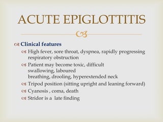
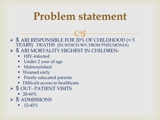
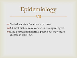
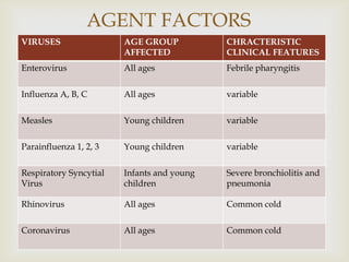
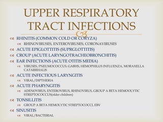
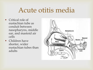
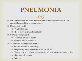
Acute respiratory infections (ARI) are responsible for 20% of childhood deaths under 5 years of age, with pneumonia accounting for 90% of ARI mortality. Children under 2 years old, malnourished children, children with HIV, and those with poor access to healthcare or poorly educated parents are most at risk. ARIs are a common cause of outpatient visits (20-60%) and admissions (12-45%) in children. Common respiratory infections affecting children include the common cold, acute epiglottitis, croup, ear infections, tonsillitis, sinusitis, and pneumonia. Bacteria and viruses can cause ARIs, with symptoms and severity depending on the specific infectious agent.
Measles is a highly contagious viral infection.
It is exanthematous disease with fewer, cough, coryza (rhinitis) and conjunctivitis.
Before the widespread use of measles vaccines, it was estimated that measles caused between 5 million and 8 million deaths worldwide each year.
Acute respiratory infection in children, etiology, clinical features, diagnosis, treatment. Common infections in children including common cold, tonsillitis, LTB, Croup, Epiglottitis etc.
Pulmonary TB is a bacterial infection of the lungs that can cause a range of symptoms, including chest pain, breathlessness, and severe coughing. Pulmonary TB can be life-threatening if a person does not receive treatment. People with active TB can spread the bacteria through the air.
Acute Respiratory Infection is an important topic that one must know about in the health care field. This is just a short presentation of a very vast topic.
Heritage of any nation is best represented by its culture, beliefs and traditions. A subset of these that has given India, a profound identity for India is Art. Art forms in India exist since time immemorial. Over the past century, Indian Art has undergone through vast and diverse change in their forms that exist in the present. For instance, the themes chosen by the traditional painters were societal. But later with the emergence of modernists followed by the contemporaries, the facets of Indian painting were changed radically.
Evaluation And Management Of Upper Respiratory Tract Infections In Children Dawood Al nasser
Evaluation And Management Of Upper Respiratory Tract Infections In Children
This presentation offers helpful comparison tables, please note that some recommendation might have changed since preparation and publication of this material.
A variety of viruses and bacteria can cause upper respiratory tract infections. These cause a variety of patient diseases including acute bronchitis, the common cold, influenza, and respiratory distress syndromes. Defining most of these patient diseases is difficult because the presentations connected with upper respiratory tract infections (URIs) commonly overlap and their causes are similar. Upper respiratory tract infections can be defined as self-limited irritation and swelling of the upper airways with associated cough with no proof of pneumonia, lacking a separate condition to account for the patient symptoms, or with no history of COPD/emphysema/chronic bronchitis. Upper respiratory tract infections involve the nose, sinuses, pharynx, larynx, and the large airways.
Pertussis : Highly contagious respiratory infection caused by Bordetella pertussis
Outbreaks first described in 16th century
Bordetella pertussis isolated in 1906
Estimated >300,000 deaths annually worldwide
Before the availability of pertussis vaccine in the 1940s, public health experts reported more than 200,000 cases of pertussis annually.
Since widespread use of the vaccine began, incidence has decreased more than 75% compared with the pre-vaccine era.
In 2012, the last peak year, CDC reported 48,277 cases of pertussis.
Extremely contagious-attack rate 100%
Immunity is never complete
Protection begins to wane in 3-5 yrs after vaccination
The prostate is an exocrine gland of the male mammalian reproductive system
It is a walnut-sized gland that forms part of the male reproductive system and is located in front of the rectum and just below the urinary bladder
Function is to store and secrete a clear, slightly alkaline fluid that constitutes 10-30% of the volume of the seminal fluid that along with the spermatozoa, constitutes semen
A healthy human prostate measures (4cm-vertical, by 3cm-horizontal, 2cm ant-post ).
It surrounds the urethra just below the urinary bladder. It has anterior, median, posterior and two lateral lobes
It’s work is regulated by androgens which are responsible for male sex characteristics
Generalised disease of the prostate due to hormonal derangement which leads to non malignant enlargement of the gland (increase in the number of epithelial cells and stromal tissue)to cause compression of the urethra leading to symptoms (LUTS
NVBDCP.pptx Nation vector borne disease control programSapna Thakur
NVBDCP was launched in 2003-2004 . Vector-Borne Disease: Disease that results from an infection transmitted to humans and other animals by blood-feeding arthropods, such as mosquitoes, ticks, and fleas. Examples of vector-borne diseases include Dengue fever, West Nile Virus, Lyme disease, and malaria.
These simplified slides by Dr. Sidra Arshad present an overview of the non-respiratory functions of the respiratory tract.
Learning objectives:
1. Enlist the non-respiratory functions of the respiratory tract
2. Briefly explain how these functions are carried out
3. Discuss the significance of dead space
4. Differentiate between minute ventilation and alveolar ventilation
5. Describe the cough and sneeze reflexes
Study Resources:
1. Chapter 39, Guyton and Hall Textbook of Medical Physiology, 14th edition
2. Chapter 34, Ganong’s Review of Medical Physiology, 26th edition
3. Chapter 17, Human Physiology by Lauralee Sherwood, 9th edition
4. Non-respiratory functions of the lungs https://academic.oup.com/bjaed/article/13/3/98/278874
Title: Sense of Taste
Presenter: Dr. Faiza, Assistant Professor of Physiology
Qualifications:
MBBS (Best Graduate, AIMC Lahore)
FCPS Physiology
ICMT, CHPE, DHPE (STMU)
MPH (GC University, Faisalabad)
MBA (Virtual University of Pakistan)
Learning Objectives:
Describe the structure and function of taste buds.
Describe the relationship between the taste threshold and taste index of common substances.
Explain the chemical basis and signal transduction of taste perception for each type of primary taste sensation.
Recognize different abnormalities of taste perception and their causes.
Key Topics:
Significance of Taste Sensation:
Differentiation between pleasant and harmful food
Influence on behavior
Selection of food based on metabolic needs
Receptors of Taste:
Taste buds on the tongue
Influence of sense of smell, texture of food, and pain stimulation (e.g., by pepper)
Primary and Secondary Taste Sensations:
Primary taste sensations: Sweet, Sour, Salty, Bitter, Umami
Chemical basis and signal transduction mechanisms for each taste
Taste Threshold and Index:
Taste threshold values for Sweet (sucrose), Salty (NaCl), Sour (HCl), and Bitter (Quinine)
Taste index relationship: Inversely proportional to taste threshold
Taste Blindness:
Inability to taste certain substances, particularly thiourea compounds
Example: Phenylthiocarbamide
Structure and Function of Taste Buds:
Composition: Epithelial cells, Sustentacular/Supporting cells, Taste cells, Basal cells
Features: Taste pores, Taste hairs/microvilli, and Taste nerve fibers
Location of Taste Buds:
Found in papillae of the tongue (Fungiform, Circumvallate, Foliate)
Also present on the palate, tonsillar pillars, epiglottis, and proximal esophagus
Mechanism of Taste Stimulation:
Interaction of taste substances with receptors on microvilli
Signal transduction pathways for Umami, Sweet, Bitter, Sour, and Salty tastes
Taste Sensitivity and Adaptation:
Decrease in sensitivity with age
Rapid adaptation of taste sensation
Role of Saliva in Taste:
Dissolution of tastants to reach receptors
Washing away the stimulus
Taste Preferences and Aversions:
Mechanisms behind taste preference and aversion
Influence of receptors and neural pathways
Impact of Sensory Nerve Damage:
Degeneration of taste buds if the sensory nerve fiber is cut
Abnormalities of Taste Detection:
Conditions: Ageusia, Hypogeusia, Dysgeusia (parageusia)
Causes: Nerve damage, neurological disorders, infections, poor oral hygiene, adverse drug effects, deficiencies, aging, tobacco use, altered neurotransmitter levels
Neurotransmitters and Taste Threshold:
Effects of serotonin (5-HT) and norepinephrine (NE) on taste sensitivity
Supertasters:
25% of the population with heightened sensitivity to taste, especially bitterness
Increased number of fungiform papillae
Knee anatomy and clinical tests 2024.pdfvimalpl1234
This includes all relevant anatomy and clinical tests compiled from standard textbooks, Campbell,netter etc..It is comprehensive and best suited for orthopaedicians and orthopaedic residents.
These lecture slides, by Dr Sidra Arshad, offer a quick overview of physiological basis of a normal electrocardiogram.
Learning objectives:
1. Define an electrocardiogram (ECG) and electrocardiography
2. Describe how dipoles generated by the heart produce the waveforms of the ECG
3. Describe the components of a normal electrocardiogram of a typical bipolar leads (limb II)
4. Differentiate between intervals and segments
5. Enlist some common indications for obtaining an ECG
Study Resources:
1. Chapter 11, Guyton and Hall Textbook of Medical Physiology, 14th edition
2. Chapter 9, Human Physiology - From Cells to Systems, Lauralee Sherwood, 9th edition
3. Chapter 29, Ganong’s Review of Medical Physiology, 26th edition
4. Electrocardiogram, StatPearls - https://www.ncbi.nlm.nih.gov/books/NBK549803/
5. ECG in Medical Practice by ABM Abdullah, 4th edition
6. ECG Basics, http://www.nataliescasebook.com/tag/e-c-g-basics
Prix Galien International 2024 Forum ProgramLevi Shapiro
June 20, 2024, Prix Galien International and Jerusalem Ethics Forum in ROME. Detailed agenda including panels:
- ADVANCES IN CARDIOLOGY: A NEW PARADIGM IS COMING
- WOMEN’S HEALTH: FERTILITY PRESERVATION
- WHAT’S NEW IN THE TREATMENT OF INFECTIOUS,
ONCOLOGICAL AND INFLAMMATORY SKIN DISEASES?
- ARTIFICIAL INTELLIGENCE AND ETHICS
- GENE THERAPY
- BEYOND BORDERS: GLOBAL INITIATIVES FOR DEMOCRATIZING LIFE SCIENCE TECHNOLOGIES AND PROMOTING ACCESS TO HEALTHCARE
- ETHICAL CHALLENGES IN LIFE SCIENCES
- Prix Galien International Awards Ceremony
TEST BANK for Operations Management, 14th Edition by William J. Stevenson, Ve...kevinkariuki227
TEST BANK for Operations Management, 14th Edition by William J. Stevenson, Verified Chapters 1 - 19, Complete Newest Version.pdf
ARTIFICIAL INTELLIGENCE IN HEALTHCARE.pdfAnujkumaranit
Artificial intelligence (AI) refers to the simulation of human intelligence processes by machines, especially computer systems. It encompasses tasks such as learning, reasoning, problem-solving, perception, and language understanding. AI technologies are revolutionizing various fields, from healthcare to finance, by enabling machines to perform tasks that typically require human intelligence.
micro teaching on communication m.sc nursing.pdfAnurag Sharma
Microteaching is a unique model of practice teaching. It is a viable instrument for the. desired change in the teaching behavior or the behavior potential which, in specified types of real. classroom situations, tends to facilitate the achievement of specified types of objectives.
Couples presenting to the infertility clinic- Do they really have infertility...Sujoy Dasgupta
Dr Sujoy Dasgupta presented the study on "Couples presenting to the infertility clinic- Do they really have infertility? – The unexplored stories of non-consummation" in the 13th Congress of the Asia Pacific Initiative on Reproduction (ASPIRE 2024) at Manila on 24 May, 2024.
2.
Problem statement
Š ARI RESPONSIBLE FOR 20% OF CHILDHOOD (< 5
YEARS) DEATHS (IN WHICH 90% FROM PNEUMONIA)
Š ARI MORTALITY HIGHEST IN CHILDREN-
HIV-infected
Under 2 year of age
Malnourished
Weaned early
Poorly educated parents
Difficult access to healthcare
Š OUT- PATIENT VISITS
20-60%
Š ADMISSIONS
12-45%
3.
Children with ARI presenting in OPD
Place % of children
London (UK) 35.0
Herston (Australia) 34
Ethiopia (Whole country) 25.5
Sau aulo (Brazil) 41.8
India 38.9
Nepal 37.6
4.
Varied agents – Bacteria and viruses
Clinical picture may vary with etiological agent
May be present in normal people but may cause
disease in only few.
Epidemiology
5.
Infections of the respiratory tract are described in a number of
different ways according to the general areas of involvement
in the more common infections. The upper respiratory tract or
upper airway consists of primarily of the nose and pharynx.
The lower respiratory tract consists of bronchi and
bronchioles, which constitute the reactive protein of the
airway because of their smooth muscle content and ability to
constrict the alveoli.
ACUTE RESPIRATORY
INFECTIONS
6. May cause the inflammation of respiratory tract
anywhere from nose to alveoli.
May be classified as –
AURI – Acute Upper Respiratory Infection
(common cold, pharyngitis, epiglottitis, & otitis media etc.)
or
ALRI – Acute Lower Respiratory Infection
(laryngitis, layngotracheitis, bronchitis, bronchiolitis & pneumonia)
ACUTE RESPIRATORY
INFECTIONS(ARI)
8.
AGENT FACTORS
BACTERIA AGE GROUP
AFFECTED
CHRACTERISTIC
CLINICAL FEATURES
Bordetella pertussis Infants & young children Poroxysmal cough
Corynebacterium
diphtheriae
Children diphtheria
Hemophilus influenzae Adults
Children
Acute ex of ch bronchitis
Acute epiglottitis
Klebsiella pneumoniae Adults Lobar pneumonia
Legionella pneumophila Adults Pneumonia
Staph. pyogenes All ages Lobar and
bronchopneumonia
Strep. pneumoniae All ages Pneumonia
Strep. pyogenes All ages Acute pharyngitis and
tonsillitis
9.
VIRUSES AGE GROUP
AFFECTED
CHRACTERISTIC
CLINICAL FEATURES
Enterovirus All ages Febrile pharyngitis
Influenza A, B, C All ages variable
Measles Young children variable
Parainfluenza 1, 2, 3 Young children variable
Respiratory Syncytial
Virus
Infants and young
children
Severe bronchiolitis and
pneumonia
Rhinovirus All ages Common cold
Coronavirus All ages Common cold
AGENT FACTORS
11. Nature of infectious agent: The respiratory tract is
subjected to a wide variety of infectious agents.
Size and frequency of dose: The larger the dose and
the more frequent the exposure, the greater the
likelihood of a significant infection.
Age of child: Children of preschool and school age
are more often exposed to infectious agents generally
after 3 months of age infants have less resistance to
infections.
Size of child: Airways are smaller in young children
and more subjected to considerable narrowing from
edema.
12. Ability to resist invading organisms: School
age children have greater resistance to infection
than infants and young children.
Presence of great conditions:
Malnutrition, anemia, fatigue, chilling of the
body and immune deficiencies decrease normal
resistance to infection.
Presence of disorders affecting respiratory
tract: Allergies, cardiac abnormalities and cystic
fibrosis weaken respiratory defense mechanism.
Seasons: The most common respiratory tract
pathogens appear in epidemics during winter
and spring months.
14. Children average 8 episodes per year, adults 3
episodes per year
Etiologies :
Rhinoviruses 30 to 35%
Coronaviruses about 10%
Miscellaneous known viruses about 20%
Influenza and adenovirus-30%
Presumed undiscovered viruses up to 35%
Group A streptococci 5% to 10%
Parainfluenza was the first respiratory virus
isolated (1955)
Seasonal variation
Rhinovirus early fall
Coronavirus- winter
The Common Cold
15.
Common symptoms are sore throat, runny
nose, nasal congestion, sneezing,
Sometimes accompanied by
conjunctivitis, myalgias, fatigue
Sinusitis often present by CT scan;
“rhinosinusitis” might be a better term
Common Cold
17.
Direct contact is the most efficient means of
transmission: 40% to 90% recovery from hands.
Infectious droplet nuclei
Brief exposure (e.g., handshake) transmits in less
than 10% of instances
Kissing does not seem to be a common mode of
transmission.
Transmission of rhinoviruses
18.
Incubation period 12-72 hours
Nasal obstruction, drainage, sneezing, scratchy
throat
Median duration 1 week but 25% can last 2 weeks
Pharyngeal erythema is commoner with adenovirus
than with rhino or coronavirus
Clinical characteristics
19.
Main challenge is to distinguish between
uncomplicated cold and streptococcal pharyngitis or
bacterial sinusitis
Good examination
Marked exudate or pharyngeal erythema suggests
Streptococcal infection
Adenovirus
Diphtheria
Rapid antigen tests for group A streptococcus
Rapid techniques for influenza, RSV, parainfluenza
Treat with NSAIDs and whatever else your
grandmother advises
Diagnosis and treatment
20.
Life-threatning infection of the epiglottis, the
aryepiglottic folds and arytenoid soft tissue
Occurs mostly in winters
Peak incidence :- 1 – 6 years
Male affected more
bacterial infection (Hemophilus influenza type b)
Concomitant bacteremia, pneumonia, otitis
media, arthritis and other invasive infections caused by
H.influenza type b may be present
ACUTE EPIGLOTTITIS
21.
Clinical features
High fever, sore throat, dyspnea, rapidly progressing
respiratory obstruction
Patient may become toxic, difficult
swallowing, laboured
breathing, drooling, hyperextended neck
Tripod position (sitting upright and leaning forward)
Cyanosis , coma, death
Stridor is a late finding
ACUTE EPIGLOTTITIS
22.
Do not examine the throat
Assessment of severity
Degree of stridor
Respiratory rate
Heart rate
Level of consciousness
Pulse oximetry
EXAMINATION
23.
DIAGNOSIS:
“CHERRY RED”APPEARANCE OF EPIGLOTTIS ON
LARYNGOSCOPY
THUMB SIGN ON LATERAL NECK RADIOGRAPH
ACUTE EPIGLOTTITIS
27.
NEED TO BE MANAGED IN ICU WITH
ENDOTRACHEAL INTUBATION
HELP FROM ANAESTHETIST AND ENT SURGEON
BLOOD CULTURES
FLUID AND ELECTROLYTE SUPPORT
INTRAVENOUS AMPLICILLIN 100 mg/kg/day OR
CEFTRIAXONE 100 mg/kg/day .
OTHER OPTIONS
(CEFUROXIME OR CEFOTAXIME) TOTAL TREATMENT
:-7-10 DAYS
CHOLRAMPHENICOL 50-75 mg/kg/day IV
RIFAMPICIN PROPHYLAXIS TO CLOSE CONTACTS
TREATMENT
(ACUTE EPIGLOTTITIS)
28.
VIRAL INFECTION LEADING TO MUCOSAL
INFLAMMATION OF THE GLOTTIC AND
SUBGLOTTIC REGIONS
COMMONLY DUE TO INFLUENZA (TYPE A),
PARAINFLUENZA(1, 2, 3) AND RSV
AGE :- 6 MONTHS – 6 YEARS
ACUTE
LARYNGOTRACHEOBRONCHITIS
(VIRAL CROUP)
29.
CLINICAL FEATURES
INITIAL :- RHINORRHEA, MILD COUGH, FEVER(LOW
GRADE)
LATER (24-48 HOURS) :-
BARKING COUGH
HOARSENESS OF VOICE
NOISY BREATHING (MAINLY ON INSPIRATION)
SYMPTOMS WORSEN AT NIGHT AND ON LYING
DOWN
CHILDREN PREFER TO BE HELD UPRIGHT OR SIT IN
BED
SYMPTOMS RESOLVE WITHIN A WEEK
ACUTE LTB
30.
CLINICAL EXAMINATION
HOARSE VOICE
NORMAL TO MODERATELY INFLAMMED
PHARYNX
SLIGHTLY INCREASED RESP RATE WITH
PROLONGED INSPIRATION AND INSPIRATORY
STRIDOR
ACUTE LTB
33.
TREATMENT
MOIST OR HUMIDIFIED AIR
STEROIDS
REDUCE THE SEVERITY AND DURATION / NEED
FOR ENDOTRACHEAL INTUBATION
PREDNISOLONE PO 2mg/kg/day FOR 3 DAYS
NEBULIZED BUDESONIDE 2mg STAT
NEBULIZED ADRENALINE (EPINEPHRINE)
ACUTE LTB
34.
Tonsillitis is a viral or bacterial infection in the throat that
causes inflammation of the tonsils. Tonsils are small glands
(lymphoid tissue) in the pharyngeal cavity.
In the first six months of life tonsils provide a useful defense
against infections. Tonsillitis is one of the most common
ailments in pre-school children, but it can also occur at any
age.
Children are most often affected from around the age of three
or four, when they start nursery or school and come into
contact with many new infections.
A child may have tonsillitis if he/she has a sore throat, a fever
and is off food.
Tonsillitis
36.
Tonsillitis is caused by a variety of contagious viral
and bacterial infections.
It is spread by close contact with other individuals
and occurs more during winter periods.
The most common bacterium causing tonsillitis is
streptococcus.
Causes of tonsilitis
37.
Encourage bed rest.
Introduce soft liquid diet according to the child's
preferences.
Provide cool mist atmosphere to keep the mucous
membranes moist during periods of mouth
breathing.
Warm saline gargles & paracetamol are useful to
promote comfort.
If antibiotics are prescribed, counsel the child's
parents regarding the necessity of completing the
treatment period
Advice and treatment:
38.
The controversy of tonsillectomy:
Surgical removal of chronic tonsillitis (tonsillectomy)
is controversial. Generally, tonsils should not
removed before 3 or 4 yrs of age, because of the
problem of excessive blood loss & the possibility of
re-growth or hypertrophy of lymphoid tissue, in
young children.
Management:
39.
Community acquired bacterial sinusitis
S.pneumoniae
H. influenzae
S. pyogenes
Nosocomial sinusitis
Seen in critically ill, mechanically ventilated
S. aureus
Pseudomonas aeruginosa
Serratia marcescens
fungal
Sinusitis
40.
41.
42.
Clinical features
Sneezing
Nasal discharge
Facial pressure
Fever
Purulent drainage
Headache
Sinus imaging not routinely recommended
Clinical features
43.
Maxillary: usually uncomplicated
Ethmoid: cavernous sinus thrombosis-serious
Frontal: osteomyelitis of frontal bone; cavernous
sinus thrombosis; epidural, subdural, or
intracerebral abscess; orbital extension
Sphenoid: Rare; extension to internal carotid
artery, cavernous sinuses, pituitary, optic nerves;
common misdiagnoses include ophthalmic
migraine, aseptic meningitis, trigeminal
neuralgia, cavernous sinus thrombosis
Acute sinusitis:
complications
44. Otitis externa
Acute, localized: often S.
aureus, S. epidermidis or S.
pyogenes
Acute diffuse (swimmer’s ear):
gram-negative rods, especially
Ps. Aeruginosa ; Rx: topical
quinolones
Chronic: mainly with chronic
otitis media
Malignant: life-threatening
infection in
diabetics, elderly, immunecom
promised
45.
S. pneumoniae and H. influenzae the leading
causes in all age groups (most H. flu is from
non-typable strains and not “B”)
Moraxella catarrhalis: 10% of cases
Some cases may be viral
(RSV, influenza, enteroviruses)
Mycoplasma pneumoniae: inflammation of the
tympanic membrane (“bullous myringitis”)
Acute otitis media
46. Acute otitis media
Critical role of
eustachian tube as
conduit between
nasopharynx, middle
ear, and mastoid air
cells
Children have
shorter, wider
eustachian tubes than
adults
47.
Presence of fluid in the middle ear AND
Ear pain, drainage, hearing loss
The fluid may take weeks to resolve
Amoxicillin remains the drug of choice
Beta-lactamase producing strains of H. influenza will
need amoxicillin/clavulanic acid or cephalosporins
Diagnosis and treatment
49.
Inflammatory syndrome of the pharynx
Most cases are viral
Most important bacterial cause is Streptococcus
pyogenes (15-20%)
Presents with sore or scratchy throat
In severe bacterial cases there may be
odynophagia, fever, headache
Acute pharyngitis
50.
Viral: edema and hyperemia of tonsils and
pharyngeal mucosa
Streptococcal: exudate and hemorrhage involving
tonsils and pharyngeal walls
Epstein-Barr virus (infectious mono): may also
cause exudate, with nasopharyngeal lymphoid
hyperplasia
Acute pharyngitis:
physical examination
51.
Adenoviral pharyngitis
Pharyngeal erythema and exudate
may mimic streptococcal pharyngitis
Conjunctivitis (follicular) present in
1/3 to 1/2 of cases; commonly
unilateral but bilateral in 1/4 of cases
Pharyngoconjuntival fever
52.
Herpangina
Uncommon
Due to coxsackieviruss
Small, 1-2 mm vesicles on the soft palate, uvula, and
anterior tonsillar pillars which rupture to form small
white ulcers
Occurs mainly in children
Also think of Herpes simplex virus when you
see vesicular lesions
Vesicular lesions
53.
Vincent’s angina: anaerobic pharyngitis (exudate;
foul odor to breath)
Ludwig’s angina- cellulitis of dental origin
Quinsy: peritonsillitis/peritonsillar abscess. Medial
displacement of the tonsil; often spread of infection
to carotid sheath
Vincent’s angina
and Quinsy
54.
55.
Classic diphtheria (Corynebacterium diphtheriae):
slow onset, then marked toxicity
Arcanobacterium hemolyticum (formerly
Cornyebacterium hemolyticum): exudative
pharyngitis in adolescents and young adults with
diffuse, sometimes pruritic maculopapular rash on
trunk and extremities
Diphtheria
59.
Inflammatory disease of the bronchioles
Peak age of onset : 6 months
Most common agent :- rsv
Male : female :- 2:1
Occurs mostly in winter/spring
BRONCHIOLITIS
60.
Coryza with cough followed by worsening
breathlessness
Vomiting
Irritability
Wheeze
Feeding difficulty
Episodes of apnoea
CLINICAL FEATURES
63. A chest X-ray demonstrating lung hyperinflation with a
flattened diaphragm and bilateral atelectasis in the right
apical and left basal regions in a 16-day-old infant with
severe bronchiolitis
65.
TREATMENT
Mainly supportive
Prop up (30 – 40 degrees)
Oxygen inhalation (achieve o2 >92%)
If tachypneic, limit the oral feeds and use a ng tube for
feeding
Parenteral fluids to limit dehydration
Correct resp acidosis and electrolyte imbalance
Bronchodilators for wheeze (nebulized adrenaline)
Mechanical ventilation (severe resp distress or apnoea)
BRONCHIOLITIS
66. Inflammation of the lung parenchyma and is associated with the
consolidation of the alveolar spaces
Developed world
Viral infections
Low morbidity and mortality
Š Developing world
Common cause of death
Bacteria and PCP in 65%
Š ARI case management WHO
84% reduction in mortality
Respiratory rate, recession, ability to drink
Cheap, oral and effective antibiotics, Co-trimoxazole, amoxycillin
Maternal education
Referral
PNEUMONIA
67.
Š Vary according to
Age, immune status, where contracted
Š Community acquired (CAP)
Developing countries
S. pneumoniae, H. influenzae, S aureus
Viruses 40%
Other: Mycoplasma, Chlamydia, Moraxella
Developed countries
Viruses: RSV, Adenovirus, Parainfluenza, Influenza
Mycoplasma pneumoniae and Chlamydia pneumoniae
Bacteria: 5-10%
Etiology
68.
ETIOLOGY ACCORDING TO AGE
AGE GROUP CAUSATIVE ORGANISM
NEONATES GROUP B STREPTOCOCCUS
E.COLI
KLEBSIELLA
STAPH AUREUS
INFANTS PNEUMOCOCCUS
CHLAMYDIA
RSV
H.INFLUENZA TYPE b
CHILDREN 1 TO 5 YRS RESPIRATORY VIRUSES
PNEUMOCOCCUS
H.INFLUENZA TYPE b
C.TRACHOMATIS
M.PNEUMONIAE
S.AUREUS
GP A STREPTOCOCCUS
CHILDREN 5 TO 18 YRS M.PNEUMONIAE
PNEUMOCOCCUS
C.PNEUMONIAE
H.INFLUENZA TYPE b
69.
WHO Classification and management
NO PNEUMONIA COUGH
NO TACHYPNEA
-HOME CARE
-SOOTHE THE THROAT AND RELIEVE
COUGH
-ADVISE MOTHER WHEN TO RETURN
-FOLLOWUP IN 5 DAYS IF NOT
IMPROVING
PNEUMONIA -COUGH
-TACHYPNEA
-NO RIB OR STERNAL
RETRACTION
-ABLE TO DRINK
- NO CYANOSIS
-HOME CARE
-ANTIBIOTICS FOR 5 DAYS
-SOOTHE THE THROAT AND RELIEVE
COUGH
-ADVISE MOTHER WHEN TO RETURN
-FOLLOWUP IN 2 DAYS
SEVERE PNEUMONIA -COUGH
-TACHYPNEA
-RIB AND STERNAL
RETRACTION
-ABLE TO DRINK
-NO CYANOSIS
-ADMIT IN HOSPITAL
-GIVE RECOMMENDED ANTIBIOTICS
-MANAGE AIRWAY
-TREAT FEVER IF PRESENT
VERY SEVERE
PNEUMONIA
-COUGH
-TACHYPNOEA
-CHEST WALL RETRACTION
-UNABLE TO DRINK
-CENTRAL CYANOSIS
-ADMIT IN HOSPITAL
-GIVE RECOMMENDED ANTIBIOTICS
-OXYGEN
-MANAGE AIRWAY
-TREAT FEVER IF PRESENT
70.
Significant risk factors are younger age (2-6 months), low parental
education, smoking at home, prematurity, low birth weight,
weaning from breast milk at < 6 months, a negative history of
diphtheria, pertussis and tetanus vaccination, anaemia,
malnutrition and overcrowding.
Infection rate higher in siblings of school children who introduce
infection in the household.
Other risk factors
Congenital lung cysts
Chronic lung disease
Immunodeficiency
Cystic fibrosis
Sickle cell disease
Tracheostomy in situ
HIGH RISK CHILDREN FOR
PNEUMONIA
71.
Š Sign of respiratory distress; nasal flaring & chest
indrawing
Younger than 2 months
Decreased level of consciousness
Stridor when calm
Severe malnutrition
Associated symptomatic HIV/AIDS
Danger Signs (IMCI)
76.
Š Clues to other specific
organisms
Staphylococcus – areas of
break-down
Klebsiella, anaerobes, H.
influenza or TB –cavitating
or expansile pneumonia
TB, S. aureus, H. influenza
pleural effusion and
empyema
Radiology
77.
White cell count and CRP
>15,000 – 40,000/mm3 neutrophil predominance
Blood cultures
25% positive
NASOPHARYNGEAL ASPIRATE
Viral immunoflorescence in infants
Sputum specimen
Gram staining
Acid fast bacilli
Pleural fluid examination (if present)
ASO titer (in case of streptococcal pneumonia)
Tuberculin skin test
Viral Titres
culture
antigen
Diagnosis
79.
Š Antibiotics
Under 5 yrs
First line treatment :- amoxicillin
Alternatives : coamoxiclav, cefaclor,(for typical)
macrolides (for atypical)
Over 5 yrs
First line treatment :- amoxicillin or macrolides
Alternatives :- macrolide or flucloxacillin + amoxicillin
Severe pneumonia
Co-amoxiclav, cefotaxime or cefuroxime
Special categories (as per the suspected organism)
Treatment
80. Treatment in special groups
GROUP ORGANISMS ANTIBIOTICS
IMMUNOCOMPROMISED -GRAM NEGATIVE
-S. AUREUS
-OPPORTUNISTIC
PNEUMOCYSTIS
JIROVECI
-M. TUBERCULOSIS
AMPICILLIN +
CLOXACILLIN +
AMINOGLYCOSIDE
LESS THAN 3 MONTHS -GRAM NEGATIVE
-GROUP B
STREPTOCOCCUS
-S.AUREUS
AMPICILLIN +
AMINOGLYCOSIDE
HOSPITAL ACQUIRED
PNEUMONIA
-GRAM NEGATIVE
-METHICILLIN
RESISTANT S.
AUREUS
AMINOGLYCOSIDE +
VANCOMYCIN +
CEPHALOSPORIN
(3RD GENERATION)
81.
Š Oxygen
intranasaly
Š Hydration
50 – 80ml/kg/day
Š Temperature control
Š Airway obstruction management
Chest drain :- for fluid or pus collection in chest
(empyema)
Treatment (contd.)
82.
Š Most children recover without residual damage
Š Incorrect treatment leads to tissue destruction and
bronchiectasis
Š Half of children with pneumonia secondary to
measles or adenovirus have persistent airway
obstruction
Prognosis
83.
Early diagnosis of pneumonia and the warning signs
of severe disease and prompt management – key
factors which determine the outcome of disease
Guidelines have been given by WHO regarding
management and use of antibiotics.
Recent changes – Management as per the IMNCI
protocol
PREVENTION AND CONTROL
OF ARIs
84. as per the “Integrated Management of
Neonate & Child Illnesses” (IMNCI)
protocol
85.
History taking and clinical assessment very
important
Age of the child, for how long the child has been
coughing, whether the child is able to drink, has the
young infant stopped feeding well, does the child
have fever, is the child drowsy or difficult to wake,
did the child have convulsions, is there irregular
breathing, any history of treatment.
Clinical Assessment
86.
1. COUNTING THE NUMBER OF BREATHS IN ONE
MINUTE - to assess fast breathing
Respiratory rate cut-offs:
>/= 60 breaths per minute in a child less than 2 months
>/=50 breaths per minute in child aged 2month upto
12 months
>/=40 breaths per minute in child aged 12 months upto
5 years
Physical examination
87.
2. LOOK FOR CHEST INDRAWING
when the child breathes IN
3. LOOK AND LISTEN FOR STRIDOR
when the child breathes IN
4. LOOK FOR WHEEZE
when the child breathes out
5. FEEL FEVER OR LOW BODY TEMPERATURE
6. CHECK FOR SEVERE MALNUTRITION
7. CHECK FOR CYANOSIS
Physical examination cont.
88.
CHILD BELOW 2 MONTHS
Very severe disease
Severe pneumonia
No pneumonia
CHILD AGED 2 MONTHS UPTO 5 YEARS
Very severe disease
Severe pneumonia
Pneumonia
No pneumonia (cold & cough)
CLASSIFICATION OF DISEASE
89. SIGNS
STOPPED
FEEDING WELL
CONVULSIONS
ABN. SLEEPY
STIDOR IN
CALM CHILD
WHEEZE
FEVER/LOW
BODY TEMP.
SEVERE CHEST
IDRAWING
FAST
BREATHING
NO SEVERE
CHEST
INDRAWING
NO FAST
BREATHING
CLASSIFY AS VERY SEVERE
DISEASE
SEVERE
PNEUMONIA
NO PNEUMONIA
TREATMENT REFER URGENTLY
KEEP WARM
GIVE FIRST DOSE
OF ANTIBIOTIC
REFER URGENTLY
KEEP WARM
GIVE FIRST DOSE
OF ANTIBIOTIC
ADVICE FOR
HOME CARE
EXPLAIN DANGER
SIGNS
MANAGEMENT OF ARI
CHILDREN BELOW 2 MONTHS
90. MANAGEMENT OF ARI
CHILD AGED 2 MONTHS UPTO 5 YEARS
SIGNS
NOT ABLE TO
DRINK
CONVULSIONS
ABNORMALLY
SLEEPY OR
DIFFICULT TO
WAKE
STRIDOR IN A
CALM CHILD
SEVERE
MALNUTRITION
FAST
BREATHING
CHEST
INDRAWING
NASALFLARI
NG
GRUNTING
FAST
BREATHING
ONLY
NO CHEST
INDRAWING
NO FAST
BREATHING
NO CHEST
INDRAWING
CLASSIFY AS VERY SEVERE
DISEASE
SEVERE
PNEUMONIA
PNEUMONIA NO
PNEUMONIA/
COLD & COUGH
TREATMENT REFER URGENTLY
GIVE FIRST DOSE OF
ANTIBIOTIC
TREAT FEVER, IF
PRESENT
TREAT WHEEZE, IF
PRESENT
REFER
URGENTLY
GIVE FIRST DOSE
OF ANTIBOTIC
TREAT FEVER
TREAT WHEEZE
ADVICE FOR
HOME CARE
GIVE
ANTIBIOTIC
TREAT FEVER
TREAT WHEEZE
ASSESS AND
TREAT EAR
PROBLEM/ SORE
THROAT
TREAT FEVER
TREAT WHEEZE