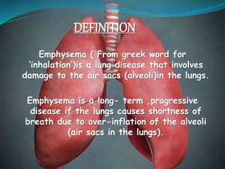
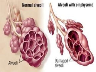
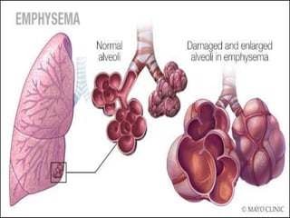
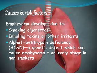
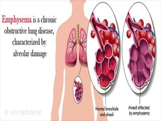
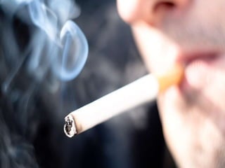
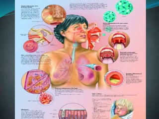
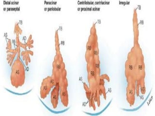
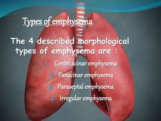
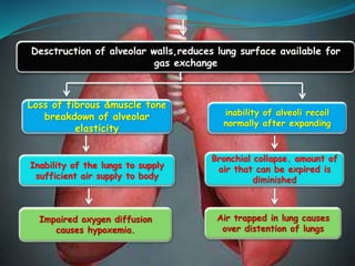
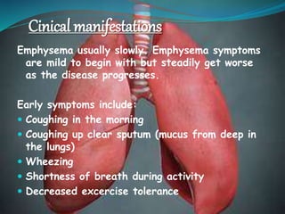
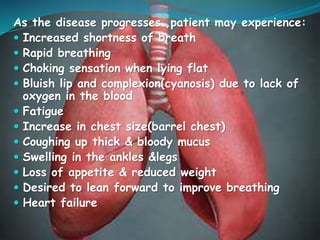
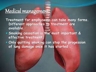
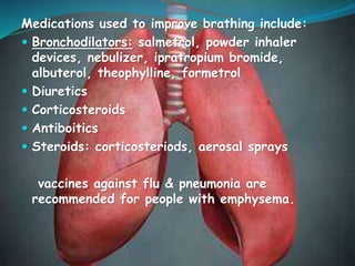
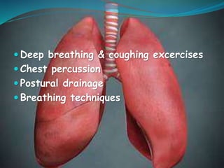
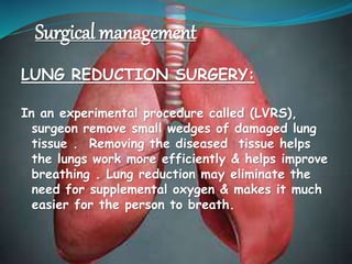
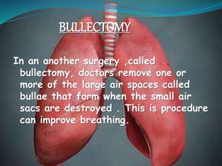
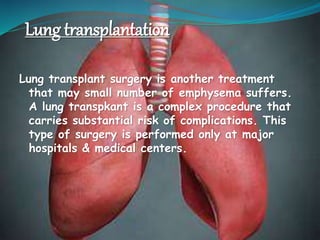
DEFINITION
Emphysema ( From greek word for
‘inhalation’)is a lung disease that involves
damage to the air sacs (alveoli)in the lungs.
Emphysema is a long- term ,progressive
disease if the lungs causes shortness of
breath due to over-inflation of the alveoli
(air sacs in the lungs).
Causes & risk factors
Emphysema develops due to:
 Smoking cigarettes
 Inhaling toxins or other irritants
 Alpha1-antitrypsin deficiency
(A1AD)—a genetic defect which can
cause emphysema t an early stage in
non smokers.
These factors increase the chance of developing
emphysema:
 Smoking cigarettes & long term second hand
or passive smoke exposure.
 Family members with emphysema
 Exposure to pollutants at work
(chemicals,fumes or dust)
 Exposure to indoor and outdoor pollution
 Frequent lower respiratory tract infections
History of frequent childhood
lung infections
Connective tissue
disorders(ehlers-danlos
syndrome,marfan syndrome)
HIV infection
Types of emphysema
The 4 described morphological
types of emphysema are :
1. Centri acinar emphysema
2. Panacinar emphysema
3. Paraseptal emphysema
4. Irregular emphysema
1. Centriacinar
 Centri acinar emphysema begins in the
respiratory bronchiole and spreads to
peripherally.
 Also termed centrilobular emphysema, this
form is associated with long standing
cigarette smoking predominantly involves
destruction of bronchioles in the upper lobe
of lungs but the alveolar sac remain in
contact.
Centri acinar
2. panacinar
 In panacinar or panlobular emphysem there is a
destruction of the respiratory bronchioles,
alveolar duct & alveoli in the lower half of the
lungs.
 Panacinar emphysema is generally is observed in
patients with homozygous alpha1-
antitrypsin(AAT) deficiency.
 In people who spoke , focal panacinar
emphysemaat the lung bases may accompany
centriacinar emphysema.
panacinar
3. Paraseptal emphysema
 Paraseptal emphysema,also known as distal
acinar emphysema, preferentially involves
the distal airways structures ,alveolar
ducts,and alveolar sacs .
 The process is localized around the septea
of the lungs or pleura.
 Although the airflow frequently is preserved
,the apical bullae may lead to spontaneous
pneumothorax.
 Giant bullae occassionally cause severe
compression of adjacent lung tissue.
Distal acinar emphysema
4. Irregular emphysema
Irregular emphysema refers to
the any part of the bronchiole
or alveoli or ducts of alveoli .
Irregular emphysema
pathophysiology
Due to etiological factors
Inherited α-1 anti-trypsin
deficiency
Inflammation of airway
epithelium
Neutrophils&macrophages
release elastage
Infiltration of inflammatory cells
& release of cytokines
Α-1 anti-trypsin deficiency
leads to failure of elastage
inactivation
Increased protease activity with breakdown of elastin in
connective tissue of lungs
Loss of elastic recoil of bronchial wall from breakdown of elastin
by the elastage
lungs become damaged because of reaction of irritants entering
airways & alveoli
Irreversible enlargement of the air spaces distal to the terminal
bronchioles
Collapse of terminal bronchioles causing airway obstruction
Desctruction of alveolar walls,reduces lung surface available for
gas exchange
Loss of fibrous &muscle tone
breakdown of alveolar
elasticity
Inability of the lungs to supply
sufficient air supply to body
Impaired oxygen diffusion
causes hypoxemia.
inability of alveoli recoil
normally after expanding
Bronchial collapse. amount of
air that can be expired is
diminished
Air trapped in lung causes
over distention of lungs
Cinical manifestations
Emphysema usually slowly. Emphysema symptoms
are mild to begin with but steadily get worse
as the disease progresses.
Early symptoms include:
 Coughing in the morning
 Coughing up clear sputum (mucus from deep in
the lungs)
 Wheezing
 Shortness of breath during activity
 Decreased excercise tolerance
As the disease progresses ,patient may experience:
 Increased shortness of breath
 Rapid breathing
 Choking sensation when lying flat
 Bluish lip and complexion(cyanosis) due to lack of
oxygen in the blood
 Fatigue
 Increase in chest size(barrel chest)
 Coughing up thick & bloody mucus
 Swelling in the ankles &legs
 Loss of appetite & reduced weight
 Desired to lean forward to improve breathing
 Heart failure
Diagnostic evaluations
 Health history & physical examination
 Spirometry & other pulmonary function tests
 Chest x-ray &/ or CT of the chest
 ABG analysis
 CBC
 Pulse oximetry
 Sputum examination
 Pulmonary ventilation/perfusion scans
 Testing for alpha-1 antitrypsin deficiency(AAT
deficiency)
management
1.Medical management
2.Surgical management
3.Nursing management
Medical management
Treatment for emphysema can take many forms.
Different approaches to treatment are
available.
 Smoking cessation is the most important &
effective treatment.
 Only quitting smoking can stop the progession
of lung damage once it has started .
Medications used to improve brathing include:
 Bronchodilators: salmetrol, powder inhaler
devices, nebulizer, ipratropium bromide,
albuterol, theophylline, formetrol
 Diuretics
 Corticosteroids
 Antiboitics
 Steroids: corticosteriods, aerosal sprays
vaccines against flu & pneumonia are
recommended for people with emphysema.
 Deep breathing & coughing excercises
 Chest percussion
 Postural drainage
 Breathing techniques
Surgical management
LUNG REDUCTION SURGERY:
In an experimental procedure called (LVRS),
surgeon remove small wedges of damaged lung
tissue . Removing the diseased tissue helps
the lungs work more efficiently & helps improve
breathing . Lung reduction may eliminate the
need for supplemental oxygen & makes it much
easier for the person to breath.
In an another surgery ,called
bullectomy, doctors remove one or
more of the large air spaces called
bullae that form when the small air
sacs are destroyed . This is procedure
can improve breathing.
BULLECTOMY
Lung transplantation
Lung transplant surgery is another treatment
that may small number of emphysema suffers.
A lung transpkant is a complex procedure that
carries substantial risk of complications. This
type of surgery is performed only at major
hospitals & medical centers.
Pulmonary rehabilitation
pulmonary rehabilitation is probably the most
effective therapy for COPD patients with
emphysema . It improves quality of life and
decreases hospitalization .
Pulmonary rehabilitation includes education ,
nutrition, counselling, learning special breathing
techniques, help with quitting smoking and starting
an exercise regimen. Because people with
emphysema are often physically limited , they may
avoid any kind of physical activity .
Nursing management
 Assess for signs & symptoms of hypoxia and
hypercapnia.
 Auscultation chest to listen to breath sounds
every hour .
 Increase or decrease in PaO2 PaCO2.
 Encourage the patient to stop smoking and
avoid air pollution including second hand
cigarette smoke.
 Drink plenty of water
 Adequate nutritional intake
Emphysema ppt
Emphysema ppt
Emphysema ppt
Emphysema ppt
Emphysema ppt
Emphysema ppt
Emphysema ppt
Emphysema ppt
Emphysema ppt
Emphysema ppt
Emphysema ppt
Emphysema ppt
Emphysema ppt
Emphysema ppt

Emphysema ppt

  • 2.
    DEFINITION Emphysema ( Fromgreek word for ‘inhalation’)is a lung disease that involves damage to the air sacs (alveoli)in the lungs. Emphysema is a long- term ,progressive disease if the lungs causes shortness of breath due to over-inflation of the alveoli (air sacs in the lungs).
  • 5.
    Causes & riskfactors Emphysema develops due to:  Smoking cigarettes  Inhaling toxins or other irritants  Alpha1-antitrypsin deficiency (A1AD)—a genetic defect which can cause emphysema t an early stage in non smokers.
  • 6.
    These factors increasethe chance of developing emphysema:  Smoking cigarettes & long term second hand or passive smoke exposure.  Family members with emphysema  Exposure to pollutants at work (chemicals,fumes or dust)  Exposure to indoor and outdoor pollution  Frequent lower respiratory tract infections
  • 7.
    History of frequentchildhood lung infections Connective tissue disorders(ehlers-danlos syndrome,marfan syndrome) HIV infection
  • 15.
    Types of emphysema The4 described morphological types of emphysema are : 1. Centri acinar emphysema 2. Panacinar emphysema 3. Paraseptal emphysema 4. Irregular emphysema
  • 16.
    1. Centriacinar  Centriacinar emphysema begins in the respiratory bronchiole and spreads to peripherally.  Also termed centrilobular emphysema, this form is associated with long standing cigarette smoking predominantly involves destruction of bronchioles in the upper lobe of lungs but the alveolar sac remain in contact.
  • 17.
  • 18.
    2. panacinar  Inpanacinar or panlobular emphysem there is a destruction of the respiratory bronchioles, alveolar duct & alveoli in the lower half of the lungs.  Panacinar emphysema is generally is observed in patients with homozygous alpha1- antitrypsin(AAT) deficiency.  In people who spoke , focal panacinar emphysemaat the lung bases may accompany centriacinar emphysema.
  • 19.
  • 20.
    3. Paraseptal emphysema Paraseptal emphysema,also known as distal acinar emphysema, preferentially involves the distal airways structures ,alveolar ducts,and alveolar sacs .  The process is localized around the septea of the lungs or pleura.  Although the airflow frequently is preserved ,the apical bullae may lead to spontaneous pneumothorax.  Giant bullae occassionally cause severe compression of adjacent lung tissue.
  • 21.
  • 22.
    4. Irregular emphysema Irregularemphysema refers to the any part of the bronchiole or alveoli or ducts of alveoli .
  • 23.
  • 25.
    pathophysiology Due to etiologicalfactors Inherited α-1 anti-trypsin deficiency Inflammation of airway epithelium Neutrophils&macrophages release elastage Infiltration of inflammatory cells & release of cytokines Α-1 anti-trypsin deficiency leads to failure of elastage inactivation Increased protease activity with breakdown of elastin in connective tissue of lungs
  • 26.
    Loss of elasticrecoil of bronchial wall from breakdown of elastin by the elastage lungs become damaged because of reaction of irritants entering airways & alveoli Irreversible enlargement of the air spaces distal to the terminal bronchioles Collapse of terminal bronchioles causing airway obstruction
  • 27.
    Desctruction of alveolarwalls,reduces lung surface available for gas exchange Loss of fibrous &muscle tone breakdown of alveolar elasticity Inability of the lungs to supply sufficient air supply to body Impaired oxygen diffusion causes hypoxemia. inability of alveoli recoil normally after expanding Bronchial collapse. amount of air that can be expired is diminished Air trapped in lung causes over distention of lungs
  • 28.
    Cinical manifestations Emphysema usuallyslowly. Emphysema symptoms are mild to begin with but steadily get worse as the disease progresses. Early symptoms include:  Coughing in the morning  Coughing up clear sputum (mucus from deep in the lungs)  Wheezing  Shortness of breath during activity  Decreased excercise tolerance
  • 29.
    As the diseaseprogresses ,patient may experience:  Increased shortness of breath  Rapid breathing  Choking sensation when lying flat  Bluish lip and complexion(cyanosis) due to lack of oxygen in the blood  Fatigue  Increase in chest size(barrel chest)  Coughing up thick & bloody mucus  Swelling in the ankles &legs  Loss of appetite & reduced weight  Desired to lean forward to improve breathing  Heart failure
  • 30.
    Diagnostic evaluations  Healthhistory & physical examination  Spirometry & other pulmonary function tests  Chest x-ray &/ or CT of the chest  ABG analysis  CBC  Pulse oximetry  Sputum examination  Pulmonary ventilation/perfusion scans  Testing for alpha-1 antitrypsin deficiency(AAT deficiency)
  • 31.
  • 32.
    Medical management Treatment foremphysema can take many forms. Different approaches to treatment are available.  Smoking cessation is the most important & effective treatment.  Only quitting smoking can stop the progession of lung damage once it has started .
  • 33.
    Medications used toimprove brathing include:  Bronchodilators: salmetrol, powder inhaler devices, nebulizer, ipratropium bromide, albuterol, theophylline, formetrol  Diuretics  Corticosteroids  Antiboitics  Steroids: corticosteriods, aerosal sprays vaccines against flu & pneumonia are recommended for people with emphysema.
  • 34.
     Deep breathing& coughing excercises  Chest percussion  Postural drainage  Breathing techniques
  • 35.
    Surgical management LUNG REDUCTIONSURGERY: In an experimental procedure called (LVRS), surgeon remove small wedges of damaged lung tissue . Removing the diseased tissue helps the lungs work more efficiently & helps improve breathing . Lung reduction may eliminate the need for supplemental oxygen & makes it much easier for the person to breath.
  • 36.
    In an anothersurgery ,called bullectomy, doctors remove one or more of the large air spaces called bullae that form when the small air sacs are destroyed . This is procedure can improve breathing. BULLECTOMY
  • 37.
    Lung transplantation Lung transplantsurgery is another treatment that may small number of emphysema suffers. A lung transpkant is a complex procedure that carries substantial risk of complications. This type of surgery is performed only at major hospitals & medical centers.
  • 38.
    Pulmonary rehabilitation pulmonary rehabilitationis probably the most effective therapy for COPD patients with emphysema . It improves quality of life and decreases hospitalization . Pulmonary rehabilitation includes education , nutrition, counselling, learning special breathing techniques, help with quitting smoking and starting an exercise regimen. Because people with emphysema are often physically limited , they may avoid any kind of physical activity .
  • 39.
    Nursing management  Assessfor signs & symptoms of hypoxia and hypercapnia.  Auscultation chest to listen to breath sounds every hour .  Increase or decrease in PaO2 PaCO2.  Encourage the patient to stop smoking and avoid air pollution including second hand cigarette smoke.  Drink plenty of water  Adequate nutritional intake