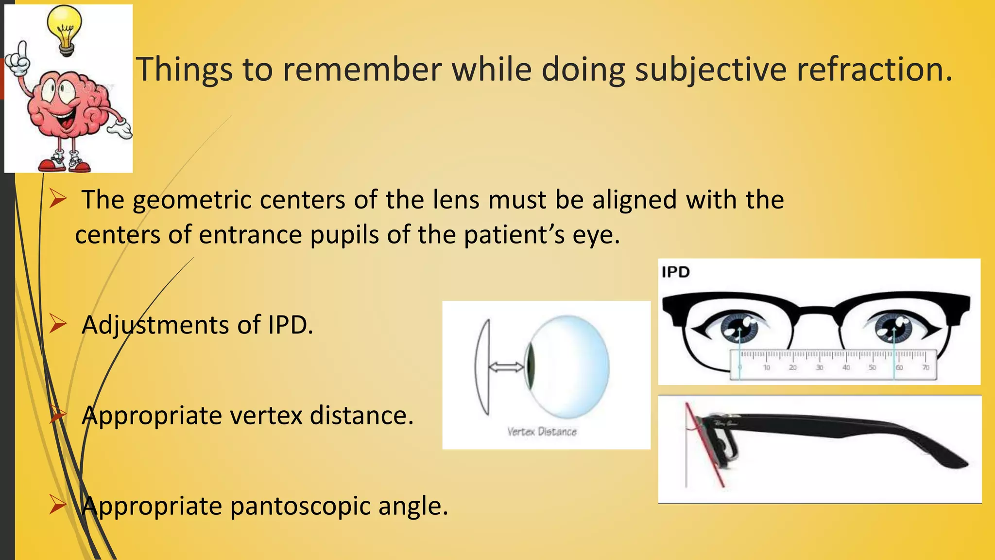
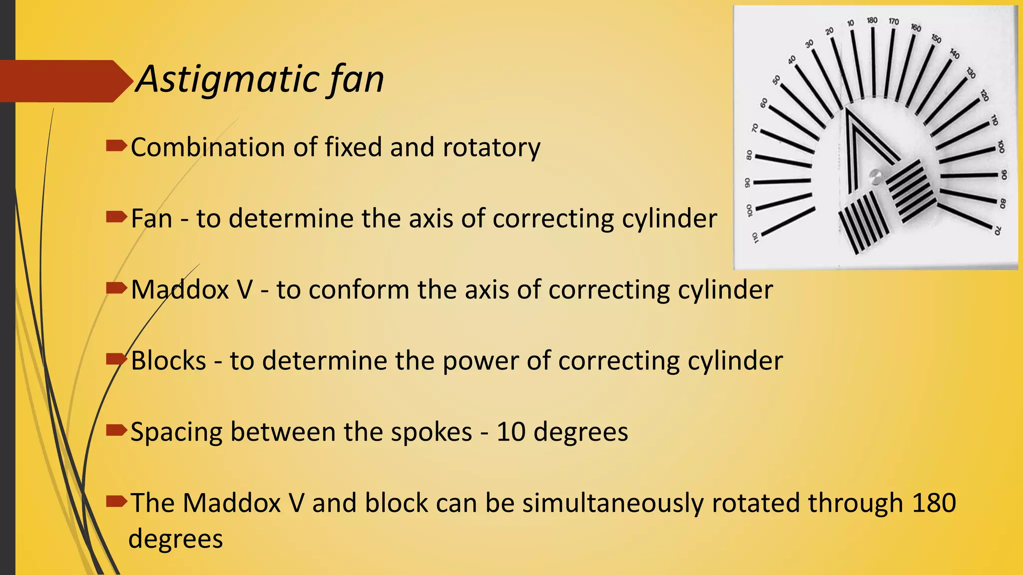
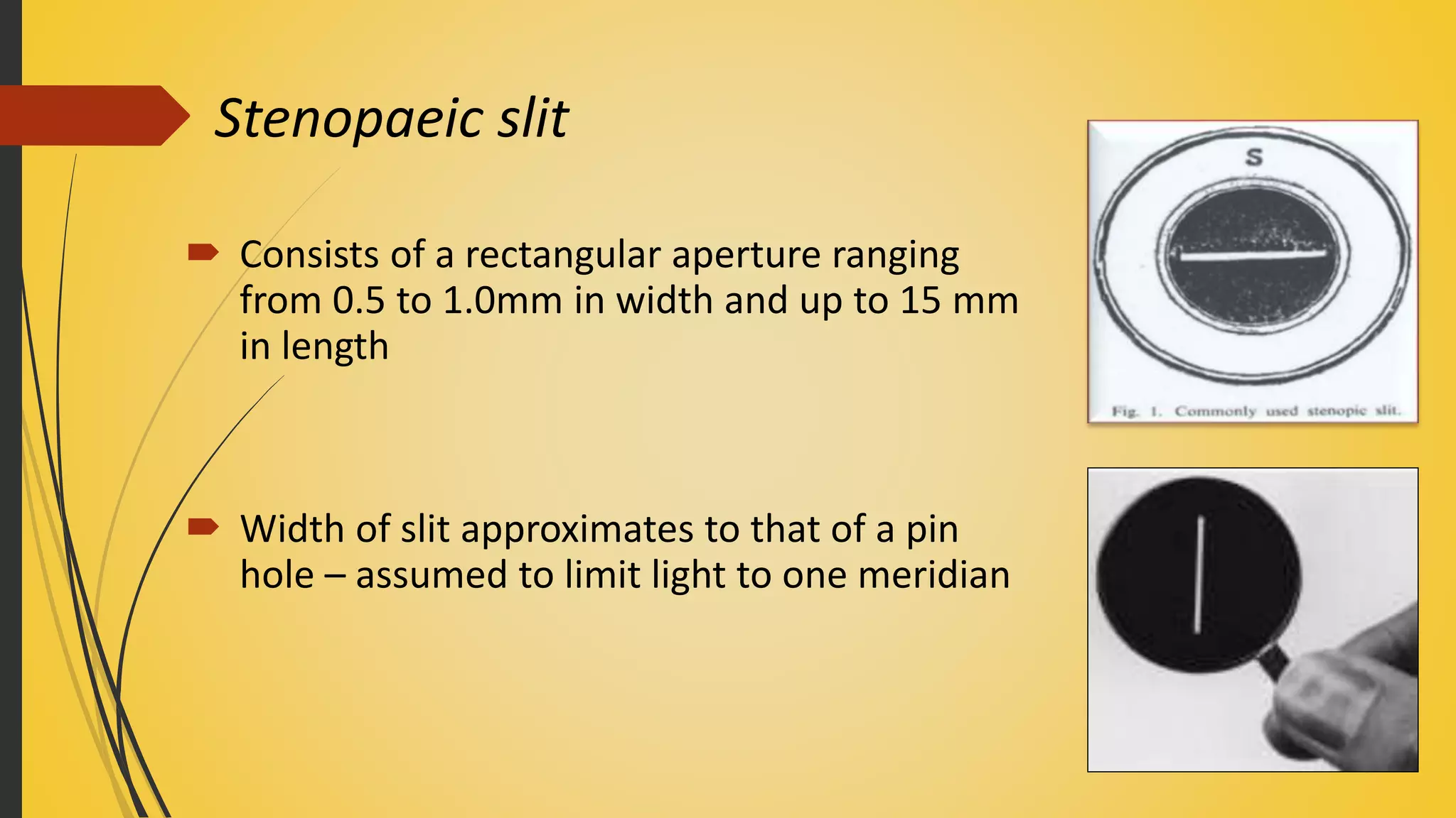
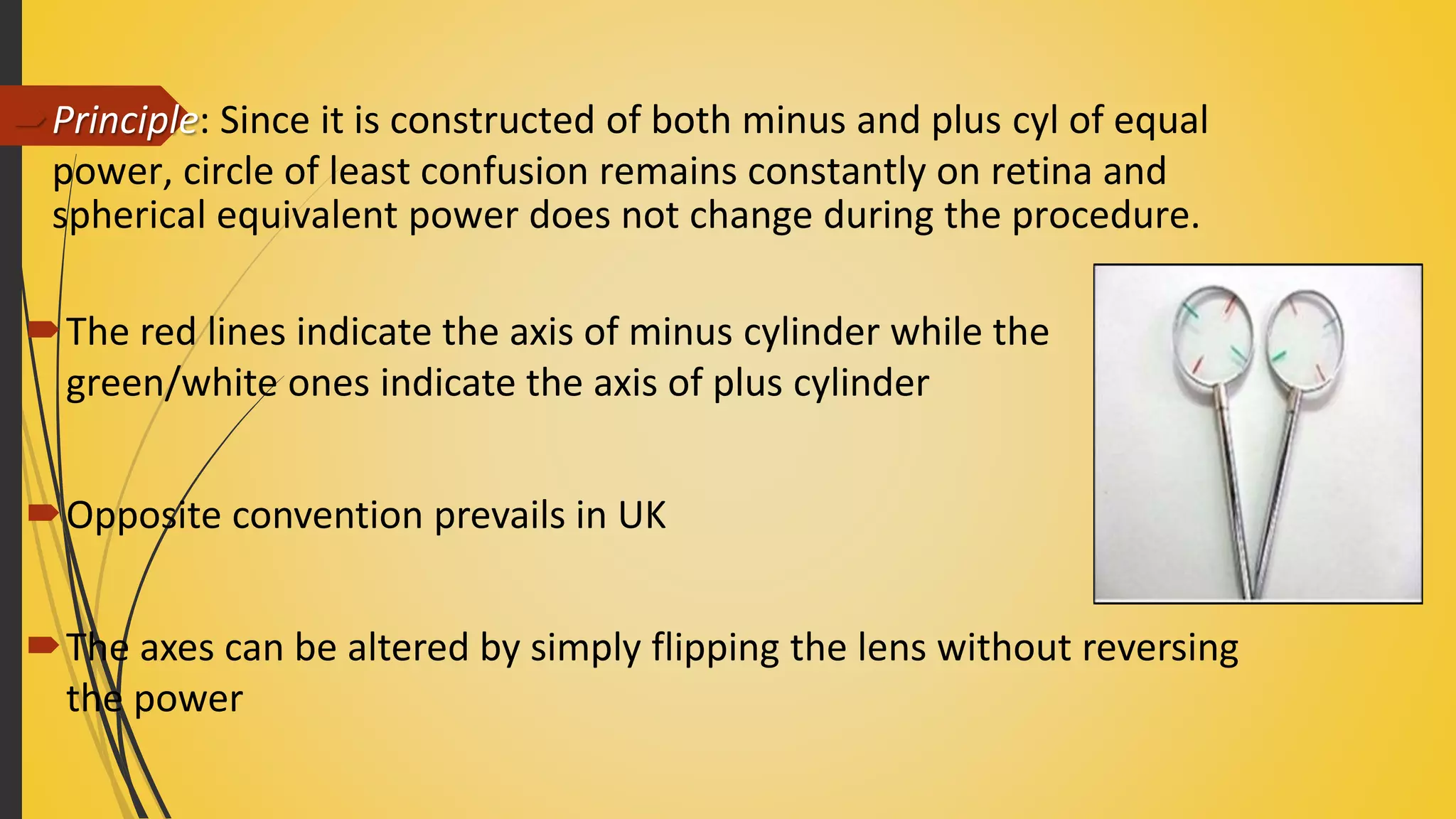
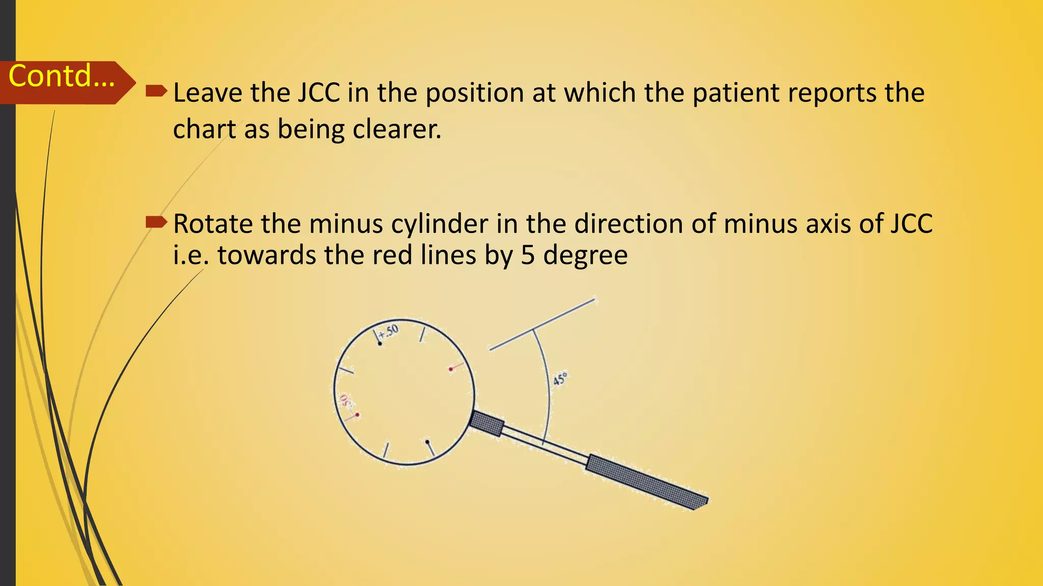
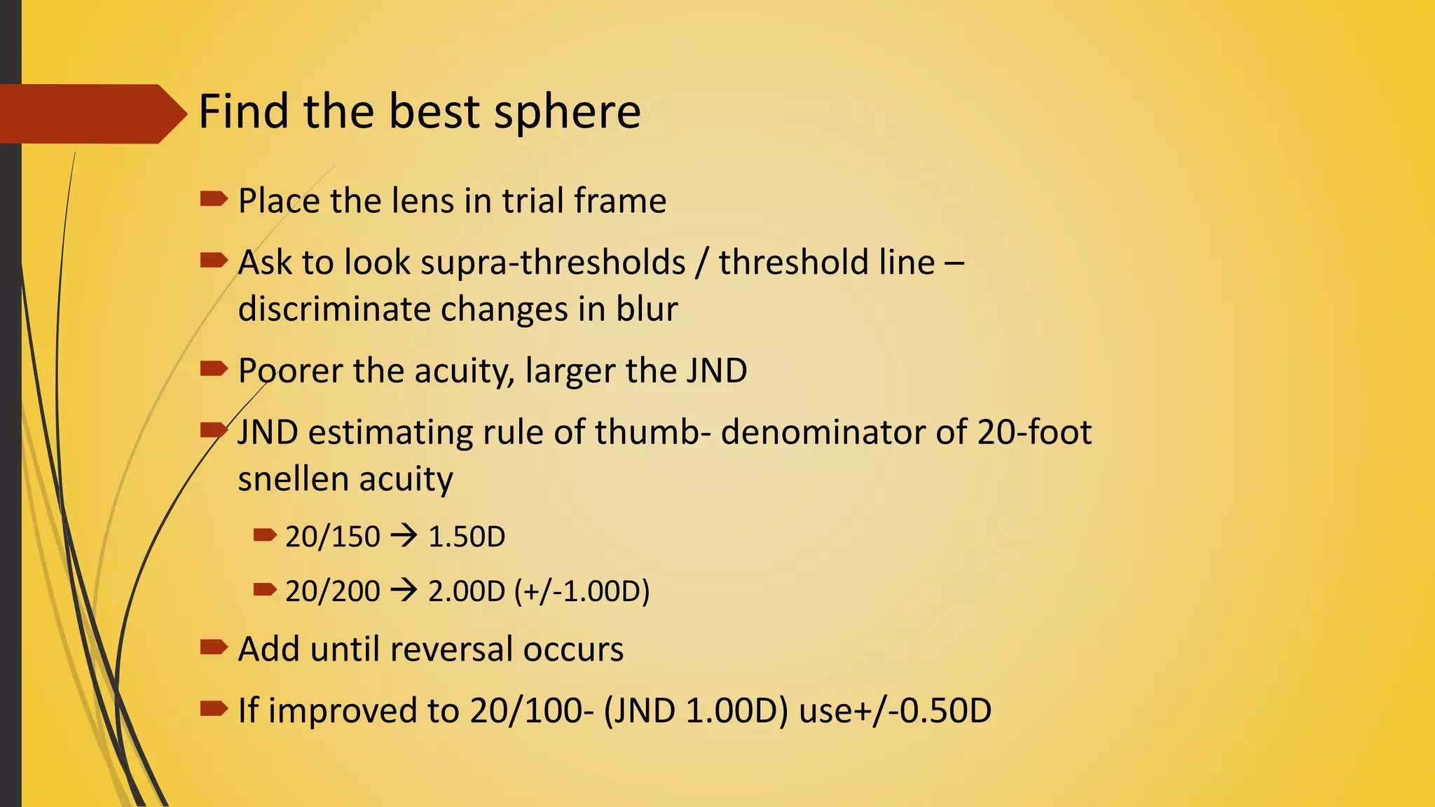
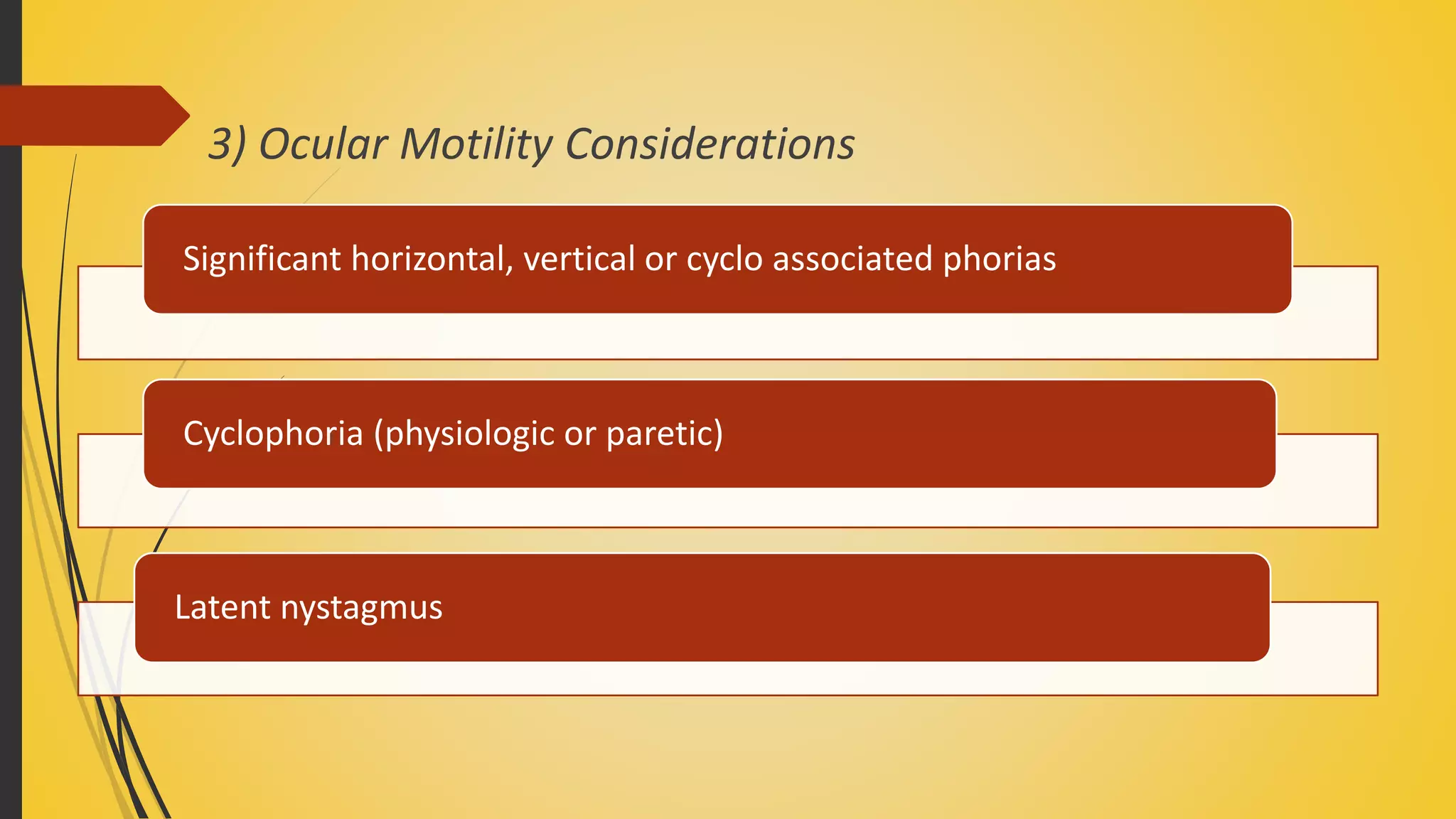
Subjective Refraction in Astigmatism:-
Various Modalities & Test Procedures.
Guided by :- Presenters:-
Sanjeev Kumar Mishra Bipin Koirala
Jenisha Bhattarai
IOM, MMC
Quote of the day…..
Contents:-
Astigmatism and its types
Introduction to subjective refraction.
Principle
Monocular Subjective Refraction
 Monocular best sphere
 Cylindrical correction by astigmatic charts
 Cylindrical correction by Jackson cross cylinder
 Monocular spherical end point
 Spherical equalization
Astigmatism:-
Type of refractive error in which power of eye varies in different meridian.
Types of astigmatism
Regular astigmatism:- Refractive power changes uniformly from one meridian
to next i.e. (two principal meridian are present )
Irregular astigmatism :- Irregular change of refractive power in different
meridian i.e. (multiple meridians are present )
Astigmatism
Regular Irregular
Regular astigmatism
Classification based on etiology :-
They are following 3 types :-
Regular
astigmatism
Corneal
Astigmatism Lenticular
astigmatism
Retinal
astigmatism
Curvatural Positional Index
Classification based on angle between two principal meridian:-
They are following 4 types :-
Regular
Astigmatism
With the rule
Astigmatism
Against the rule
Astigmatism
Oblique
Astigmatism
Bi-oblique
Astigmatism
With the rule astigmatism
 Principal meridians are at right angle to each other .
 Vertical meridian is more curved than horizontal
 Call with the rule because such type of astigmatic error exists normally.
 Concave cylinder at (180+/- 20 ) & convex cylinder at (90 +/- 20) needed for
correcting error.
Against the rule astigmatism
 Principal meridians at right angle to each other.
 Horizontal meridian more curved than vertical meridian.
 Concave cylinder at (90+/-20 ) & convex cylinder at (180+/-20) needed for
correcting error.
Oblique astigmatism
 Principal meridians are aligned at right angle to each other
 But axes are not horizontal and vertical
 Examples : one meridians at 45 degree and next aligned at
135 degree
Bi-oblique astigmatism
 Principal meridians are not at right angles to each other.
 Example : one meridian at 30 degree and next at 100
degree
Classification based on Refractive type:-
During refraction of light in astigmatic eye two focal lines are formed with
respect to retina.
Depending upon the position of two focal lines types of regular
astigmatism are as follows.
Regular
Astigmatism
Simple
Astigmatism
Compound
Astigmatism
Mixed
Astigmatism
Simple astigmatism
 Rays from one meridian focused at retina
 And ray from next meridian focused either in front or behind retina.
 Simple myopic :- one at retina , next in front of retina
 Simple hyperopic :- one at retina , next behind retina
Compound astigmatism
 Rays from both meridian are focused either in front or behind
retina.
 Compound myopic :- light from both meridian focused in front
of retina.
 Compound hyperopic:- light from both meridian focused behind
retina
Mixed astigmatism
 Light in one meridian focused in front of retina and next
behind retina.
 Thus in one meridian eye is myopic and in next hyperopic
hence called mixed.
Patient with mixed astigmatism are comparatively less
symptomatic as compared to compound astigmatism
becoz circle of least diffusion is formed near to the retina
Irregular astigmatism
Irregular change in refractive error in different meridian.
No principal meridian present.
 Types based on etiology
Irregular
astigmatism
Corneal
Lenticular
Retinal
Examples of some conditions in which irregular astigmatism is seen
Corneal irregular Lenticular irregular Retinal irregular
1. Keratoconus
2. limbal dermoid
3. Corneal scars .
1. Variation of
refractive index in
different parts.
2. Matured cataract
1. Distortion of
macular area due
to scarring or
tumors of retina or
choroid.
Did you know????
Age changes in astigmatism
 Child is born with Against the rule astigmatism.
 preschool child :- Against rule decreases
 School age :- Small amount of with the rule starts.
 After 30s :- Again gradually changes towards against rule.
Mnemonics
AWA
Image formation in an astigmatic eye
Strum’s conoid :- It is the configuration of light rays refracted from an
astigmatic surface.
Circle of least diffusion :- A point in strums conoid in which divergence of
rays of one meridian is exactly equal to convergence of rays from another
principal meridian.
Interval of strum :- Distance between two focal points is called interval of
strum.
Strum’s conoid
Subjective refraction
Subjective refraction is the method of determining the most suitable lens
to be prescribed, with the proper response of patient.
It is the technique of comparing one lens against another, using changes
in vision as the criterion, to arrive the dioptric lens combination that
results in maximum visual acuity.(polasky 1991)
The purpose is to find the strongest plus lens or the smallest minus lens
which allows the patient to obtain the best possible visual acuity.
Principle:-
Subjective determination of the combination of sphere and
cylindrical lens that artificially places the far point of each eye of
patient at infinity.
This is the combination of lenses that provides best VA with
accomodation relaxed.
Types:-
1) Monocular subjective refraction:
Performed with contralateral eye (not being
tested) under occlusion.
2) Binocular subjective refraction:
Performed with both eyes viewing a single
target while one of the eyes tested.
Monocular subjective refraction:-
Steps:-
1) Starting point
2) Control of accomodation
3) Monocular best sphere
4) Cylindrical component of refractive correction
testing under fog
 testing without fog
5) Monocular spherical end point
6) Spherical equalization
1) Starting point
 Objective refraction
 Cycloplegic refraction
 Habitual spectacle correction or results of
previous subjective refraction.
 Sometimes random choice can be done in
absence of auto refractors and retinoscope
Things to remember while doing subjective refraction.
 The geometric centers of the lens must be aligned with the
centers of entrance pupils of the patient’s eye.
 Adjustments of IPD.
 Appropriate vertex distance.
 Appropriate pantoscopic angle.
2) Control of accomodation
It is an important step since ophthalmic asthenopia is largely
caused due to overactive cilliary muscle contraction.
Refractive status is represented by focal position of eye
relative to outer limiting membrane of retina with
accomodation at rest.
Two classic means of keeping accomodation at rest are:
 Cycloplegia
Fogging
Cycloplegia:
Cycloplegia causes paralysis of accomodation.
Advantageous in case of accommodative spasm, latent
hyperopia, convergent strabismus due to over
accomodation.
These conditions tend to resist routine technique for
accomodative relaxation.
However, cycloplegia also inhibits normal cilliary tonus.
So subjective refraction is done by reducing that amount
of cilliary tonus.
Fogging
Accomodation activity is best controlled by placing the focus of distant
target in front of retina i.e. making the eye artificially myopic- fogging
technique.
Activation of accomodative system  moves the focus forward and
backward from retina Vision deteriorates
Optimal vision is only attainable by moving the focus towards the
retina by interchanging of lenses while accomodation remains
inactive- unfogging
For hypermetropes/compound hyperopic astigmatism:
Add sufficient plus to ensure that both primary meridians are focused
in front of retina.
 For myopes/compound myopic astigmatism:
 Both meridians are already in front of retina.
 High myopes; minus lenses may be added, but weaker than that of
any meridian.
 For mixed astigmatism:
Plus lens of sufficient strength to place hyperopic meridian in front of
retina.
Classic fogging technique
Fogging applied to uncorrected eye.
Initial objective- to blur the eye by sufficient plus(or reduce minus)
to reduce snellen acuity to 20/100(6/30) or worse.
Reduce plus power(or add minus) in 0.25D steps until VA is
improved to a point that patient can distinguish lines in a card at
distance for a astigmatic correction (unfogging).
Contd..
In hyperopia, the distance VA is not lessened (or even
improves) with plus lens.
In such cases, progerssively stronger plus lens are placed until
vision blurs to 20/100(6/30).
In myopia, distant vision blurs with small plus lens.
3.Monocular best sphere
A patient without astigmatism should be fully corrected by this step
alone.
However, if astigmatism is present, the aim of this step is to position
the two focal lines at retina.
Two methods:
 VA method
 Bichrome method
VA method:-
Patient is asked to read the smallest letters possible on the chart with
objective reading.
plus sphere power(or reduce minus) is added in 0.25D until the patient
reports the blurring to relax the accomodation.
Now plus power(or add minus) is reduced in 0.25 steps until any
decrease in plus power gives no improvement or makes vision worse.
 Check for myopic overcorrection(if the patient reports the letters
appearing smaller and darker)
Bichrome or duochrome test:-
Based on eyes natural chromatic aberration.
Duochrome chart is a distance VA chart with black letters,
split equally into two equal halves.
Letters on one half on red background and others on
green background.
Contd..
Since the red and green foci are nearly equally spaced about
yellow, an emmetrope should see black objects on the two
backgrounds equally clear.
In myopes red are clearer.
In hypermetropes, letters on green are clearer.
Emmetropic
Myopic
Hyperopic
Procedure:-
Dim illumination preferable for better accuracy.
Target: 20/20 letters on red and green background.
Steps:
Add plus power to the objective reading till the letters on red are clearer.
 Reduce the plus power till the letters on the red and green sides are
equally clear.
 Or, the last power with which the patient reports the letters on red side
to be clearer is the best sphere.
Demerits of bichrome test
If ametropia is larger than 1D, the test will be unreliable since the patterns
on both colors will be grossly out of focus.
With older patients the crystalline lens becomes markedly yellow, blue
green light being partially absorbed and scattered. This gives red bias to the
test.
Difficult in protanopic patient; since the red background will appear much
dimmer than green.
Contd….
However subject is instructed to emphasize on clarity of letters not
the background.
Thus, duo chrome test is also useful in color deficient person.
Testing under fog
 Clock dial
 Sunburst dial
 Rotatory T
 Astigmatic fan and block
 Stenopaeic slit
Testing without fog
 Jackson cross cylinder
Fixed astigmatic dials
Rotatory astigmatic dial
Combined dial
4. Cylindrical component of refractive correction
Why exact axis is needed?
To correct patient’s refractive error, the axis of correcting
cylinder must be exact.
If the axis is off slightly, the correcting cylinder in refractor
combines with the uncorrected astigmatism to form
resultant cylinder in new axis.
Testing under fog
 In astigmatic eye under the fog, the two principal meridians will focus in
front of the retina at different distance; the difference being the
cylindrical component.
 While unfogging, both meridians are moved back towards the retina
until the point of greatest contrast is reached.
 The spherical component corresponds to this power, and minus cylinder
is required to bring the other meridian’s shorter focus to the retina.
Fixed astigmatic dials
Consist of fixed, radial dials which present in the form of lines radiating
away from a center.
Principle : the point image along any line would be extended in a
direction determined by the axis of astigmatic error
The meridian parallel with the faintest line or that perpendicular to the
darkest line denotes the axis of the minus cylinder
For example : if axis of minus astigmatic error is 1800, the vertical line
would appear more clear and prominent while the horizontal line would
appear fainter and fuzzy.
Clock dial
Introduced by John Green 1868
Presents meridians spaced at 30° intervals that coincide with the
positions of hours on a clock face.
Each radiation is represented by a set of three lines, spaced to be
distinguished at VA equivalent to 20/25 (6/7.5) or (20/30)6/9
Procedure:-
Pt is fogged, as indicated earlier, then unfogged until some or all of the
radial lines are apparent.
(Pt. is asked whether 3 lines can be seen in any or all of the spokes)
During unfogging, subject is asked to compare the sharpness and darkness
of the lines in various directions.
Indicate when the differences between the blackest/sharpest and
faintest/fuzziest line is maximum. This is the point of greatest contrast.
Contd…
Unfogging is discontinued when the next reduction in fog makes little or
no difference between the contrast.
For axis of correcting cylinder, multiply the lesser hour of the most
prominent line by 30
E.g : 12-6 0’clock distinct
6 x 30 = 180°
Contd…
The cylinder power can be determined by adding cylindrical lens in -
0.25D step till the most prominent line and the faint line set at right
angles to it are equally clear.
Procedure is referred as “collapsing the conoid of sturm”
 vertical and horizontal lines are replaced by point image
Contd…
The cylinder may be further refined by refogging in increments of +0.25Ds.
(Check test)
If astigmatism is corrected properly, all lines appear equally clear/blurred
under the fogging lens
If the power is insufficient : the original prominent line will appear first
If the power is too great : the line sets reverse in prominence
Disadvantages of clock dial
Relatively large gaps between the orientation of adjacent spokes.
For e.g. if the axis of astigmatic error is between 30 and 60 degrees,
(say 45),
 the prominence of two lines might be similar
difficulty in subjective differentiation
Sunburst dial
Consists of a fixed protractor in which single lines radiate away
from the hub, spaced at 10 degree angles
Procedure similar to clock dial
More critical identification of the darkest line
Demerit : difficulty to inform the precise location of
darkest or faintest line
Rotatory astigmatic dials
Adjustable line targets which can be rotated to align with the primary
meridians of astigmatic eye more exactly
More clinically understandable
Greater reliability and precision
Basic construction : two groups of parallel lines at
right angles to each other
Rotatory T
Presented to fogged eye with the lines in
the 090 and 180 meridians.
Unfogged till the patient reports visibility of the lines.
Pt is asked whether one set of lines appears more prominent than the
other.
If both are equally distinct, the chart is rotated slowly
-the subject is asked about the position at which one set
of lines appear blacker or more visible than the line in
opposite direction.
If difference is reported either at the original placement or a newly found
position, the axis is refined by continuing to rotate the chart until both
crossed lines appear of equal prominence to the subject.
This represents the position exactly 45° from the positions of the principle
meridians of the eye.
Contd…
 To check the position, the chart is then rotated in the opposite
direction until a point of equality is reached again.
(The chart is now in a meridian perpendicular to the first bracketed
setting)
The numerical average of the two bracketed meridians is the
meridional position of the minus cylinder axis.
 E.g. firstly equal clarity at 45° and secondly at 130° then, position
of minus cylinder axis = (45+130)/2=87.5°
Contd…
Astigmatic fan
Combination of fixed and rotatory
Fan - to determine the axis of correcting cylinder
Maddox V - to conform the axis of correcting cylinder
Blocks - to determine the power of correcting cylinder
Spacing between the spokes - 10 degrees
The Maddox V and block can be simultaneously rotated through 180
degrees
Procedure
Obtain the best VA putting the best spherical power in frame
 Astigmatism error is estimated and half of this is added as positive sphere
so as to bring the eye into a state of simple myopic astigmatism
Pt. asked to locate clearest lines in the fan chart; this gives the approximat
direction of astigmatic error
Check test by refogging
Contd…
The tip of maddox arrow is rotated to the point at the clear group
Directed the attention to limb of maddox arrow
- rotated away from its blacker limb until both limbs
become clear
- gives the axis of astigmatism
- ensure that pt’s head is upright
Contd…
Directed the attention to the blocks
Negative cylinder added at the appropriate axis until the both blocks
appear equally clear
Check test by refogging
Stenopaeic slit
 Consists of a rectangular aperture ranging
from 0.5 to 1.0mm in width and up to 15 mm
in length
 Width of slit approximates to that of a pin
hole – assumed to limit light to one meridian
Procedure
 Rotate the slit till pt. reports most clear vision through the slit.
 It’s one of the principal meridians – add spherical lenses to obtain best
VA
 Next meridian is at right angle to the first meridian – rotates slit by 90
degree – again add lenses to get best VA in that meridian
 This gives Rx of two meridians – spherocylindrical correction can be
calculated
Jackson Cross Cylinder(JCC) / flip cross technique
 Major technique used today
 Refinement of cylinder axis and power
 Consists of
- A pair of equal powered plano cyl of opposite sign
- ground on different sides of the same lens
- with their axes at right angle
 ±0.25 D, ±0.37 D, ±0.50 D,
±1.00D
Testing without fog
Principle: Since it is constructed of both minus and plus cyl of equal
power, circle of least confusion remains constantly on retina and
spherical equivalent power does not change during the procedure.
The red lines indicate the axis of minus cylinder while the
green/white ones indicate the axis of plus cylinder
Opposite convention prevails in UK
The axes can be altered by simply flipping the lens without reversing
the power
If a correcting cylinder of minus power is used in refractor, the axis
of minus cylinder of JCC is the axis of reference.
Plus component can be used as reference if correcting cylinder is
plus.
Procedure:-
Determining the axis
Handle of cross cylinder is made parallel to axis
of correcting cylinder so that power meridian
of JCC is 450 away from principal meridian of
correcting cylinder.
Now, lens is flipped and patient is asked to
report which of two position is more distinct.
Combination of minus cylinder in refractor and JCC results in net
correcting axis and power.
If the eye’s actual minus cylinder error is in clockwise direction,
application of JCC with its minus axis in clockwise direction should
increase the clarity.
Leave the JCC in the position at which the patient reports the
chart as being clearer.
Rotate the minus cylinder in the direction of minus axis of JCC
i.e. towards the red lines by 5 degree
Contd…
While rotating the refractor cylinder, the JCC lens must also be
rotated an equal amount so the red and white lines remain exactly
45 degree away from the axis of the refractor cyl.
With the correcting cyl in new position, flip the lens again.
Repeat the above procedure until the patient can no longer tell any
difference in the appearance of the chart when JCC is flipped.
Contd…
Determining the power
Axis of JCC is placed parallel to that of correcting cylinder
The lens is then flipped in front of pt’s eye
Patient is asked to report the position which permits better visual acuity
Contd… In one position, minus axis of JCC (red lines) is aligned with the
axis of minus correcting cylinder
While in the other, plus axis of JCC (green lines) coincides with the
axis of minus correcting cylinder
 If the patient prefers the 1st position, then the minus cylinder
power is increased until equality is obtained.
 If the patient prefers the 2nd position, then the minus cylinder
power is decreased until equality is reported.
Contd…
Common sources of error in JCC
1. Not keeping the circle of least confusion on the retina
Starting with the wrong sphere power
Forgetting to change sphere power if cyl is changed by 0.50DC or more
2. Assuming the axis is correct if the patient says “they look the same”
without checking
Could be no astigmatism at all
Could be 900 off
3. Incorrect presentation time – esp. too quick
4. Poor alignment of JCC and trial frame axis
Spherical power
When correcting cylinder power is added, only one end of
astigmatic interval is affected.
Therefore, change in sphere power is required to recenter
the interval on retina.
For each 0.50D change in cylinder power, there must be a
0.25D change in sphere power.
5. Monocular Spherical end point
Traditional spherical end points
Duochrome/Bichrome method
Traditional spherical end points
 The residually spherical eyes are fogged until several previously
visible lines of acuity are blurred for each eye
 Then, unfogged in 0.25D steps until spherical lens providing
maximum visual acuity is reached
 End point – VA can no longer be enhanced by addition of minus or
reduction of plus
Duochrome or bichrome method
Duochrome chart is presented in dim illumination
Patient is asked whether the letters are equally
prominent in both the background or in either
one
Slightly fog the vision by adding plus sphere in
0.25D until letters on red background stand out
Contd..
Reduce plus sphere until letters in two charts appears equally
clear.
OR
Until the reduction of only -0.25 makes letters on green
background more distinct. i.e. first green(appropriate end points
for young population with active accomodation).
6. Spherical equalization
The purpose of spherical equalization (binocular balancing) is not to balance
the visual acuity but to balance the state of accommodation of the two
eyes.
If the corrected VA is same in both eye, the balancing procedure may consist
of comparison of the visual acuity for the two eyes.
If the corrected VA is not same in both eyes(aniso-oxyopia), then a method
not based on visual acuity must be used.
Unqual accommodative response between the two eyes
Inequality in the clarity or size of the retinal images
Reduce stereo acuity or fusional amplitudes
discomfort and visual inefficiency.
Common techniques of spherical equalization are :
1. Alternate occlusion method
2. Prism dissociation method
Alternate occlusion
 Both eyes are alternately occluded repeatedly while patient views VA
chart at distance through the spherocylindrical correction determined
monocularly.
 Performed with handheld cover paddle.
 Patient compares and informs which eye resolves more letters on chart.
 The examiner then adjusts the spherical balance so as to produce
equality between the eyes.
 Not helpful in aniso-oxyopia and amblyopia
 Subject compares a visible object with a previous one remembered
but no longer visible.
 Must be repetitive and slow enough for patient to recognize and
accurately state the eye with clearer vision
 Also be fast enough so that patient cannot accommodate for
residually hyperopic eye while the other is occluded
Prism dissociation
 Placing 3 of base-down prism in front of the right eye and 3 of base-
up prism in front of the left eye .
 The charts gets separated vertically, the upper chart being seen by the
RE and lower by the LE
 Patient is asked to report whether the letters are more distinct or easier
to read in the upper chart or lower chart.
 If the two 20/25 lines are equally distinct for the two eyes, the
accommodative state of the two eyes is considered to be balanced.
 If the patient reports a difference in clarity of the letters for the two eyes.
 +0.25 D is added in front of the eye with the better vision and the test is
repeated.
 Once the patients VA is balanced at 20/25, the patient is defogged
binocularly to the criterion.
 Endpoint- balance at equality of acuity.
 When end point of balance is reached, remove prism and find binocular
spherical end point.
Subjective refraction in low vision patients.
Find the best sphere
Test for astigmatism
Re-test for the “best sphere”
Find the best sphere
Place the lens in trial frame
Ask to look supra-thresholds / threshold line –
discriminate changes in blur
Poorer the acuity, larger the JND
JND estimating rule of thumb- denominator of 20-foot
snellen acuity
20/150  1.50D
20/200  2.00D (+/-1.00D)
Add until reversal occurs
If improved to 20/100- (JND 1.00D) use+/-0.50D
Find the best cylinder
Place the lens by K-reading or retinoscopy
Refine the axis first and then power by JCC
Poorer the acuity, higher the JCC power
JCC set required:
+/- 0.25D for normal vision
+/- 0.50D for 20/30 to 20/50
+/- 0.75 for 20/50 to 20/100
+/- 1.00 for 20.100 & worse
In low vision, use strong JCC to determine astigmatic
component
Re-test for best sphere
Test again for best sphere using JND lenses
Binocular subjective refraction
Clinical procedure in which the subjective refraction is
performed monocularly under binocular viewing
conditions.
Component procedures described monocularly are
performed in same or similar manner
Except that
Both eyes are open
Unoccluded
 Views a common target
Advantage over monocular refraction
Accomodation, convergence and light adaptation more
constant.
Refractive status evaluated in more nearly normal
environment.
Detection of suppression
Measurement of stereopsis
Measurement of fixation disparity.
Indications for Binocular Refraction
1)Refractive Considerations
Hyperopic anisometropia
Antimetropia
Latent hyperopia
Pseudo myopia
2) Visual Acuity Considerations
Anisooxyopia ( unequal acuities between two eyes)
Unilateral amblyopia
Unilateral reduced acuity as a result of ocular disease
3) Ocular Motility Considerations
Significant horizontal, vertical or cyclo associated phorias
Cyclophoria (physiologic or paretic)
Latent nystagmus
Difficulties with subjective refraction testing……
Intelligence, past experience, co-operation
Size of pupil : small pupil increased depth of
focus
Testing factors : target & room illumination
testing distance
retinal adaptation
Unability to discriminate
between lens choices
References
Borish’s clinical refraction, William J. Benjamin
Primary Care Optometry, Theodore Grosvenor
Clinical Visual Optics, Bennett and Rabbetts
Clinical Procedures in Optometry, J.D Bartlett
Internet
Real subjective refraction in astigmatism

Real subjective refraction in astigmatism

  • 1.
    Subjective Refraction inAstigmatism:- Various Modalities & Test Procedures. Guided by :- Presenters:- Sanjeev Kumar Mishra Bipin Koirala Jenisha Bhattarai IOM, MMC
  • 2.
    Quote of theday…..
  • 3.
    Contents:- Astigmatism and itstypes Introduction to subjective refraction. Principle Monocular Subjective Refraction  Monocular best sphere  Cylindrical correction by astigmatic charts  Cylindrical correction by Jackson cross cylinder  Monocular spherical end point  Spherical equalization
  • 4.
    Astigmatism:- Type of refractiveerror in which power of eye varies in different meridian. Types of astigmatism Regular astigmatism:- Refractive power changes uniformly from one meridian to next i.e. (two principal meridian are present ) Irregular astigmatism :- Irregular change of refractive power in different meridian i.e. (multiple meridians are present ) Astigmatism Regular Irregular
  • 5.
    Regular astigmatism Classification basedon etiology :- They are following 3 types :- Regular astigmatism Corneal Astigmatism Lenticular astigmatism Retinal astigmatism Curvatural Positional Index
  • 6.
    Classification based onangle between two principal meridian:- They are following 4 types :- Regular Astigmatism With the rule Astigmatism Against the rule Astigmatism Oblique Astigmatism Bi-oblique Astigmatism
  • 7.
    With the ruleastigmatism  Principal meridians are at right angle to each other .  Vertical meridian is more curved than horizontal  Call with the rule because such type of astigmatic error exists normally.  Concave cylinder at (180+/- 20 ) & convex cylinder at (90 +/- 20) needed for correcting error. Against the rule astigmatism  Principal meridians at right angle to each other.  Horizontal meridian more curved than vertical meridian.  Concave cylinder at (90+/-20 ) & convex cylinder at (180+/-20) needed for correcting error.
  • 8.
    Oblique astigmatism  Principalmeridians are aligned at right angle to each other  But axes are not horizontal and vertical  Examples : one meridians at 45 degree and next aligned at 135 degree Bi-oblique astigmatism  Principal meridians are not at right angles to each other.  Example : one meridian at 30 degree and next at 100 degree
  • 9.
    Classification based onRefractive type:- During refraction of light in astigmatic eye two focal lines are formed with respect to retina. Depending upon the position of two focal lines types of regular astigmatism are as follows. Regular Astigmatism Simple Astigmatism Compound Astigmatism Mixed Astigmatism
  • 10.
    Simple astigmatism  Raysfrom one meridian focused at retina  And ray from next meridian focused either in front or behind retina.  Simple myopic :- one at retina , next in front of retina  Simple hyperopic :- one at retina , next behind retina
  • 11.
    Compound astigmatism  Raysfrom both meridian are focused either in front or behind retina.  Compound myopic :- light from both meridian focused in front of retina.  Compound hyperopic:- light from both meridian focused behind retina
  • 12.
    Mixed astigmatism  Lightin one meridian focused in front of retina and next behind retina.  Thus in one meridian eye is myopic and in next hyperopic hence called mixed. Patient with mixed astigmatism are comparatively less symptomatic as compared to compound astigmatism becoz circle of least diffusion is formed near to the retina
  • 13.
    Irregular astigmatism Irregular changein refractive error in different meridian. No principal meridian present.  Types based on etiology Irregular astigmatism Corneal Lenticular Retinal
  • 14.
    Examples of someconditions in which irregular astigmatism is seen Corneal irregular Lenticular irregular Retinal irregular 1. Keratoconus 2. limbal dermoid 3. Corneal scars . 1. Variation of refractive index in different parts. 2. Matured cataract 1. Distortion of macular area due to scarring or tumors of retina or choroid.
  • 15.
    Did you know???? Agechanges in astigmatism  Child is born with Against the rule astigmatism.  preschool child :- Against rule decreases  School age :- Small amount of with the rule starts.  After 30s :- Again gradually changes towards against rule. Mnemonics AWA
  • 16.
    Image formation inan astigmatic eye Strum’s conoid :- It is the configuration of light rays refracted from an astigmatic surface. Circle of least diffusion :- A point in strums conoid in which divergence of rays of one meridian is exactly equal to convergence of rays from another principal meridian. Interval of strum :- Distance between two focal points is called interval of strum.
  • 17.
  • 18.
    Subjective refraction Subjective refractionis the method of determining the most suitable lens to be prescribed, with the proper response of patient. It is the technique of comparing one lens against another, using changes in vision as the criterion, to arrive the dioptric lens combination that results in maximum visual acuity.(polasky 1991) The purpose is to find the strongest plus lens or the smallest minus lens which allows the patient to obtain the best possible visual acuity.
  • 19.
    Principle:- Subjective determination ofthe combination of sphere and cylindrical lens that artificially places the far point of each eye of patient at infinity. This is the combination of lenses that provides best VA with accomodation relaxed.
  • 20.
    Types:- 1) Monocular subjectiverefraction: Performed with contralateral eye (not being tested) under occlusion. 2) Binocular subjective refraction: Performed with both eyes viewing a single target while one of the eyes tested.
  • 21.
    Monocular subjective refraction:- Steps:- 1)Starting point 2) Control of accomodation 3) Monocular best sphere 4) Cylindrical component of refractive correction testing under fog  testing without fog 5) Monocular spherical end point 6) Spherical equalization
  • 22.
    1) Starting point Objective refraction  Cycloplegic refraction  Habitual spectacle correction or results of previous subjective refraction.  Sometimes random choice can be done in absence of auto refractors and retinoscope
  • 23.
    Things to rememberwhile doing subjective refraction.  The geometric centers of the lens must be aligned with the centers of entrance pupils of the patient’s eye.  Adjustments of IPD.  Appropriate vertex distance.  Appropriate pantoscopic angle.
  • 24.
    2) Control ofaccomodation It is an important step since ophthalmic asthenopia is largely caused due to overactive cilliary muscle contraction. Refractive status is represented by focal position of eye relative to outer limiting membrane of retina with accomodation at rest. Two classic means of keeping accomodation at rest are:  Cycloplegia Fogging
  • 25.
    Cycloplegia: Cycloplegia causes paralysisof accomodation. Advantageous in case of accommodative spasm, latent hyperopia, convergent strabismus due to over accomodation. These conditions tend to resist routine technique for accomodative relaxation. However, cycloplegia also inhibits normal cilliary tonus. So subjective refraction is done by reducing that amount of cilliary tonus.
  • 26.
    Fogging Accomodation activity isbest controlled by placing the focus of distant target in front of retina i.e. making the eye artificially myopic- fogging technique. Activation of accomodative system  moves the focus forward and backward from retina Vision deteriorates Optimal vision is only attainable by moving the focus towards the retina by interchanging of lenses while accomodation remains inactive- unfogging
  • 27.
    For hypermetropes/compound hyperopicastigmatism: Add sufficient plus to ensure that both primary meridians are focused in front of retina.  For myopes/compound myopic astigmatism:  Both meridians are already in front of retina.  High myopes; minus lenses may be added, but weaker than that of any meridian.  For mixed astigmatism: Plus lens of sufficient strength to place hyperopic meridian in front of retina.
  • 28.
    Classic fogging technique Foggingapplied to uncorrected eye. Initial objective- to blur the eye by sufficient plus(or reduce minus) to reduce snellen acuity to 20/100(6/30) or worse. Reduce plus power(or add minus) in 0.25D steps until VA is improved to a point that patient can distinguish lines in a card at distance for a astigmatic correction (unfogging).
  • 29.
    Contd.. In hyperopia, thedistance VA is not lessened (or even improves) with plus lens. In such cases, progerssively stronger plus lens are placed until vision blurs to 20/100(6/30). In myopia, distant vision blurs with small plus lens.
  • 30.
    3.Monocular best sphere Apatient without astigmatism should be fully corrected by this step alone. However, if astigmatism is present, the aim of this step is to position the two focal lines at retina. Two methods:  VA method  Bichrome method
  • 31.
    VA method:- Patient isasked to read the smallest letters possible on the chart with objective reading. plus sphere power(or reduce minus) is added in 0.25D until the patient reports the blurring to relax the accomodation. Now plus power(or add minus) is reduced in 0.25 steps until any decrease in plus power gives no improvement or makes vision worse.  Check for myopic overcorrection(if the patient reports the letters appearing smaller and darker)
  • 32.
    Bichrome or duochrometest:- Based on eyes natural chromatic aberration. Duochrome chart is a distance VA chart with black letters, split equally into two equal halves. Letters on one half on red background and others on green background.
  • 33.
    Contd.. Since the redand green foci are nearly equally spaced about yellow, an emmetrope should see black objects on the two backgrounds equally clear. In myopes red are clearer. In hypermetropes, letters on green are clearer.
  • 34.
  • 35.
    Procedure:- Dim illumination preferablefor better accuracy. Target: 20/20 letters on red and green background. Steps: Add plus power to the objective reading till the letters on red are clearer.  Reduce the plus power till the letters on the red and green sides are equally clear.  Or, the last power with which the patient reports the letters on red side to be clearer is the best sphere.
  • 36.
    Demerits of bichrometest If ametropia is larger than 1D, the test will be unreliable since the patterns on both colors will be grossly out of focus. With older patients the crystalline lens becomes markedly yellow, blue green light being partially absorbed and scattered. This gives red bias to the test. Difficult in protanopic patient; since the red background will appear much dimmer than green.
  • 37.
    Contd…. However subject isinstructed to emphasize on clarity of letters not the background. Thus, duo chrome test is also useful in color deficient person.
  • 38.
    Testing under fog Clock dial  Sunburst dial  Rotatory T  Astigmatic fan and block  Stenopaeic slit Testing without fog  Jackson cross cylinder Fixed astigmatic dials Rotatory astigmatic dial Combined dial 4. Cylindrical component of refractive correction
  • 39.
    Why exact axisis needed? To correct patient’s refractive error, the axis of correcting cylinder must be exact. If the axis is off slightly, the correcting cylinder in refractor combines with the uncorrected astigmatism to form resultant cylinder in new axis.
  • 40.
    Testing under fog In astigmatic eye under the fog, the two principal meridians will focus in front of the retina at different distance; the difference being the cylindrical component.  While unfogging, both meridians are moved back towards the retina until the point of greatest contrast is reached.  The spherical component corresponds to this power, and minus cylinder is required to bring the other meridian’s shorter focus to the retina.
  • 41.
    Fixed astigmatic dials Consistof fixed, radial dials which present in the form of lines radiating away from a center. Principle : the point image along any line would be extended in a direction determined by the axis of astigmatic error The meridian parallel with the faintest line or that perpendicular to the darkest line denotes the axis of the minus cylinder For example : if axis of minus astigmatic error is 1800, the vertical line would appear more clear and prominent while the horizontal line would appear fainter and fuzzy.
  • 42.
    Clock dial Introduced byJohn Green 1868 Presents meridians spaced at 30° intervals that coincide with the positions of hours on a clock face. Each radiation is represented by a set of three lines, spaced to be distinguished at VA equivalent to 20/25 (6/7.5) or (20/30)6/9
  • 43.
    Procedure:- Pt is fogged,as indicated earlier, then unfogged until some or all of the radial lines are apparent. (Pt. is asked whether 3 lines can be seen in any or all of the spokes) During unfogging, subject is asked to compare the sharpness and darkness of the lines in various directions. Indicate when the differences between the blackest/sharpest and faintest/fuzziest line is maximum. This is the point of greatest contrast.
  • 44.
    Contd… Unfogging is discontinuedwhen the next reduction in fog makes little or no difference between the contrast. For axis of correcting cylinder, multiply the lesser hour of the most prominent line by 30 E.g : 12-6 0’clock distinct 6 x 30 = 180°
  • 45.
    Contd… The cylinder powercan be determined by adding cylindrical lens in - 0.25D step till the most prominent line and the faint line set at right angles to it are equally clear. Procedure is referred as “collapsing the conoid of sturm”  vertical and horizontal lines are replaced by point image
  • 46.
    Contd… The cylinder maybe further refined by refogging in increments of +0.25Ds. (Check test) If astigmatism is corrected properly, all lines appear equally clear/blurred under the fogging lens If the power is insufficient : the original prominent line will appear first If the power is too great : the line sets reverse in prominence
  • 47.
    Disadvantages of clockdial Relatively large gaps between the orientation of adjacent spokes. For e.g. if the axis of astigmatic error is between 30 and 60 degrees, (say 45),  the prominence of two lines might be similar difficulty in subjective differentiation
  • 48.
    Sunburst dial Consists ofa fixed protractor in which single lines radiate away from the hub, spaced at 10 degree angles Procedure similar to clock dial More critical identification of the darkest line Demerit : difficulty to inform the precise location of darkest or faintest line
  • 49.
    Rotatory astigmatic dials Adjustableline targets which can be rotated to align with the primary meridians of astigmatic eye more exactly More clinically understandable Greater reliability and precision Basic construction : two groups of parallel lines at right angles to each other
  • 50.
    Rotatory T Presented tofogged eye with the lines in the 090 and 180 meridians. Unfogged till the patient reports visibility of the lines. Pt is asked whether one set of lines appears more prominent than the other. If both are equally distinct, the chart is rotated slowly -the subject is asked about the position at which one set of lines appear blacker or more visible than the line in opposite direction.
  • 51.
    If difference isreported either at the original placement or a newly found position, the axis is refined by continuing to rotate the chart until both crossed lines appear of equal prominence to the subject. This represents the position exactly 45° from the positions of the principle meridians of the eye. Contd…
  • 52.
     To checkthe position, the chart is then rotated in the opposite direction until a point of equality is reached again. (The chart is now in a meridian perpendicular to the first bracketed setting) The numerical average of the two bracketed meridians is the meridional position of the minus cylinder axis.  E.g. firstly equal clarity at 45° and secondly at 130° then, position of minus cylinder axis = (45+130)/2=87.5° Contd…
  • 53.
    Astigmatic fan Combination offixed and rotatory Fan - to determine the axis of correcting cylinder Maddox V - to conform the axis of correcting cylinder Blocks - to determine the power of correcting cylinder Spacing between the spokes - 10 degrees The Maddox V and block can be simultaneously rotated through 180 degrees
  • 54.
    Procedure Obtain the bestVA putting the best spherical power in frame  Astigmatism error is estimated and half of this is added as positive sphere so as to bring the eye into a state of simple myopic astigmatism Pt. asked to locate clearest lines in the fan chart; this gives the approximat direction of astigmatic error Check test by refogging
  • 55.
    Contd… The tip ofmaddox arrow is rotated to the point at the clear group Directed the attention to limb of maddox arrow - rotated away from its blacker limb until both limbs become clear - gives the axis of astigmatism - ensure that pt’s head is upright
  • 56.
    Contd… Directed the attentionto the blocks Negative cylinder added at the appropriate axis until the both blocks appear equally clear Check test by refogging
  • 57.
    Stenopaeic slit  Consistsof a rectangular aperture ranging from 0.5 to 1.0mm in width and up to 15 mm in length  Width of slit approximates to that of a pin hole – assumed to limit light to one meridian
  • 58.
    Procedure  Rotate theslit till pt. reports most clear vision through the slit.  It’s one of the principal meridians – add spherical lenses to obtain best VA  Next meridian is at right angle to the first meridian – rotates slit by 90 degree – again add lenses to get best VA in that meridian  This gives Rx of two meridians – spherocylindrical correction can be calculated
  • 59.
    Jackson Cross Cylinder(JCC)/ flip cross technique  Major technique used today  Refinement of cylinder axis and power  Consists of - A pair of equal powered plano cyl of opposite sign - ground on different sides of the same lens - with their axes at right angle  ±0.25 D, ±0.37 D, ±0.50 D, ±1.00D Testing without fog
  • 60.
    Principle: Since itis constructed of both minus and plus cyl of equal power, circle of least confusion remains constantly on retina and spherical equivalent power does not change during the procedure. The red lines indicate the axis of minus cylinder while the green/white ones indicate the axis of plus cylinder Opposite convention prevails in UK The axes can be altered by simply flipping the lens without reversing the power
  • 61.
    If a correctingcylinder of minus power is used in refractor, the axis of minus cylinder of JCC is the axis of reference. Plus component can be used as reference if correcting cylinder is plus. Procedure:-
  • 62.
    Determining the axis Handleof cross cylinder is made parallel to axis of correcting cylinder so that power meridian of JCC is 450 away from principal meridian of correcting cylinder. Now, lens is flipped and patient is asked to report which of two position is more distinct.
  • 63.
    Combination of minuscylinder in refractor and JCC results in net correcting axis and power. If the eye’s actual minus cylinder error is in clockwise direction, application of JCC with its minus axis in clockwise direction should increase the clarity.
  • 64.
    Leave the JCCin the position at which the patient reports the chart as being clearer. Rotate the minus cylinder in the direction of minus axis of JCC i.e. towards the red lines by 5 degree Contd…
  • 65.
    While rotating therefractor cylinder, the JCC lens must also be rotated an equal amount so the red and white lines remain exactly 45 degree away from the axis of the refractor cyl. With the correcting cyl in new position, flip the lens again. Repeat the above procedure until the patient can no longer tell any difference in the appearance of the chart when JCC is flipped. Contd…
  • 66.
    Determining the power Axisof JCC is placed parallel to that of correcting cylinder The lens is then flipped in front of pt’s eye Patient is asked to report the position which permits better visual acuity
  • 67.
    Contd… In oneposition, minus axis of JCC (red lines) is aligned with the axis of minus correcting cylinder While in the other, plus axis of JCC (green lines) coincides with the axis of minus correcting cylinder
  • 68.
     If thepatient prefers the 1st position, then the minus cylinder power is increased until equality is obtained.  If the patient prefers the 2nd position, then the minus cylinder power is decreased until equality is reported. Contd…
  • 69.
    Common sources oferror in JCC 1. Not keeping the circle of least confusion on the retina Starting with the wrong sphere power Forgetting to change sphere power if cyl is changed by 0.50DC or more 2. Assuming the axis is correct if the patient says “they look the same” without checking Could be no astigmatism at all Could be 900 off 3. Incorrect presentation time – esp. too quick 4. Poor alignment of JCC and trial frame axis
  • 70.
    Spherical power When correctingcylinder power is added, only one end of astigmatic interval is affected. Therefore, change in sphere power is required to recenter the interval on retina. For each 0.50D change in cylinder power, there must be a 0.25D change in sphere power.
  • 71.
    5. Monocular Sphericalend point Traditional spherical end points Duochrome/Bichrome method
  • 72.
    Traditional spherical endpoints  The residually spherical eyes are fogged until several previously visible lines of acuity are blurred for each eye  Then, unfogged in 0.25D steps until spherical lens providing maximum visual acuity is reached  End point – VA can no longer be enhanced by addition of minus or reduction of plus
  • 73.
    Duochrome or bichromemethod Duochrome chart is presented in dim illumination Patient is asked whether the letters are equally prominent in both the background or in either one Slightly fog the vision by adding plus sphere in 0.25D until letters on red background stand out
  • 74.
    Contd.. Reduce plus sphereuntil letters in two charts appears equally clear. OR Until the reduction of only -0.25 makes letters on green background more distinct. i.e. first green(appropriate end points for young population with active accomodation).
  • 75.
    6. Spherical equalization Thepurpose of spherical equalization (binocular balancing) is not to balance the visual acuity but to balance the state of accommodation of the two eyes. If the corrected VA is same in both eye, the balancing procedure may consist of comparison of the visual acuity for the two eyes. If the corrected VA is not same in both eyes(aniso-oxyopia), then a method not based on visual acuity must be used.
  • 76.
    Unqual accommodative responsebetween the two eyes Inequality in the clarity or size of the retinal images Reduce stereo acuity or fusional amplitudes discomfort and visual inefficiency.
  • 77.
    Common techniques ofspherical equalization are : 1. Alternate occlusion method 2. Prism dissociation method
  • 78.
    Alternate occlusion  Botheyes are alternately occluded repeatedly while patient views VA chart at distance through the spherocylindrical correction determined monocularly.  Performed with handheld cover paddle.  Patient compares and informs which eye resolves more letters on chart.  The examiner then adjusts the spherical balance so as to produce equality between the eyes.
  • 79.
     Not helpfulin aniso-oxyopia and amblyopia  Subject compares a visible object with a previous one remembered but no longer visible.  Must be repetitive and slow enough for patient to recognize and accurately state the eye with clearer vision  Also be fast enough so that patient cannot accommodate for residually hyperopic eye while the other is occluded
  • 80.
    Prism dissociation  Placing3 of base-down prism in front of the right eye and 3 of base- up prism in front of the left eye .  The charts gets separated vertically, the upper chart being seen by the RE and lower by the LE  Patient is asked to report whether the letters are more distinct or easier to read in the upper chart or lower chart.  If the two 20/25 lines are equally distinct for the two eyes, the accommodative state of the two eyes is considered to be balanced.
  • 81.
     If thepatient reports a difference in clarity of the letters for the two eyes.  +0.25 D is added in front of the eye with the better vision and the test is repeated.  Once the patients VA is balanced at 20/25, the patient is defogged binocularly to the criterion.  Endpoint- balance at equality of acuity.  When end point of balance is reached, remove prism and find binocular spherical end point.
  • 82.
    Subjective refraction inlow vision patients. Find the best sphere Test for astigmatism Re-test for the “best sphere”
  • 83.
    Find the bestsphere Place the lens in trial frame Ask to look supra-thresholds / threshold line – discriminate changes in blur Poorer the acuity, larger the JND JND estimating rule of thumb- denominator of 20-foot snellen acuity 20/150  1.50D 20/200  2.00D (+/-1.00D) Add until reversal occurs If improved to 20/100- (JND 1.00D) use+/-0.50D
  • 84.
    Find the bestcylinder Place the lens by K-reading or retinoscopy Refine the axis first and then power by JCC Poorer the acuity, higher the JCC power JCC set required: +/- 0.25D for normal vision +/- 0.50D for 20/30 to 20/50 +/- 0.75 for 20/50 to 20/100 +/- 1.00 for 20.100 & worse In low vision, use strong JCC to determine astigmatic component
  • 85.
    Re-test for bestsphere Test again for best sphere using JND lenses
  • 86.
    Binocular subjective refraction Clinicalprocedure in which the subjective refraction is performed monocularly under binocular viewing conditions. Component procedures described monocularly are performed in same or similar manner Except that Both eyes are open Unoccluded  Views a common target
  • 87.
    Advantage over monocularrefraction Accomodation, convergence and light adaptation more constant. Refractive status evaluated in more nearly normal environment. Detection of suppression Measurement of stereopsis Measurement of fixation disparity.
  • 88.
    Indications for BinocularRefraction 1)Refractive Considerations Hyperopic anisometropia Antimetropia Latent hyperopia Pseudo myopia
  • 89.
    2) Visual AcuityConsiderations Anisooxyopia ( unequal acuities between two eyes) Unilateral amblyopia Unilateral reduced acuity as a result of ocular disease
  • 90.
    3) Ocular MotilityConsiderations Significant horizontal, vertical or cyclo associated phorias Cyclophoria (physiologic or paretic) Latent nystagmus
  • 91.
    Difficulties with subjectiverefraction testing…… Intelligence, past experience, co-operation Size of pupil : small pupil increased depth of focus Testing factors : target & room illumination testing distance retinal adaptation Unability to discriminate between lens choices
  • 92.
    References Borish’s clinical refraction,William J. Benjamin Primary Care Optometry, Theodore Grosvenor Clinical Visual Optics, Bennett and Rabbetts Clinical Procedures in Optometry, J.D Bartlett Internet

Editor's Notes

  • #8 With the rule probably due to pressure of eye lid .
  • #16 Againsst the rule during birth is due to sagging of cornea of ill developed globe . Small amownt of with thew rule aastigmatism developed during school age dur to continuous pressure of upper rigid tarsus on cornea. On the middle age rigidity of tarsus tends to reduce and again against the rule astigmatism starts to develop .
  • #23 Cycloplegic refraction  After 3 to 4 days (cyclopentolate/homatropine)  After 14 days (atropine)
  • #25 However that is a traditional concept.. Forcing plus lens beyond that which is advisable may result in new source of asthenopia.
  • #32  (Note: with each reduction, pt should read the next smaller line; having the chart to look “better” is not sufficient justification to give more minus
  • #35 Since the technique utilizes chromatic aberration, the amount of aberration will be greater with a large pupil. Therefore reducing room illumination will enhance performance of the test.
  • #38 While performing CVD, ask if letters are clearer on right or left.
  • #41 Retina i.e. outer limiting membrane
  • #44 When all radial lines are apparent, indicates absence of cylindrical component
  • #46 However, this point image is no on the retina….the image on the retina is a blur circle
  • #53 Equality means equal prominence of both set of lines
  • #54 Blocks of mutually perpendicular lines
  • #55 Check test to assure that eye is in the state of SMA…on fogging,both lines should blur if the eye is in the state of SMA, if doesn’t blur, fog more…if the black lines reverse, reduce the amount of fog
  • #57 Fogging, both blocks should blur equally…. If original line blackens again, undercorrecrted…if blackest line reverse, over corrected
  • #60 Depending upon VA.. cross cylinder is selected 6/9 or better 0.25…..6/12 or worse 0.50……6/24 or worse 0.75
  • #62 Customary process is to use minus correcting cylinder
  • #75 Presbyopes should be left at “last red “ so as to preserve accomodation.