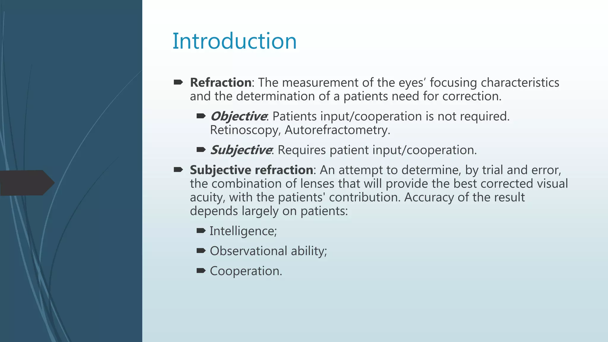
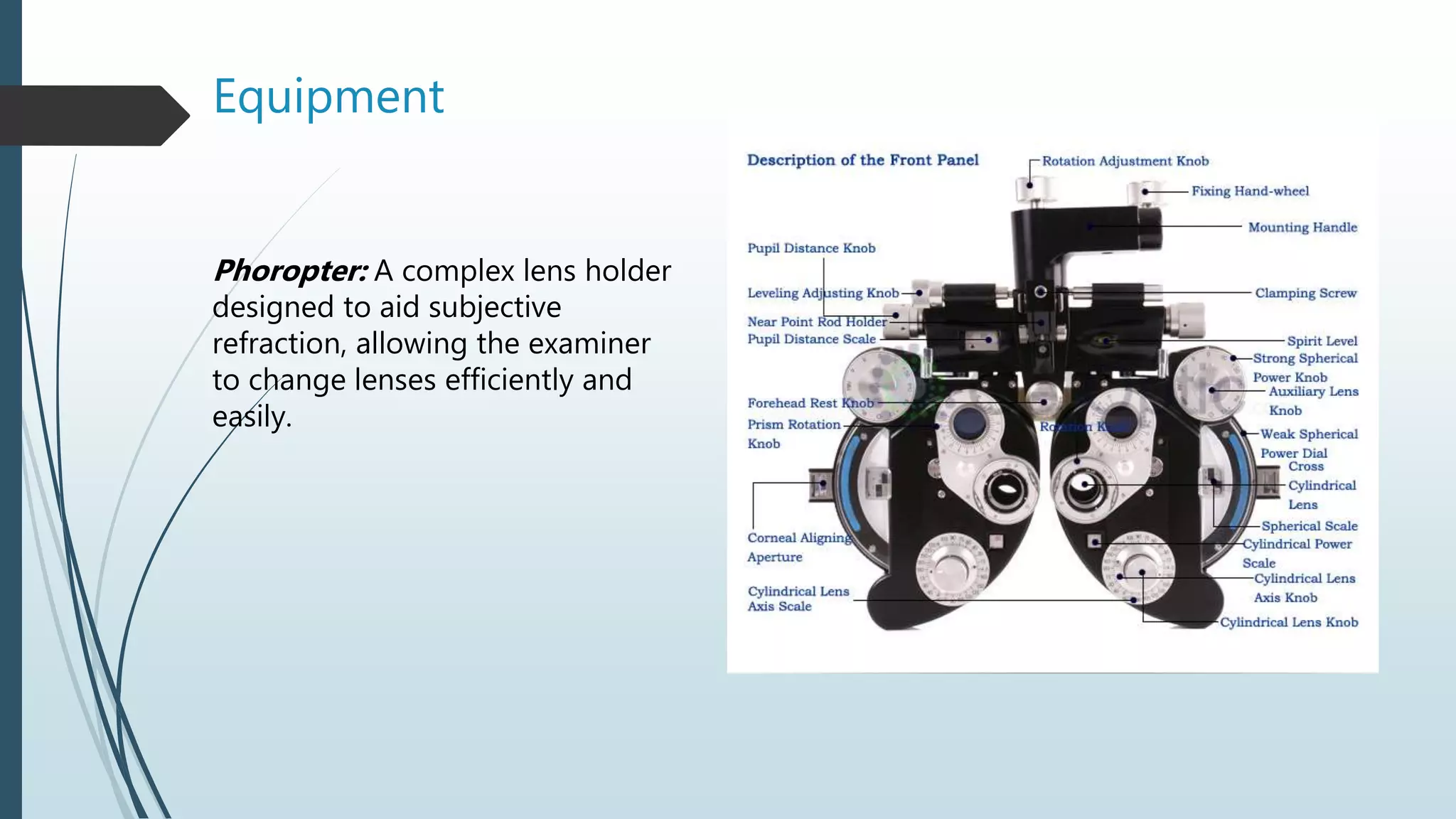
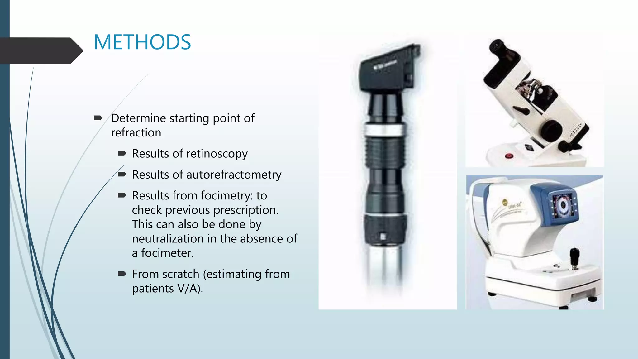
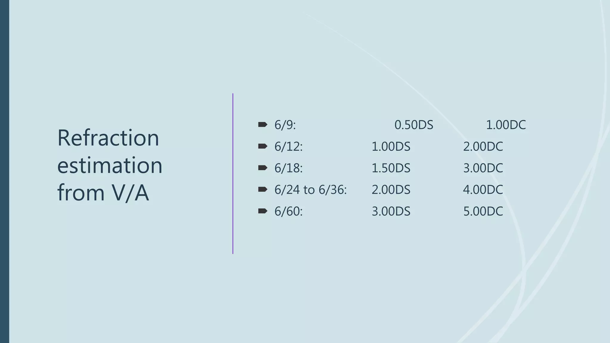
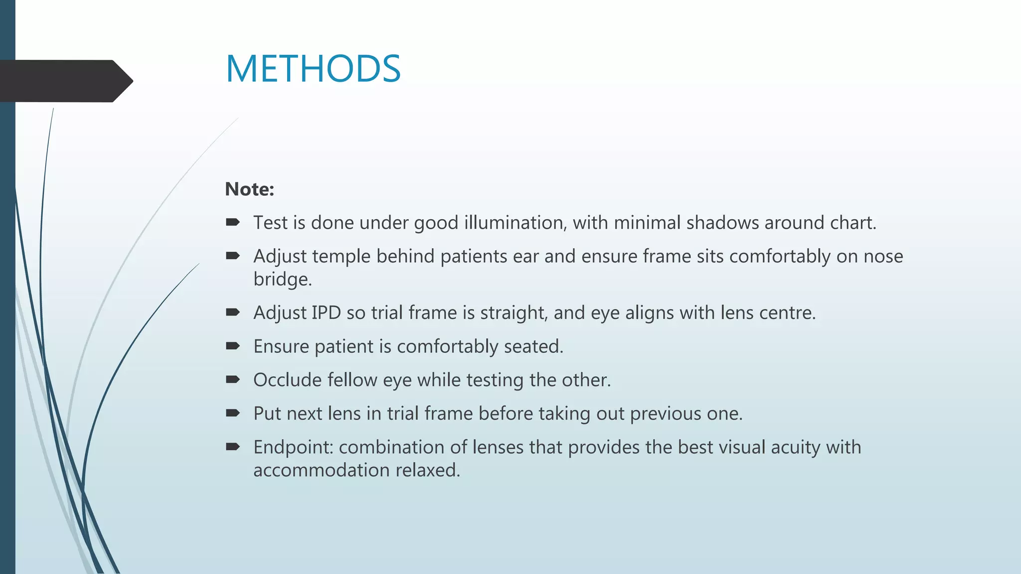
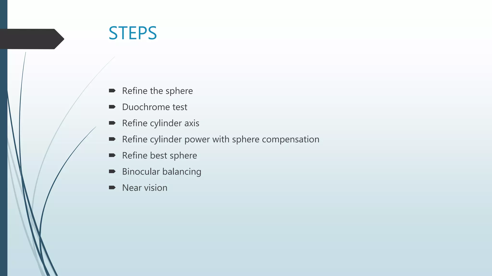
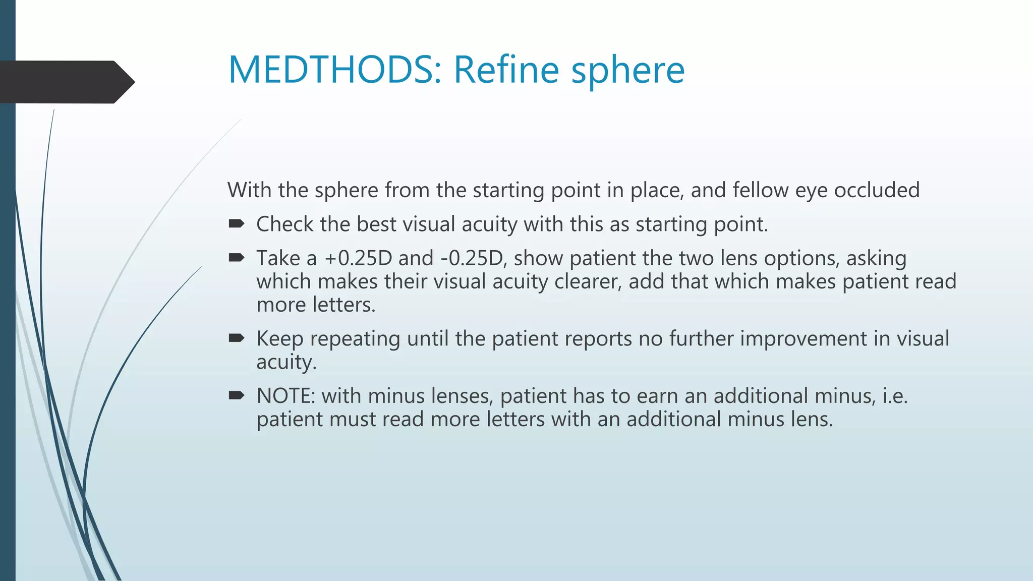
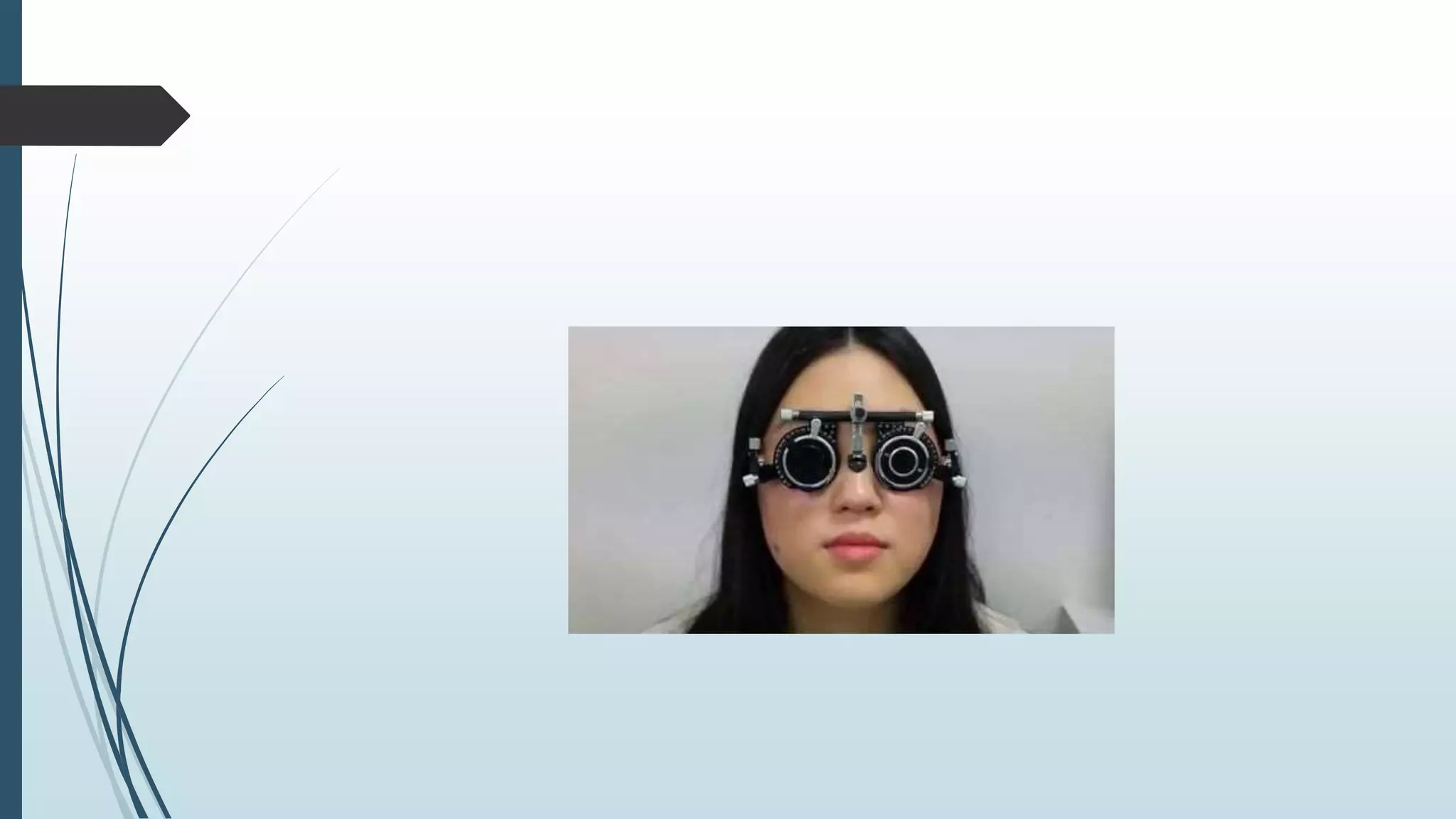
Methods of
Subjective
Refraction
Dr. Fashola Maryam B.
Outline
 Introduction
 Refraction
 Subjective refraction
 Equipments
 Steps
 Considerations
 Methods
Introduction
 Refraction: The measurement of the eyes’ focusing characteristics
and the determination of a patients need for correction.
 Objective: Patients input/cooperation is not required.
Retinoscopy, Autorefractometry.
 Subjective: Requires patient input/cooperation.
 Subjective refraction: An attempt to determine, by trial and error,
the combination of lenses that will provide the best corrected visual
acuity, with the patients' contribution. Accuracy of the result
depends largely on patients:
 Intelligence;
 Observational ability;
 Cooperation.
Equipment
Phoropter: A complex lens holder
designed to aid subjective
refraction, allowing the examiner
to change lenses efficiently and
easily.
Equipment
Visual acuity chart: Usually a
Snellen chart, used to test visual
acuity at 6metres OR at 3metres
using a mirror and a reverse chart.
Visual acuity is tested in a well-
lighted room.
Alternatives: logMAR chart, Landolt
C, tumbling E chart, Jaeger chart,
Lea test chart.
Equipment
Trial lens box set:
 Trial lenses
 Trial frame
 Accessories:
 Plano lens
 Occluder
 Pinhole
 Near vision chart
 Red and green filter
 Prisms, etc.
Equipment
Trial frame: A frame designed to
permit insertion of different lenses
used in refraction.
It has knobs that allows for
adjustment of IPD, nose bridge
height, temple tilt and cylinder axis
scale.
It also comes with four compartments
for several lens types while refracting.
Types: Full aperture, Reduced
aperture and Half-eye
Equipment
 Occluder: Opaque plastic disc used
to close one eye while testing the
other.
 Plano lens: has no power, can be
used to check malingering.
Equipment
 Pinhole: It blocks peripheral rays of
light allowing only the principal rays
fall on the retina, thereby reducing
the blur circle.
 V/A improves; vision can be
improved with refraction.
 V/A stays the same or worsens;
search for possible macular
causes of poor vision or
eccentric fixation.
Equipment
 Spherical lenses: come as pairs of
plus and minus lenses, with same
power in all meridians. Used to
correct spherical errors.
 Cylindrical lenses: plus or minus
cylinders, with power in one
meridian and axis of cylinders
marked on the rim of the lenses.
Used to correct astigmatism.
Equipment
 Near vision chart: held at a
distance of 33cms. Used to
detect people with near vision
difficulties (uncorrected
hyperopia, presbyopia).
Types:
 Roman test types;
 Snellen near vision chart;
 Jaeger chart.
Equipment
 Jackson Cross Cylinder (JCC): A
spherocylindrical lens in which the
power of the cylinder is twice that of the
sphere and of opposite signs. It is
usually mounted on a handle which is
450 to the axis of the cylinder and is
used for astigmatic refinement.
 The commonly used cross cylinders are
+/-0.25 and +/-0.50.
Equipment
 Alternatives: Astigmatic fan chart,
Astigmatic clock dial.
 Stenopaeic slit: Opaque disc with a
1mm wide and 15mm long slit
aperture in its centre. It allows rays
of light pass through only one
meridian and thus can be used to
determine astigmatic axis.
Equipment
Duochrome chart: Used to verify the final
refraction and to prevent over- or under-
correction.
 It is based on the principle of chromatic
aberration i.e. shorter wavelengths of light
(i.e., green) is refracted more by the eye
than longer wavelengths of light (i.e., red).
 In emmetropes, yellow light (570mm) is
focused on the retina, green light
(535mm) is focused in front of the retina
(1.50D) and red light (620mm) is focused
behind the retina (0.50D).
 Can be used in colour blind as it’s not
based on colour discrimination.
METHODS
 History:
 Patients age, occupation and hobbies;
 Blurred vision for near or far;
 Eye ache, eye fatigue, frontal headache;
 Jumping lines while reading;
 Previous eye exam;
 Intervention e.g. Spectacle use;
 History of systemic illnesses (Hypertension,
diabetes etc);
 Family history of cataract, glaucoma or blindness.
METHODS
 General examination: To rule out systemic illness(es).
 Visual acuity
 Uncorrected visual acuity: to know the current refractive
status of the eye.
 Visual acuity with correction: gives information as to the
possibilities for improvement.
 Visual acuity with pinhole: helps establish if vision may
be improved with refraction.
 Near visual acuity: tested at 33 – 40cm.
METHODS
Ocular examination:
 Lid: Ptosis, Large or multiple chalazia inducing astigmatism.
 Globe: Strabismus, Large globe suggesting myopia.
 Conjunctiva: Pterygium inducing astigmatism.
 Cornea: Keratoconus, Corneal oedema.
 Anterior chamber: Shallow in hyperopes.
 Pupil: RAPD, miosis, mydriasis.
 Lens: Cataract, Aphakia, Pseudophakia.
 Fundus: Small disc in hyperopes, large disc in myopes, oval
disc in astigmats; could reveal other pathologies.
METHODS
 Determine starting point of
refraction
 Results of retinoscopy
 Results of autorefractometry
 Results from focimetry: to
check previous prescription.
This can also be done by
neutralization in the absence of
a focimeter.
 From scratch (estimating from
patients V/A).
Refraction
estimation
from V/A
 6/9: 0.50DS 1.00DC
 6/12: 1.00DS 2.00DC
 6/18: 1.50DS 3.00DC
 6/24 to 6/36: 2.00DS 4.00DC
 6/60: 3.00DS 5.00DC
METHODS
Note:
 Test is done under good illumination, with minimal shadows around chart.
 Adjust temple behind patients ear and ensure frame sits comfortably on nose
bridge.
 Adjust IPD so trial frame is straight, and eye aligns with lens centre.
 Ensure patient is comfortably seated.
 Occlude fellow eye while testing the other.
 Put next lens in trial frame before taking out previous one.
 Endpoint: combination of lenses that provides the best visual acuity with
accommodation relaxed.
STEPS
 Refine the sphere
 Duochrome test
 Refine cylinder axis
 Refine cylinder power with sphere compensation
 Refine best sphere
 Binocular balancing
 Near vision
MEDTHODS: Refine sphere
With the sphere from the starting point in place, and fellow eye occluded
 Check the best visual acuity with this as starting point.
 Take a +0.25D and -0.25D, show patient the two lens options, asking
which makes their visual acuity clearer, add that which makes patient read
more letters.
 Keep repeating until the patient reports no further improvement in visual
acuity.
 NOTE: with minus lenses, patient has to earn an additional minus, i.e.
patient must read more letters with an additional minus lens.
Duochrome test
 This is done to verify the sphere
correction
 Green clearer (under-corrected
for hyperopia or over-corrected
for myopia), add +0.25DS; Red
clearer (under-corrected for
myopia or over-corrected for
hyperopia), add -0.25DS.
 NOTE: Loose red/green lens
filters can be used in the
absence of a chart.
METHODS: Refine Cylinder axis
Ask patient to look at a line one step larger than current V/A, with the objective
cylinder in place
 Hold the JCC with its handle straddling the axis of the objective cylinder.
 Flip the JCC in position 1 and 2, asking the patient in what position object appears
clearer/sharper (Always better to use a letter like O or D).
 Rotate the axis of the cylinder 50 or 100 clockwisely or anti-clockwisely in the
direction of the red mark on the JCC.
 Repeat this step, and keep rotating until patient reports both views as equally
sharp/dark.
 NOTE: If patient’s preference was in the direction of last axis, rotate back by half
the amount rotated the last time.
Refine Cylinder power
 Align the red /power dots on the JCC with axis of the cylinder in trial
frame.
 Flip the JCC such that both power options are made available to the
patient, and ask which is preferable.
 If patient responds that both views are the same, cylinder power in
frame is correct.
 If patient prefers the red(minus) dots, add -0.25DC, and continue until
patient responds to both views being the same.
Note: For every -0.50DC added to the trial lens, maintain spherical
equivalent by changing the sphere half as much in the opposite direction
i.e., add +0.25DS to the initial sphere.
Refine cylinder power
 If patient prefers the white/positive view,
remove -0.25DC, repeat until patient
finds no difference between the two
views.
 Note: For every -0.50DC removed from
the trial lens cylinder, maintain spherical
equivalent by changing the sphere half
as much in the opposite direction i.e.,
remove +0.25DS from the initial sphere.
Note: If you have to change your power by
more than -0.75DC, recheck the axis or
recheck the objective refraction.
Binocular balancing
To ensure both eyes not accommodating,
 Have both eyes open.
 Add +1.00 D over the eye not being tested (left) & Check visual acuity is
decreased to 6/12 or by about 4lines from their BCVA.
 Compare +0.25 to -0.25 over the eye being tested (right); ask the patient
which lens is clearer. If one lens is clearer adjust the sphere in that
direction.
 Repeat the comparison until equal. Repeat step for other eye.
 Repeat step for other eye.
 Alternatively, repeat Duochrome test.
Near vision
 Check near visual acuity with
best correction in place, at
patient’s regular reading
distance.
 Add +0.25DS to bring patient’s
reading distance closer.
 Add -0.25DS to take reading
distance farther.
References
 Clinical optics by A.R. Elkington, H.J. Frank, and M.J. Greaney.
 A Basic guide to practical refraction for new residents in ophthalmology by Chimdi
M. Chuka-Okosa.
 Cybersight online resources.
Methods of subjective refraction

Methods of subjective refraction

  • 1.
  • 2.
    Outline  Introduction  Refraction Subjective refraction  Equipments  Steps  Considerations  Methods
  • 3.
    Introduction  Refraction: Themeasurement of the eyes’ focusing characteristics and the determination of a patients need for correction.  Objective: Patients input/cooperation is not required. Retinoscopy, Autorefractometry.  Subjective: Requires patient input/cooperation.  Subjective refraction: An attempt to determine, by trial and error, the combination of lenses that will provide the best corrected visual acuity, with the patients' contribution. Accuracy of the result depends largely on patients:  Intelligence;  Observational ability;  Cooperation.
  • 4.
    Equipment Phoropter: A complexlens holder designed to aid subjective refraction, allowing the examiner to change lenses efficiently and easily.
  • 5.
    Equipment Visual acuity chart:Usually a Snellen chart, used to test visual acuity at 6metres OR at 3metres using a mirror and a reverse chart. Visual acuity is tested in a well- lighted room. Alternatives: logMAR chart, Landolt C, tumbling E chart, Jaeger chart, Lea test chart.
  • 6.
    Equipment Trial lens boxset:  Trial lenses  Trial frame  Accessories:  Plano lens  Occluder  Pinhole  Near vision chart  Red and green filter  Prisms, etc.
  • 7.
    Equipment Trial frame: Aframe designed to permit insertion of different lenses used in refraction. It has knobs that allows for adjustment of IPD, nose bridge height, temple tilt and cylinder axis scale. It also comes with four compartments for several lens types while refracting. Types: Full aperture, Reduced aperture and Half-eye
  • 8.
    Equipment  Occluder: Opaqueplastic disc used to close one eye while testing the other.  Plano lens: has no power, can be used to check malingering.
  • 9.
    Equipment  Pinhole: Itblocks peripheral rays of light allowing only the principal rays fall on the retina, thereby reducing the blur circle.  V/A improves; vision can be improved with refraction.  V/A stays the same or worsens; search for possible macular causes of poor vision or eccentric fixation.
  • 10.
    Equipment  Spherical lenses:come as pairs of plus and minus lenses, with same power in all meridians. Used to correct spherical errors.  Cylindrical lenses: plus or minus cylinders, with power in one meridian and axis of cylinders marked on the rim of the lenses. Used to correct astigmatism.
  • 11.
    Equipment  Near visionchart: held at a distance of 33cms. Used to detect people with near vision difficulties (uncorrected hyperopia, presbyopia). Types:  Roman test types;  Snellen near vision chart;  Jaeger chart.
  • 12.
    Equipment  Jackson CrossCylinder (JCC): A spherocylindrical lens in which the power of the cylinder is twice that of the sphere and of opposite signs. It is usually mounted on a handle which is 450 to the axis of the cylinder and is used for astigmatic refinement.  The commonly used cross cylinders are +/-0.25 and +/-0.50.
  • 13.
    Equipment  Alternatives: Astigmaticfan chart, Astigmatic clock dial.  Stenopaeic slit: Opaque disc with a 1mm wide and 15mm long slit aperture in its centre. It allows rays of light pass through only one meridian and thus can be used to determine astigmatic axis.
  • 14.
    Equipment Duochrome chart: Usedto verify the final refraction and to prevent over- or under- correction.  It is based on the principle of chromatic aberration i.e. shorter wavelengths of light (i.e., green) is refracted more by the eye than longer wavelengths of light (i.e., red).  In emmetropes, yellow light (570mm) is focused on the retina, green light (535mm) is focused in front of the retina (1.50D) and red light (620mm) is focused behind the retina (0.50D).  Can be used in colour blind as it’s not based on colour discrimination.
  • 15.
    METHODS  History:  Patientsage, occupation and hobbies;  Blurred vision for near or far;  Eye ache, eye fatigue, frontal headache;  Jumping lines while reading;  Previous eye exam;  Intervention e.g. Spectacle use;  History of systemic illnesses (Hypertension, diabetes etc);  Family history of cataract, glaucoma or blindness.
  • 16.
    METHODS  General examination:To rule out systemic illness(es).  Visual acuity  Uncorrected visual acuity: to know the current refractive status of the eye.  Visual acuity with correction: gives information as to the possibilities for improvement.  Visual acuity with pinhole: helps establish if vision may be improved with refraction.  Near visual acuity: tested at 33 – 40cm.
  • 17.
    METHODS Ocular examination:  Lid:Ptosis, Large or multiple chalazia inducing astigmatism.  Globe: Strabismus, Large globe suggesting myopia.  Conjunctiva: Pterygium inducing astigmatism.  Cornea: Keratoconus, Corneal oedema.  Anterior chamber: Shallow in hyperopes.  Pupil: RAPD, miosis, mydriasis.  Lens: Cataract, Aphakia, Pseudophakia.  Fundus: Small disc in hyperopes, large disc in myopes, oval disc in astigmats; could reveal other pathologies.
  • 18.
    METHODS  Determine startingpoint of refraction  Results of retinoscopy  Results of autorefractometry  Results from focimetry: to check previous prescription. This can also be done by neutralization in the absence of a focimeter.  From scratch (estimating from patients V/A).
  • 19.
    Refraction estimation from V/A  6/9:0.50DS 1.00DC  6/12: 1.00DS 2.00DC  6/18: 1.50DS 3.00DC  6/24 to 6/36: 2.00DS 4.00DC  6/60: 3.00DS 5.00DC
  • 20.
    METHODS Note:  Test isdone under good illumination, with minimal shadows around chart.  Adjust temple behind patients ear and ensure frame sits comfortably on nose bridge.  Adjust IPD so trial frame is straight, and eye aligns with lens centre.  Ensure patient is comfortably seated.  Occlude fellow eye while testing the other.  Put next lens in trial frame before taking out previous one.  Endpoint: combination of lenses that provides the best visual acuity with accommodation relaxed.
  • 21.
    STEPS  Refine thesphere  Duochrome test  Refine cylinder axis  Refine cylinder power with sphere compensation  Refine best sphere  Binocular balancing  Near vision
  • 22.
    MEDTHODS: Refine sphere Withthe sphere from the starting point in place, and fellow eye occluded  Check the best visual acuity with this as starting point.  Take a +0.25D and -0.25D, show patient the two lens options, asking which makes their visual acuity clearer, add that which makes patient read more letters.  Keep repeating until the patient reports no further improvement in visual acuity.  NOTE: with minus lenses, patient has to earn an additional minus, i.e. patient must read more letters with an additional minus lens.
  • 23.
    Duochrome test  Thisis done to verify the sphere correction  Green clearer (under-corrected for hyperopia or over-corrected for myopia), add +0.25DS; Red clearer (under-corrected for myopia or over-corrected for hyperopia), add -0.25DS.  NOTE: Loose red/green lens filters can be used in the absence of a chart.
  • 24.
    METHODS: Refine Cylinderaxis Ask patient to look at a line one step larger than current V/A, with the objective cylinder in place  Hold the JCC with its handle straddling the axis of the objective cylinder.  Flip the JCC in position 1 and 2, asking the patient in what position object appears clearer/sharper (Always better to use a letter like O or D).  Rotate the axis of the cylinder 50 or 100 clockwisely or anti-clockwisely in the direction of the red mark on the JCC.  Repeat this step, and keep rotating until patient reports both views as equally sharp/dark.  NOTE: If patient’s preference was in the direction of last axis, rotate back by half the amount rotated the last time.
  • 25.
    Refine Cylinder power Align the red /power dots on the JCC with axis of the cylinder in trial frame.  Flip the JCC such that both power options are made available to the patient, and ask which is preferable.  If patient responds that both views are the same, cylinder power in frame is correct.  If patient prefers the red(minus) dots, add -0.25DC, and continue until patient responds to both views being the same. Note: For every -0.50DC added to the trial lens, maintain spherical equivalent by changing the sphere half as much in the opposite direction i.e., add +0.25DS to the initial sphere.
  • 26.
    Refine cylinder power If patient prefers the white/positive view, remove -0.25DC, repeat until patient finds no difference between the two views.  Note: For every -0.50DC removed from the trial lens cylinder, maintain spherical equivalent by changing the sphere half as much in the opposite direction i.e., remove +0.25DS from the initial sphere. Note: If you have to change your power by more than -0.75DC, recheck the axis or recheck the objective refraction.
  • 27.
    Binocular balancing To ensureboth eyes not accommodating,  Have both eyes open.  Add +1.00 D over the eye not being tested (left) & Check visual acuity is decreased to 6/12 or by about 4lines from their BCVA.  Compare +0.25 to -0.25 over the eye being tested (right); ask the patient which lens is clearer. If one lens is clearer adjust the sphere in that direction.  Repeat the comparison until equal. Repeat step for other eye.  Repeat step for other eye.  Alternatively, repeat Duochrome test.
  • 28.
    Near vision  Checknear visual acuity with best correction in place, at patient’s regular reading distance.  Add +0.25DS to bring patient’s reading distance closer.  Add -0.25DS to take reading distance farther.
  • 30.
    References  Clinical opticsby A.R. Elkington, H.J. Frank, and M.J. Greaney.  A Basic guide to practical refraction for new residents in ophthalmology by Chimdi M. Chuka-Okosa.  Cybersight online resources.