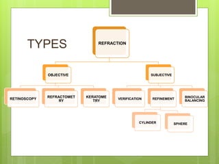
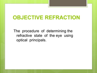
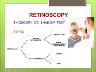
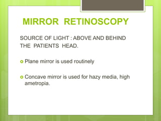
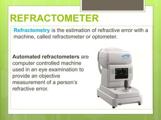
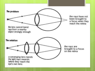
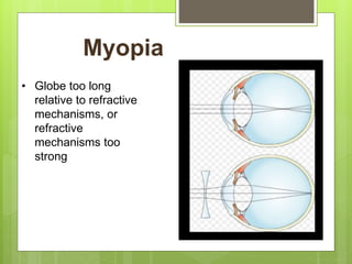
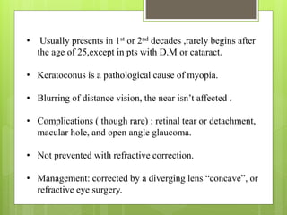
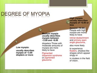
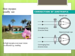
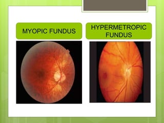
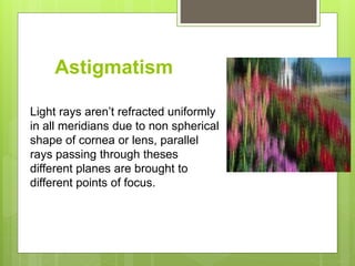
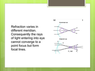
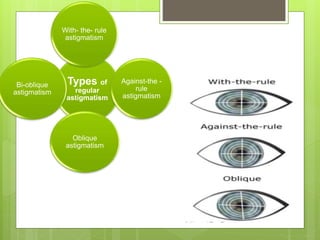
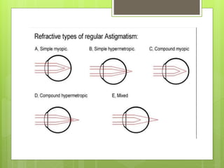
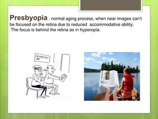
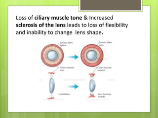
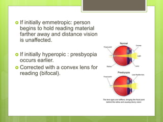
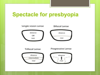
REFRACTION
DR. ARVIND KUMAR MORYA
MBBS, MS OPHTHALMOLOGY(GOLD MEDALIST), MNAMS,
CATARACT(MICS), GLAUCOMA, PAEDIATRIC OPHTHALMOLOGY,
STRABISMUS,REFRACTIVE AND MEDICAL RETINA SERVICES,
ASSOCIATE PROFESSOR AND HEAD ,
DEPARTMENT OF OPHTHALMOLOGY,
AIIMS, JODHPUR
Physical optics
• The visible part of the EM spectrum, to which the retina
is sensitive, lies between the wavelengths 390nm &
760nm.
• Light should be correctly focused on the retina for the
eye to generate accurate visual information.
Light travels in a straight line. But when it
reaches another medium it will bend. This is
called refraction.
Refraction of light
Angle of incidence is
equal to angle of
refraction.
It is measured relative
to the normal.
When moving from high
to low refractive index
light bends away from
normal
Refractive components of the eye :
• Mainly 2/3 cornea and1/3 lens
Refractive component Refractive index
Cornea 1.377
Aqueous humor 1.377
Anterior capsule of lens 1.359
Anterior cortex of lens 1.387
Nucleus of lens 1.406
Posterior cortex of lens 1.385
Vitreous 1.336
These elements converge parallel rays
because :-
1)Cornea has higher refractive index than
air.
2)lens has higher refractive index than
vitreous humor.
3)Cornea & lens are spherically convex in
shape
schematic eye
 It is a physical model of the eye that
represents the basic features of the eye
 According to this the eye forming a
homocentric complex lens system.
 Cardinal data of schematic eye-
 Principle foci F1and F2
 Principle points P1 and P2
 Nodal point N1 and N2
Schematic eye
Other physical model of eye
 Listing’s reduced eye
 Emsely’s reduced eye
 Donder’s reduced eye
 Bennett and rabbetts reduced eye.
GEOMETRIC OPTICS
* A negative lens is a diverging lens ,concave ,corrects for myopia
* A positive lens is a converging lens ,convex ,corrects for hypermetropia.
Example: +1 D lens focuses light at 1m.
+2 D lens focuses light at 0.5m.
Optical aberration of eye
 Lapse from perfection are called aberration.
 Physiological optical defects in normal eye
include the following-
 Diffraction of light
 Spherical aberration
 Chromatic aberration
 Decentring
 Oblique aberration
Diffraction of light
 Diffraction is a bending of
light caused by the edge of
an aperture or the rim of the
lens.
 Pattern- series of concentric
bright and dark ring.
 Centre of the pattern is a
bright spot ,known as airy
disc.
 In the eye with the pupil of
2mm diameter, the diameter
of the spot of the concentric
rings is about 0.01mm.
Spherical aberration
 Spherical lens reflects peripheral
rays more strongly than paraxial
rays.
 Factor that contribute in
diminishing spherical aberration
of the human eye-
 Peculiar curvature of the cornea.
 Peculiar structure of the lens.
 Iris block the peripheral rays to
enter into the eye.
Chromatic aberration
 Result because of the
index of refraction of any
transparent medium
varies with the
wavelength of the
incident light.
 The emmetropic eye is
slightly hypermetropic for
red rays and myopic for
blue and green rays.
Decentring
 The crystalline lens is slightly decentred and
tipped with respect to the axis of the cornea
and with respect to the visual axis of the eye.
Oblique aberration
 Objects in the peripheral field are
seen by the virtue of obliquely
incident narrow pencil of rays which
are limited by the pupil.
 Because of this the refracted pencil
shows oblique astigmatism.
 Much evident when more biconvex
or biconcave lenses are used and is
considerably reduced by the use of
best form of lenses such as
“periscope” or other meniscus.
Coma
 Different area of the lens will from foci in planers
other than the chief focus.
WHAT IS REFRACTION ?
Refraction is the procedure of determining and
correcting refractive errors.
TYPES REFRACTION
OBJECTIVE
RETINOSCOPY
REFRACTOMET
RY
KERATOME
TRY
SUBJECTIVE
VERIFICATION REFINEMENT
CYLINDER SPHERE
BINOCULAR
BALANCING
OBJECTIVE REFRACTION
The procedure of determining the
refractive state of the eye using
optical principals.
RETINOSCOPY
SKIOSCOPY OR SHADOW TEST
TYPES
RETINOSCOPE
MIRROR
RETINOSCOPE
SINGLE/PLANE
PLANE +
CONCAVE(PRIESTLY-
SMITH MIRROR)
SELF ILLUMINATED
STREAK
SPOT
MIRROR RETINOSCOPY
SOURCE OF LIGHT : ABOVE AND BEHIND
THE PATIENTS HEAD.
 Plane mirror is used routinely
 Concave mirror is used for hazy media, high
ametropia.
ADVANTAGES :
 Cheaper than self illuminated
DISADVANTAGE :
 Requires separate light sources.
 Glare.
 Difficult to check axis and amount of cylinder.
 Intensity and type of beam cannot be controlled.
SELF
ILLUMINATEDRETINOSCOPES
 STREAK RETINOSCOPE :
Circular beam of light is modified to produce linear
streak using planocylindrical retinoscopy
Mirror.
 SPOT RETINOSCOPE:
Round filament scoped in any meridian used for
assessment of contact lens fitting, pediatric patients,
vision programmes.
PRINCIPLE OF
RETINOSCOPY
 To locate the far point of the eye/ plane conjugate to
the retina.
 Bring far point to infinity while using appropriate
lenses.
 Accommodation at a minimum.
PRE REQUISITES
 Dark room: 6metres
 Trial box: spherical, cylindrical lenses, pin hole,
occluder and prisms.
 Trial frame
 Vision box
 Retinoscope
PRE REQUISITES
FIXATION AND FOGGING
 Patient’s accommodation should be relaxed and fixation should
be at a distance on non accommodative target
 Fogging implies adding plus power sphere during retinoscopy
to prevent accommodation
POSITIONING ANDALIGNMENT
Right for right, left for left
Optimal alignment is just off-centre
THE RETINAL REFLEX
ORIGIN:
Interface between vitreous and retinal pigment
epithelium of retina or bruch’s membrane.
CHARACTERISTICS OF RETINAL
REFLEX
 Speed: large refractive errors have slow moving reflex,
small errors have fast reflex
 Brilliance: large errors have a dull reflex, small errors
have bright reflex. Becomes brighter when neutrality
approaches
 Width: narrow and high degree and wide and lower
degree error
CORRECTING LENS
 Appropriate correcting lens to be used to neutralise the
reflex
 This brings patient’s far point to the peephole and the
reflex fills patient’s entire pupil
 Dioptric equivalent of working distance should be
subtracted from the power of the correcting lens. Eg: at a
distance of 67cms phoropters have a 1.5 D (1/0.67 m),
correction
NEUTRALITY
 Against movement: Far point is between examiner
and patient. To bring the far point to the peep hole of
retinoscope minus lenses are placed.
 With movement: plus lenses are used.
REFRACTOMETER
Refractometry is the estimation of refractive error with a
machine, called refractometer or optometer.
Automated refractometers are
computer controlled machine
used in an eye examination to
provide an objective
measurement of a person’s
refractive error.
PRINCIPLES
 SCHEINER’S PRINCIPLE
 OPTOMETER
PRINCIPLE
 Five types of AR:
1. Manual objective
2. Automatic objective without VA
3. Automatic objective with VA
4. Automated subjective
5. Remote controlled conventional
MODERN REFRACTOMETERS
 Plusoptix has designed handheld autorefractors
for babies, children and uncooperative patients that
measure the refraction of both eyes from one meter
distance within one second. Used in un-dilated pupils,
• SureSight hand held/portable
autorefractometer is designed to
measure the refractive error of both
eyes at a distance of 14”
• The Spot Vision Screener is a
handheld vision screening
device that helps users quickly
and easily detect vision issues
on patients from 6 months of
age through adult. Spot screens
both eyes at once from a
nonthreatening 3-foot distance.
KERATOMETER
 Keratometry is an objective method of estimating
corneal astigmatism by measuring the curvature of
central cornea.
 Principle: Anterior surface of cornea acts as a convex
mirror. Size of first purkinje image determines the
radius of curvature of cornea.
 Types:
1. Javal-schiotz
keratometer: it is
based on the
principle of
variable object
size and constant
image size.
Bausch and Lomb keratometer
it is based on the principle of constant object size and
variable image size.
Interpretation
horizontal oval mires is seen in with-the-rule
astigmatism.
Vertically oval mires are seen in against-the-rule
astigmatism
Irregular anterior characterized by either irregular
mires or doubling of mires
SUBJECTIVE REFRACTION
 Meant for finding out most suitable lens to be
prescribed.
 Three steps:
1. Subjective verification
2. Refinement
3. Subjective binocular balancing
VERIFICATION
 Performed by trial and error method
 Patient at a distance of 6 m from Snellen’s
 Occluder is put in front of one eye
 Trial lens determined by AR or retinoscopy is
put
REFINEMENT
 The combination of lenses chosen after
verification is refined before final prescription
 Cylinder should be refined before sphere
CYLINDER
 Refinement of cylinder can be done by
1. Jacksons cross cylinder
2. Astigmatic fan test
3. Staenopic slit
JACKSONS CROSS CYLINDER
 Combination of 2 cylinders equal strength with
opposite sign placed with their axes at right
angles
 Verification of strength
 Verification of axis
ASTIGMATIC FAN TECHNIQUE
 Consists of dials of line radiating at 10degree
interval
 Patient is asked to see the fan after fogging
+o.5 D
STENOPAEIC SLIT TEST
 1 mm wide
stenopaeic slit
allows clearest
vision when
rotated into the
axis of
astigmatism.
REFINING OF SPHERE
 Fogging
 Duochrome
 Pin-hole test
FOGGING
 +2 D SPHERE IN MYOPES
 +4D SPHERE IN HYPERMETROPES
 Unfog by reducing every time +0.25 DS till the
best snellen’s visual acuity is attained.
DUOCHROME TEST
 Based on the principle of
chromatic aberration
 Patient is asked to read red
and green letters
 Emmetropes : green in front
and red behind retina
PIN- HOLE TEST
 Helps in confirming
whether the
correction in trial
frame is correct or
not.
 If vision with pinhole
improve it indicate
incorrect correction.
BINOCULAR BALANCE
 To ensure accommodation is relaxed equally
in both the eyes
 Fogging ,prism dissociation
Prism
 Refracting medium, having two plane surface,
inclined at an angle.
 Refractive angle or apical angle of a prism is the
angle between two surface. The greater the angle
formed by two surface at the apex the stronger
the prismatic effect.
 Axis of prism is the line bisecting the apical angle.
 base of the prism refers to the surface opposite to
the apical angle.
PRISM DISSCOCIATION
 Most sensitive test of binocular balance
 With best corrected lens eyes are fogged with +1
D sphere
 vertical prisms of 4or5 dioptre is placed, base
down in RE and base up in LE.
 Then single line projected (usually 6/12)
 If patient reports difference in clarity between
upper and lower line, then as two separate image,
+0.25 DS is placed until two lines are equally
distinct for two eyes.
FOGGING
 After fogging, alternate cover test is
performed and patient is asked, which eye
showing comparatively clearer image.
 If the eyes are not in balance +0.25 added to
better seeing eye untill balance is achieved.
CYCLOPLEGIC AND NON
CYCLOPLEGIC REFRACTION
Indication and appropriate dosage depends on
1. Patient’s age
2. Accommodative amplitude
3. Refractive error
Cycloplegic refraction in
children
The younger the patient, the greater the patient’s amplitude of
accommodation and the more difficult it is to inhibit it. Because of the
powerful accommodation and the inability of young pediatric patients to
respond with accurate subjective responses, cycloplegic refraction rather
than manifest or subjective refraction usually is necessary.
It is universally accepted that refraction in children, irrespective presence or
absence of strabismus and type of refractive error, should preferably be
performed under cycloplegia
DRUG CONCENTRATION DOSAGE ONSET TOTAL
DURATION
ATROPINE 0.5, 1.0 2-3/DAY FOR
3 DAYS
1-2 HOUR 7-14 DAYS
HOMATROPINE 2.0,5.0 30-60 MIN 1-2 DAYS
SCOPOLAMINE 0.25 2 DROPS / 5
MIN
20-60 MIN 3-4 DAYS
CYCLOPENTOLAT
E
0.5 , 1.0 ,2.0 20-60 MIN 1-2 DAYS
TROPICAMIDE 0.5, 1.0 , 2.0 20-40 MIN 4-6 HOURS
 Atropine is indicated in children below the age
of 5 years.
 Hometropine is used for most of the
hypermetropic individual between 5 to 25
years of age.
 Adverse reaction
 Atropine : dryness , flushing , high fever ,
delirium
 Scopolamine : hallucination and ataxia
Refractive errors
OVERVIEW
Emmetropia :- When parallel rays of light from a distant object are
brought to focus on the retina with the eye at rest “not accommodating “.
Ammetropia :- when parallel
rays of light are not
brought to a focus on the
retina in an eye at rest. It’s
simply “refractive error”. A
change in refraction is
needed to achieve sharp
vision.
Ametropia is divided into:
1) Myopia: Near sightedness
2) Hyperopia (Hypermetropia):
= Far sightedness.
3) Astigmatism :non spherical cornea.
Hypermetropia
Hypermetropic eye is too short
for its converging ability.
The eye’s optical system is less
powerful, and/or the eye is too
short.
Rays of light converge behind
the retina, which causes
blurring of vision for near
objects ± distant objects.
Hypermetropia- pathophysiology
• The length of the eyeball is shorter than it
should be.
• stage in normal development of the eyes—at
birth eyes are hypermetropic (2.5 to 3.0
Diopters)
• When persists in adulthood it represents an
imperfectly developed eye.
• Lens changes (cataract)
Hypermetropia -symptoms
• Eye-strain (ciliary muscle is straining to maintain
accommodation) – Asthenopia
• “watering” /“redness”.
• They have difficulty seeing both near and far with
more difficulty seeing near objects
• Headaches in later part of the day.
• Complain of blurring of text.
Clinical features:
- Youth: usually don’t require glasses, they have sufficient accommodative ability to
focus the image on the retina, but may develop accommodative ET.
- 30s-40s: blurring of near vision due to decreased accommodation, may need
reading glasses.
- >50s: blurring of distance vision due to severely decreased accommodation.
corrected by adding a powerful lens, a converging (convex) lens.
Complications: Angle-closure glaucoma, particularly later in life.
Total
hypermetropia
Latent
hypermetropia
Facultative
hypermetropia
Absolute
hypermetropia
Manifest
hypermetropia
COMPONENETS OF
HYPERMETROPIA
 Total hypermetropia-total amount of refractive
error , estimated after complete cycloplegic
refraction under atropine.
 Latent hypermetropia-
 Which is normally corrected by inherent tone of
ciliary muscles.
 Manifest hypermetropia-
 Remaining portion of total hypermetropia which is
not corrected by the ciliary muscle tone
 1. facultative-corrected by patient’s
accommodative effort.
 2. Absolute- cannot be corrected by patient
accommodative effort
Myopia
• Globe too long
relative to refractive
mechanisms, or
refractive
mechanisms too
strong
• Usually presents in 1st or 2nd decades ,rarely begins after
the age of 25,except in pts with D.M or cataract.
• Keratoconus is a pathological cause of myopia.
• Blurring of distance vision, the near isn’t affected .
• Complications ( though rare) : retinal tear or detachment,
macular hole, and open angle glaucoma.
• Not prevented with refractive correction.
• Management: corrected by a diverging lens “concave”, or
refractive eye surgery.
DEGREE OF MYOPIA
Low myopia:
usually describes
myopia of −3.00
diopters or more
Medium myopia
usually describes
myopia between
−3.00 and −6.00
diopeters Those with
moderate amounts of
myopia are more
likely to have
Pigmented
dispersion syndrome
or pigmented
glaucoma
High myopia
usually describes
myopia of −6.00 or
less
(toward -10.00).
People with high
myopia are more
likely to have
retinal deatachment
and primary open
angle glaucoma,
also more likely
to experience
floaters, shadow-like
shapes which appear
singly or
in clusters in the field
of vision.
How myopes
usually see
. In high myopia even near vision
is affected e.g. reading
MYOPIC FUNDUS
HYPERMETROPIC
FUNDUS
Astigmatism
Light rays aren’t refracted uniformly
in all meridians due to non spherical
shape of cornea or lens, parallel
rays passing through theses
different planes are brought to
different points of focus.
Refraction varies in
different meridian.
Consequently the rays
of light entering into eye
cannot converge to a
point focus but form
focal lines.
Types of
regular
astigmatism
With- the- rule
astigmatism
Against-the -
rule
astigmatism
Oblique
astigmatism
Bi-oblique
astigmatism
The cause of astigmatism is
unknown. It is usually present
from birth, and often occurs
together with nearsightedness
or farsightedness.
Cylindrical lenses
Accommodation
Near focusing of the eye.
Ciliary muscle contraction……..zonules relax……..lens more spherical .
Eyes converge, pupils constrict.
Presbyopia : normal aging process, when near images can’t
be focused on the retina due to reduced accommodative ability.
The focus is behind the retina as in hyperopia.
Loss of ciliary muscle tone & Increased
sclerosis of the lens leads to loss of flexibility
and inability to change lens shape.
 If initially emmetropic: person
begins to hold reading material
farther away and distance vision
is unaffected.
 If initially hyperopic : presbyopia
occurs earlier.
 Corrected with a convex lens for
reading (bifocal).
Spectacle for presbyopia
1. Dr. Sonalika Gogia
2. Mrs Diksha Bishnoi
3. Miss Neha Sharma
ACKNOWLEDGEMENTS
Thank you

Refraction in different refractive errors and their Management

  • 1.
    REFRACTION DR. ARVIND KUMARMORYA MBBS, MS OPHTHALMOLOGY(GOLD MEDALIST), MNAMS, CATARACT(MICS), GLAUCOMA, PAEDIATRIC OPHTHALMOLOGY, STRABISMUS,REFRACTIVE AND MEDICAL RETINA SERVICES, ASSOCIATE PROFESSOR AND HEAD , DEPARTMENT OF OPHTHALMOLOGY, AIIMS, JODHPUR
  • 2.
    Physical optics • Thevisible part of the EM spectrum, to which the retina is sensitive, lies between the wavelengths 390nm & 760nm. • Light should be correctly focused on the retina for the eye to generate accurate visual information.
  • 3.
    Light travels ina straight line. But when it reaches another medium it will bend. This is called refraction. Refraction of light
  • 4.
    Angle of incidenceis equal to angle of refraction. It is measured relative to the normal. When moving from high to low refractive index light bends away from normal
  • 5.
    Refractive components ofthe eye : • Mainly 2/3 cornea and1/3 lens Refractive component Refractive index Cornea 1.377 Aqueous humor 1.377 Anterior capsule of lens 1.359 Anterior cortex of lens 1.387 Nucleus of lens 1.406 Posterior cortex of lens 1.385 Vitreous 1.336
  • 6.
    These elements convergeparallel rays because :- 1)Cornea has higher refractive index than air. 2)lens has higher refractive index than vitreous humor. 3)Cornea & lens are spherically convex in shape
  • 7.
    schematic eye  Itis a physical model of the eye that represents the basic features of the eye  According to this the eye forming a homocentric complex lens system.  Cardinal data of schematic eye-  Principle foci F1and F2  Principle points P1 and P2  Nodal point N1 and N2
  • 8.
  • 9.
    Other physical modelof eye  Listing’s reduced eye  Emsely’s reduced eye  Donder’s reduced eye  Bennett and rabbetts reduced eye.
  • 10.
  • 11.
    * A negativelens is a diverging lens ,concave ,corrects for myopia * A positive lens is a converging lens ,convex ,corrects for hypermetropia. Example: +1 D lens focuses light at 1m. +2 D lens focuses light at 0.5m.
  • 12.
    Optical aberration ofeye  Lapse from perfection are called aberration.  Physiological optical defects in normal eye include the following-  Diffraction of light  Spherical aberration  Chromatic aberration  Decentring  Oblique aberration
  • 13.
    Diffraction of light Diffraction is a bending of light caused by the edge of an aperture or the rim of the lens.  Pattern- series of concentric bright and dark ring.  Centre of the pattern is a bright spot ,known as airy disc.  In the eye with the pupil of 2mm diameter, the diameter of the spot of the concentric rings is about 0.01mm.
  • 14.
    Spherical aberration  Sphericallens reflects peripheral rays more strongly than paraxial rays.  Factor that contribute in diminishing spherical aberration of the human eye-  Peculiar curvature of the cornea.  Peculiar structure of the lens.  Iris block the peripheral rays to enter into the eye.
  • 15.
    Chromatic aberration  Resultbecause of the index of refraction of any transparent medium varies with the wavelength of the incident light.  The emmetropic eye is slightly hypermetropic for red rays and myopic for blue and green rays.
  • 16.
    Decentring  The crystallinelens is slightly decentred and tipped with respect to the axis of the cornea and with respect to the visual axis of the eye.
  • 17.
    Oblique aberration  Objectsin the peripheral field are seen by the virtue of obliquely incident narrow pencil of rays which are limited by the pupil.  Because of this the refracted pencil shows oblique astigmatism.  Much evident when more biconvex or biconcave lenses are used and is considerably reduced by the use of best form of lenses such as “periscope” or other meniscus.
  • 18.
    Coma  Different areaof the lens will from foci in planers other than the chief focus.
  • 19.
    WHAT IS REFRACTION? Refraction is the procedure of determining and correcting refractive errors.
  • 20.
  • 21.
    OBJECTIVE REFRACTION The procedureof determining the refractive state of the eye using optical principals.
  • 22.
    RETINOSCOPY SKIOSCOPY OR SHADOWTEST TYPES RETINOSCOPE MIRROR RETINOSCOPE SINGLE/PLANE PLANE + CONCAVE(PRIESTLY- SMITH MIRROR) SELF ILLUMINATED STREAK SPOT
  • 23.
    MIRROR RETINOSCOPY SOURCE OFLIGHT : ABOVE AND BEHIND THE PATIENTS HEAD.  Plane mirror is used routinely  Concave mirror is used for hazy media, high ametropia.
  • 25.
    ADVANTAGES :  Cheaperthan self illuminated DISADVANTAGE :  Requires separate light sources.  Glare.  Difficult to check axis and amount of cylinder.  Intensity and type of beam cannot be controlled.
  • 26.
    SELF ILLUMINATEDRETINOSCOPES  STREAK RETINOSCOPE: Circular beam of light is modified to produce linear streak using planocylindrical retinoscopy Mirror.  SPOT RETINOSCOPE: Round filament scoped in any meridian used for assessment of contact lens fitting, pediatric patients, vision programmes.
  • 28.
    PRINCIPLE OF RETINOSCOPY  Tolocate the far point of the eye/ plane conjugate to the retina.  Bring far point to infinity while using appropriate lenses.  Accommodation at a minimum.
  • 29.
    PRE REQUISITES  Darkroom: 6metres  Trial box: spherical, cylindrical lenses, pin hole, occluder and prisms.  Trial frame  Vision box  Retinoscope
  • 30.
  • 31.
    FIXATION AND FOGGING Patient’s accommodation should be relaxed and fixation should be at a distance on non accommodative target  Fogging implies adding plus power sphere during retinoscopy to prevent accommodation POSITIONING ANDALIGNMENT Right for right, left for left Optimal alignment is just off-centre
  • 32.
    THE RETINAL REFLEX ORIGIN: Interfacebetween vitreous and retinal pigment epithelium of retina or bruch’s membrane.
  • 33.
    CHARACTERISTICS OF RETINAL REFLEX Speed: large refractive errors have slow moving reflex, small errors have fast reflex  Brilliance: large errors have a dull reflex, small errors have bright reflex. Becomes brighter when neutrality approaches  Width: narrow and high degree and wide and lower degree error
  • 34.
    CORRECTING LENS  Appropriatecorrecting lens to be used to neutralise the reflex  This brings patient’s far point to the peephole and the reflex fills patient’s entire pupil  Dioptric equivalent of working distance should be subtracted from the power of the correcting lens. Eg: at a distance of 67cms phoropters have a 1.5 D (1/0.67 m), correction
  • 35.
    NEUTRALITY  Against movement:Far point is between examiner and patient. To bring the far point to the peep hole of retinoscope minus lenses are placed.  With movement: plus lenses are used.
  • 36.
    REFRACTOMETER Refractometry is theestimation of refractive error with a machine, called refractometer or optometer. Automated refractometers are computer controlled machine used in an eye examination to provide an objective measurement of a person’s refractive error.
  • 37.
  • 38.
     Five typesof AR: 1. Manual objective 2. Automatic objective without VA 3. Automatic objective with VA 4. Automated subjective 5. Remote controlled conventional
  • 39.
    MODERN REFRACTOMETERS  Plusoptixhas designed handheld autorefractors for babies, children and uncooperative patients that measure the refraction of both eyes from one meter distance within one second. Used in un-dilated pupils,
  • 40.
    • SureSight handheld/portable autorefractometer is designed to measure the refractive error of both eyes at a distance of 14” • The Spot Vision Screener is a handheld vision screening device that helps users quickly and easily detect vision issues on patients from 6 months of age through adult. Spot screens both eyes at once from a nonthreatening 3-foot distance.
  • 41.
    KERATOMETER  Keratometry isan objective method of estimating corneal astigmatism by measuring the curvature of central cornea.  Principle: Anterior surface of cornea acts as a convex mirror. Size of first purkinje image determines the radius of curvature of cornea.
  • 42.
     Types: 1. Javal-schiotz keratometer:it is based on the principle of variable object size and constant image size.
  • 43.
    Bausch and Lombkeratometer it is based on the principle of constant object size and variable image size.
  • 44.
    Interpretation horizontal oval miresis seen in with-the-rule astigmatism. Vertically oval mires are seen in against-the-rule astigmatism Irregular anterior characterized by either irregular mires or doubling of mires
  • 45.
    SUBJECTIVE REFRACTION  Meantfor finding out most suitable lens to be prescribed.  Three steps: 1. Subjective verification 2. Refinement 3. Subjective binocular balancing
  • 46.
    VERIFICATION  Performed bytrial and error method  Patient at a distance of 6 m from Snellen’s  Occluder is put in front of one eye  Trial lens determined by AR or retinoscopy is put
  • 47.
    REFINEMENT  The combinationof lenses chosen after verification is refined before final prescription  Cylinder should be refined before sphere
  • 48.
    CYLINDER  Refinement ofcylinder can be done by 1. Jacksons cross cylinder 2. Astigmatic fan test 3. Staenopic slit
  • 49.
    JACKSONS CROSS CYLINDER Combination of 2 cylinders equal strength with opposite sign placed with their axes at right angles  Verification of strength  Verification of axis
  • 50.
    ASTIGMATIC FAN TECHNIQUE Consists of dials of line radiating at 10degree interval  Patient is asked to see the fan after fogging +o.5 D
  • 51.
    STENOPAEIC SLIT TEST 1 mm wide stenopaeic slit allows clearest vision when rotated into the axis of astigmatism.
  • 52.
    REFINING OF SPHERE Fogging  Duochrome  Pin-hole test
  • 53.
    FOGGING  +2 DSPHERE IN MYOPES  +4D SPHERE IN HYPERMETROPES  Unfog by reducing every time +0.25 DS till the best snellen’s visual acuity is attained.
  • 54.
    DUOCHROME TEST  Basedon the principle of chromatic aberration  Patient is asked to read red and green letters  Emmetropes : green in front and red behind retina
  • 55.
    PIN- HOLE TEST Helps in confirming whether the correction in trial frame is correct or not.  If vision with pinhole improve it indicate incorrect correction.
  • 56.
    BINOCULAR BALANCE  Toensure accommodation is relaxed equally in both the eyes  Fogging ,prism dissociation
  • 57.
    Prism  Refracting medium,having two plane surface, inclined at an angle.  Refractive angle or apical angle of a prism is the angle between two surface. The greater the angle formed by two surface at the apex the stronger the prismatic effect.  Axis of prism is the line bisecting the apical angle.  base of the prism refers to the surface opposite to the apical angle.
  • 58.
    PRISM DISSCOCIATION  Mostsensitive test of binocular balance  With best corrected lens eyes are fogged with +1 D sphere  vertical prisms of 4or5 dioptre is placed, base down in RE and base up in LE.  Then single line projected (usually 6/12)  If patient reports difference in clarity between upper and lower line, then as two separate image, +0.25 DS is placed until two lines are equally distinct for two eyes.
  • 59.
    FOGGING  After fogging,alternate cover test is performed and patient is asked, which eye showing comparatively clearer image.  If the eyes are not in balance +0.25 added to better seeing eye untill balance is achieved.
  • 60.
    CYCLOPLEGIC AND NON CYCLOPLEGICREFRACTION Indication and appropriate dosage depends on 1. Patient’s age 2. Accommodative amplitude 3. Refractive error
  • 61.
    Cycloplegic refraction in children Theyounger the patient, the greater the patient’s amplitude of accommodation and the more difficult it is to inhibit it. Because of the powerful accommodation and the inability of young pediatric patients to respond with accurate subjective responses, cycloplegic refraction rather than manifest or subjective refraction usually is necessary. It is universally accepted that refraction in children, irrespective presence or absence of strabismus and type of refractive error, should preferably be performed under cycloplegia
  • 62.
    DRUG CONCENTRATION DOSAGEONSET TOTAL DURATION ATROPINE 0.5, 1.0 2-3/DAY FOR 3 DAYS 1-2 HOUR 7-14 DAYS HOMATROPINE 2.0,5.0 30-60 MIN 1-2 DAYS SCOPOLAMINE 0.25 2 DROPS / 5 MIN 20-60 MIN 3-4 DAYS CYCLOPENTOLAT E 0.5 , 1.0 ,2.0 20-60 MIN 1-2 DAYS TROPICAMIDE 0.5, 1.0 , 2.0 20-40 MIN 4-6 HOURS
  • 63.
     Atropine isindicated in children below the age of 5 years.  Hometropine is used for most of the hypermetropic individual between 5 to 25 years of age.  Adverse reaction  Atropine : dryness , flushing , high fever , delirium  Scopolamine : hallucination and ataxia
  • 64.
    Refractive errors OVERVIEW Emmetropia :-When parallel rays of light from a distant object are brought to focus on the retina with the eye at rest “not accommodating “.
  • 65.
    Ammetropia :- whenparallel rays of light are not brought to a focus on the retina in an eye at rest. It’s simply “refractive error”. A change in refraction is needed to achieve sharp vision.
  • 66.
    Ametropia is dividedinto: 1) Myopia: Near sightedness 2) Hyperopia (Hypermetropia): = Far sightedness. 3) Astigmatism :non spherical cornea.
  • 67.
    Hypermetropia Hypermetropic eye istoo short for its converging ability. The eye’s optical system is less powerful, and/or the eye is too short. Rays of light converge behind the retina, which causes blurring of vision for near objects ± distant objects.
  • 68.
    Hypermetropia- pathophysiology • Thelength of the eyeball is shorter than it should be. • stage in normal development of the eyes—at birth eyes are hypermetropic (2.5 to 3.0 Diopters) • When persists in adulthood it represents an imperfectly developed eye. • Lens changes (cataract)
  • 69.
    Hypermetropia -symptoms • Eye-strain(ciliary muscle is straining to maintain accommodation) – Asthenopia • “watering” /“redness”. • They have difficulty seeing both near and far with more difficulty seeing near objects • Headaches in later part of the day. • Complain of blurring of text.
  • 70.
    Clinical features: - Youth:usually don’t require glasses, they have sufficient accommodative ability to focus the image on the retina, but may develop accommodative ET. - 30s-40s: blurring of near vision due to decreased accommodation, may need reading glasses. - >50s: blurring of distance vision due to severely decreased accommodation. corrected by adding a powerful lens, a converging (convex) lens. Complications: Angle-closure glaucoma, particularly later in life.
  • 71.
  • 72.
     Total hypermetropia-totalamount of refractive error , estimated after complete cycloplegic refraction under atropine.  Latent hypermetropia-  Which is normally corrected by inherent tone of ciliary muscles.  Manifest hypermetropia-  Remaining portion of total hypermetropia which is not corrected by the ciliary muscle tone  1. facultative-corrected by patient’s accommodative effort.  2. Absolute- cannot be corrected by patient accommodative effort
  • 74.
    Myopia • Globe toolong relative to refractive mechanisms, or refractive mechanisms too strong
  • 75.
    • Usually presentsin 1st or 2nd decades ,rarely begins after the age of 25,except in pts with D.M or cataract. • Keratoconus is a pathological cause of myopia. • Blurring of distance vision, the near isn’t affected . • Complications ( though rare) : retinal tear or detachment, macular hole, and open angle glaucoma. • Not prevented with refractive correction. • Management: corrected by a diverging lens “concave”, or refractive eye surgery.
  • 76.
    DEGREE OF MYOPIA Lowmyopia: usually describes myopia of −3.00 diopters or more Medium myopia usually describes myopia between −3.00 and −6.00 diopeters Those with moderate amounts of myopia are more likely to have Pigmented dispersion syndrome or pigmented glaucoma High myopia usually describes myopia of −6.00 or less (toward -10.00). People with high myopia are more likely to have retinal deatachment and primary open angle glaucoma, also more likely to experience floaters, shadow-like shapes which appear singly or in clusters in the field of vision.
  • 77.
    How myopes usually see .In high myopia even near vision is affected e.g. reading
  • 78.
  • 79.
    Astigmatism Light rays aren’trefracted uniformly in all meridians due to non spherical shape of cornea or lens, parallel rays passing through theses different planes are brought to different points of focus.
  • 80.
    Refraction varies in differentmeridian. Consequently the rays of light entering into eye cannot converge to a point focus but form focal lines.
  • 81.
    Types of regular astigmatism With- the-rule astigmatism Against-the - rule astigmatism Oblique astigmatism Bi-oblique astigmatism
  • 83.
    The cause ofastigmatism is unknown. It is usually present from birth, and often occurs together with nearsightedness or farsightedness. Cylindrical lenses
  • 84.
    Accommodation Near focusing ofthe eye. Ciliary muscle contraction……..zonules relax……..lens more spherical . Eyes converge, pupils constrict.
  • 85.
    Presbyopia : normalaging process, when near images can’t be focused on the retina due to reduced accommodative ability. The focus is behind the retina as in hyperopia.
  • 86.
    Loss of ciliarymuscle tone & Increased sclerosis of the lens leads to loss of flexibility and inability to change lens shape.
  • 87.
     If initiallyemmetropic: person begins to hold reading material farther away and distance vision is unaffected.  If initially hyperopic : presbyopia occurs earlier.  Corrected with a convex lens for reading (bifocal).
  • 88.
  • 89.
    1. Dr. SonalikaGogia 2. Mrs Diksha Bishnoi 3. Miss Neha Sharma ACKNOWLEDGEMENTS
  • 90.