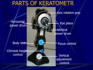
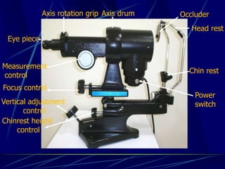
KERATOMETRY
Shishir Shukla
Optometrist, Ocularist
MGM Eye Institute
Keratometer
It is an optical instrument which is use to
measure the central corneal curvature.
Kerato = cornea
Metry = measurement
History
Christopher Scheiner in 1619.
Ramsden in 1796.
Developed in 1854 by Hermann Von Helmholtz.
Later improved by Javal Schiotz & others.
The term keratometer is a registered trademark of
Bausch & Lomb now.
Helmholtz Assumption
Helmholtz proposed that the cornea was
spherical in shape and this hypothesis was the
basis of the design of the keratometer
The central cornea is spherical and becomes
flatter towards the periphery
Principle of Keratometer
The anterior surface of cornea acts as a
convex mirror and the size of the image
formed varies with its curvature .
Principle of Keratometer
The cornea is comparable to a convex mirror in that
it reflects the illuminated object of known size, i.e.
the mire (the target), forming an image which is
virtual, small and erect.
r=radius of curvature
d= distance between object and cornea
O= object size I=image size
r = 2d x I/O
Target Size 64 mm(6.4cm) used in
keratometer.
Object distance from patient eye each 75
mm(7.5cm).
Object and image size
Fixed object size : Image size varies
h (object size)constant
Variable doubling
B & L keratometer
Variable object size : Image size constant
h’(image size)constant
Fixed doubling
Javal Schiotz ophthalmometer
Configuration of mire(object ) used in B&L keratometer
Doubling principle
•Difficult to estimate the image height
because of continuous movement of the eye
•Prisms used to split the central image
into two images
•The distance between these images are
independent of the eye movement
The Doubling Principle
+
+ +
+
+ +
+
+
+
+
+
A
B
Adjustment of prism is made to align the doubled
images
Optical design
Illumination System
An observation System
Optical system of the B +L Keratometer
Sinners disk
Skeleton of keratometer
The plus and minus signs are the extremities
of the image and acts as reference points
Plus sign – Horizontal meridian
Minus sign – Vertical meridian
Eye piece
Vertical
adjustment
control
Focus control
Chinrest height
control
Body lock
Horizontal
power drum
Vertical
power drum
Axis rotation grip
PARTS OF KERATOMETR
Eye piece
Measurement
control
Focus control
Vertical adjustment
control
Chinrest height
control
Head rest
Chin rest
OccluderAxis drum
Power
switch
Axis rotation grip
Types of keratometer
One position keratometer.
Fixed object size variable image size
Exam. B & L keratometer
Two position keratometer.
Variable object size Fixed Image size
Exam. Javal schiotz ophthalmometer .
Clinical procedure of the
keratometry
Focus the eyepiece.
Instruct the patient.
Position the patient.
Adjust the instrument.
Locate the principal meridian.
Coincide the plus sign by horizontal drum.
Coincide the minus sign by vertical drum
Record the measurements.
Preliminary adjustments
•Position a white background in front
of occluder
•Turn the eyepiece cap anticlockwise
•Switch on the instrument lamp & view
through eyepiece :a blurred cross will be seen
•Slowly turn the eye cap in clockwise
direction till the cross is well focused
Target
Configuration of mire used in B&L
keratometer
Examiner’s view before focus
Examiner’s view before
alignment
Keratometry reading
K1 46D@90
K2 45.50@180
K1 46.00@120
K2 46.50@30
Examiner’s view after
alignment
Range of Keratometer
Bausch and Lomb
36 D(9.38mm) - 52 D(6.49)
Extended Keratometry.
If > 52.0 D add +1.25Dsp lens
add +9.00 D to K reading
If < 36D add – 1.00Dsp
add – 6.00 D to K reading
Uses of keratometer
• Measurement of corneal astigmatism.
• Estimate radius of curvature of cornea which helps in contact
lens fitting.
• Assess integrity of cornea and/or tear film.
• Detection of irregular astigmatism keratoconus/
pterygium/corneal scarring.
• Assess refractive error in cases with hazy media (Rough
estimate, comparison of two eyes).
• IOL power calculation.
Sources of error in keratometry
Improper calibration.
Faulty positioning of patient.
Lack of proper fixation by patient.
Reduced visual acuity of examiner.
Accommodative fluctuation by examiner
Localized corneal distortion
Abnormal lid position
Improper focusing of the corneal image
Disadvantage
•Central 3.0 mm of cornea can be measured.
•Irregular corneal surface can not be
measured.
•Cannot measure more than 52D & less
than 36D.
•Difficult to perform for nystagmus pt.
Keratometer

Keratometer

  • 1.
  • 2.
    Keratometer It is anoptical instrument which is use to measure the central corneal curvature. Kerato = cornea Metry = measurement
  • 3.
    History Christopher Scheiner in1619. Ramsden in 1796. Developed in 1854 by Hermann Von Helmholtz. Later improved by Javal Schiotz & others. The term keratometer is a registered trademark of Bausch & Lomb now.
  • 4.
    Helmholtz Assumption Helmholtz proposedthat the cornea was spherical in shape and this hypothesis was the basis of the design of the keratometer The central cornea is spherical and becomes flatter towards the periphery
  • 5.
    Principle of Keratometer Theanterior surface of cornea acts as a convex mirror and the size of the image formed varies with its curvature .
  • 6.
    Principle of Keratometer Thecornea is comparable to a convex mirror in that it reflects the illuminated object of known size, i.e. the mire (the target), forming an image which is virtual, small and erect. r=radius of curvature d= distance between object and cornea O= object size I=image size r = 2d x I/O
  • 7.
    Target Size 64mm(6.4cm) used in keratometer. Object distance from patient eye each 75 mm(7.5cm).
  • 8.
    Object and imagesize Fixed object size : Image size varies h (object size)constant Variable doubling B & L keratometer Variable object size : Image size constant h’(image size)constant Fixed doubling Javal Schiotz ophthalmometer
  • 9.
    Configuration of mire(object) used in B&L keratometer
  • 10.
    Doubling principle •Difficult toestimate the image height because of continuous movement of the eye •Prisms used to split the central image into two images •The distance between these images are independent of the eye movement
  • 11.
    The Doubling Principle + ++ + + + + + + + + A B Adjustment of prism is made to align the doubled images
  • 12.
  • 13.
    Optical system ofthe B +L Keratometer Sinners disk
  • 14.
  • 15.
    The plus andminus signs are the extremities of the image and acts as reference points Plus sign – Horizontal meridian Minus sign – Vertical meridian
  • 16.
    Eye piece Vertical adjustment control Focus control Chinrestheight control Body lock Horizontal power drum Vertical power drum Axis rotation grip PARTS OF KERATOMETR
  • 17.
    Eye piece Measurement control Focus control Verticaladjustment control Chinrest height control Head rest Chin rest OccluderAxis drum Power switch Axis rotation grip
  • 18.
    Types of keratometer Oneposition keratometer. Fixed object size variable image size Exam. B & L keratometer Two position keratometer. Variable object size Fixed Image size Exam. Javal schiotz ophthalmometer .
  • 19.
    Clinical procedure ofthe keratometry Focus the eyepiece. Instruct the patient. Position the patient. Adjust the instrument. Locate the principal meridian. Coincide the plus sign by horizontal drum. Coincide the minus sign by vertical drum Record the measurements.
  • 20.
    Preliminary adjustments •Position awhite background in front of occluder •Turn the eyepiece cap anticlockwise •Switch on the instrument lamp & view through eyepiece :a blurred cross will be seen •Slowly turn the eye cap in clockwise direction till the cross is well focused
  • 21.
  • 22.
    Configuration of mireused in B&L keratometer
  • 23.
  • 24.
  • 25.
    Keratometry reading K1 46D@90 K245.50@180 K1 46.00@120 K2 46.50@30
  • 26.
  • 27.
    Range of Keratometer Bauschand Lomb 36 D(9.38mm) - 52 D(6.49) Extended Keratometry. If > 52.0 D add +1.25Dsp lens add +9.00 D to K reading If < 36D add – 1.00Dsp add – 6.00 D to K reading
  • 30.
    Uses of keratometer •Measurement of corneal astigmatism. • Estimate radius of curvature of cornea which helps in contact lens fitting. • Assess integrity of cornea and/or tear film. • Detection of irregular astigmatism keratoconus/ pterygium/corneal scarring. • Assess refractive error in cases with hazy media (Rough estimate, comparison of two eyes). • IOL power calculation.
  • 31.
    Sources of errorin keratometry Improper calibration. Faulty positioning of patient. Lack of proper fixation by patient. Reduced visual acuity of examiner. Accommodative fluctuation by examiner Localized corneal distortion Abnormal lid position Improper focusing of the corneal image
  • 32.
    Disadvantage •Central 3.0 mmof cornea can be measured. •Irregular corneal surface can not be measured. •Cannot measure more than 52D & less than 36D. •Difficult to perform for nystagmus pt.