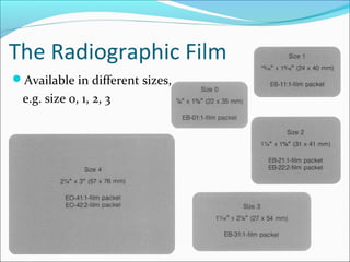
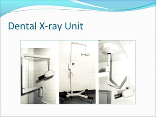
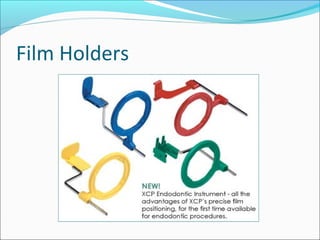
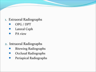
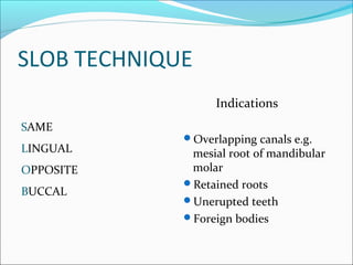
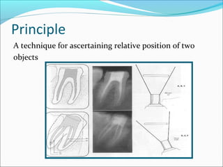
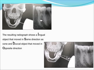
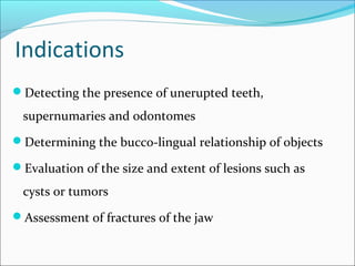
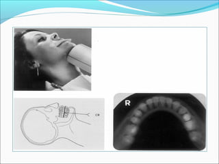
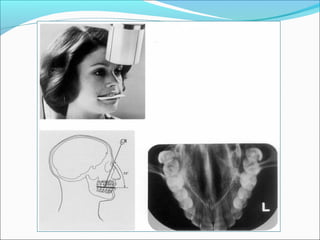
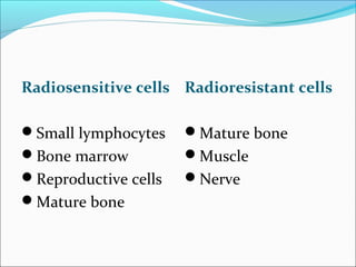
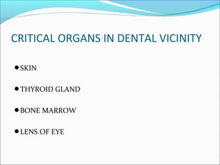
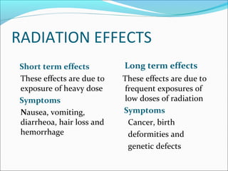
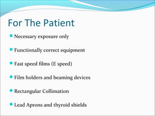
Dr.Shahbaz Ahmed
BDS, MSc (UK), FCPS (Pak)
Assistant Professor
Department of Operative Dentistry
DIKIOHS
Definition
ļ‚—Radiograph - record of an image produced by
transmission of x-rays through an object
ļ‚—Radiography – techniques involved in producing
various radiographic images
ļ‚—Radiology – interpretation of radiographic images
What Are X-Rays?
ļ‚—A form of electromagnetic radiation
ļ‚—Similar to visible light but of shorter wavelength
ļ‚—X-rays, gamma rays, radio waves and visible
light…..all electromagnetic waves
Why do we need radiographs?
ļ‚—Case selection
ļ‚—Diagnosis & Treatment Plan
ļ‚—Treatment
ļ‚—Review /Prognosis
The Radiographic Film
ļ‚—Available in different sizes,
e.g. size 0, 1, 2, 3
Dental X-ray Unit
Film Holders
Lead Aprons
Dark Room
X-ray Viewer
1. Extraoral Radiographs
ļ‚— OPG / DPT
ļ‚— Lateral Ceph
ļ‚— PA view
2. Intraoral Radiographs
ļ‚— Bitewing Radiographs
ļ‚— Occlusal Radiographs
ļ‚— Periapical Radiographs
Indications
ļ‚—Routine radiograph at regular dental checkups
ļ‚—Proximal Caries
ļ‚—Monitoring of progression of caries
ļ‚—Overhanging restorations
ļ‚—Diagnosis of Periodontal disease
Aims
ļ‚—Image of whole length of tooth & surrounding bone.
ļ‚—Allows comparison between pre & post op condition
ļ‚—To undertake procedures as swiftly as possible to
minimize patient discomfort
Indications
ļ‚—Assessment of periodontal status
ļ‚—Endodontics
ļ‚—Assessment of apical surgery
ļ‚—Detection of apical cyst
ļ‚—After dental trauma
ļ‚—Assessment of root morphology before extraction
Techniques
ļ‚—Paralleling film Or Paralleling Technique
ļ‚—Bisecting Angle Technique
ļ‚—SLOB Technique
Paralleling Technique
ļ‚—Film should be placed in the
mouth parallel to long axis of
tooth being examined
ļ‚—Central ray of X-ray beam is
directed perpendicular to
film and long axis of the
tooth
ļ‚—A film holder must be used
to keep the film parallel to
the tooth
Advantages
ļ‚—ACCURACY
Image produced has dimensional accuracy and free of
distortion
ļ‚—SIMPLICITY
Simple and easy to learn and use
ļ‚—DUPLICATION
Easy to standardize and can be accurately repeated
Disadvantages
ļ‚—FILM PLACEMENT
Requires film holder to place films which is difficult to
place in children , small mouth patients.
Anatomy sometimes make technique impossible, e.g.
shallow palate, third molar region
ļ‚— DISCOMFORT
Positioning of film packet can be un-comfortable
Bisecting Angle Technique
ļ‚—Also known as short cone
technique
ļ‚—Film placed close to the
tooth
ļ‚—Angle formed between
tooth and film bisected
ļ‚—Tube head directed at
right angles to bisecting
line
Advantages
ļ‚—This technique can be used without film holder
ļ‚—Comfortable for patient
ļ‚—Less time required
Disadvantages
ļ‚—Image distortion/magnification
ļ‚—Technique sensitive, retakes!!!
ļ‚—Not reproducible
ļ‚—Superimposition of zygomatic buttress over upper
molars
SLOB TECHNIQUE
SAME
LINGUAL
OPPOSITE
BUCCAL
Indications
ļ‚—Overlapping canals e.g.
mesial root of mandibular
molar
ļ‚—Retained roots
ļ‚—Unerupted teeth
ļ‚—Foreign bodies
ļ‚—Cone shift technique, mesial shift, Clark’s shift, Buccal object
rule
ļ‚—Reveals the third dimension
Principle
A technique for ascertaining relative position of two
objects
The resulting radiograph shows a lingual
object that moved in same direction as
cone and buccal object that moved in
opposite direction
Indications
ļ‚—Detecting the presence of unerupted teeth,
supernumaries and odontomes
ļ‚—Determining the bucco-lingual relationship of objects
ļ‚—Evaluation of the size and extent of lesions such as
cysts or tumors
ļ‚—Assessment of fractures of the jaw
Panoramic radiography
Panoramic radiography ( rotational radiography) is a
radiographic procedure that produces a single image
of the facial structures, including both maxillary and
mandibular arches and their supporting structures
Advantages
ļ‚—Broad anatomic region imaged
ļ‚—Positioning is relatively simple
ļ‚—Relatively less radiation dose (about one third of the dose from
a full mouth survey of intra-oral films)
ļ‚—View of both sides of the jaws is useful in the assessment of
fractures
ļ‚—Over all view is helpful for initial assessment of periodontal
status
ļ‚—Antral floor can be seen
ļ‚—Both condylar heads are shown on one film, allowing easy
comparison
ļ‚—Procedure can be performed in patients of limited mouth
opening
Disadvantages
ļ‚—Resultant image does not resolve the fine anatomic detail that
may be seen on intra oral peri-apical radiograph
ļ‚—Soft tissues and air shadows can overlie the required hard
structures
ļ‚—Technique is not suitable for children because of length of
exposure cycle
ļ‚—Cost of machine is almost double or four times than that of
intra-oral machine
Indications
ļ‚—Orthodontic assessment
ļ‚—Lesions like cyst,tumors and development anomalies in the
body and rami of mandible to establish the site and size
ļ‚—Fractures of mandible
ļ‚—Antral diseases, especially to asses the floor, posterior and
anterior walls of antrum
ļ‚—To investigate the quality of articular surface of the condylar
heads
ļ‚—Periodontal diseases; an overall view of alveolar bone levels
ļ‚—Assessment of the presence and position of wisdom teeth
ļ‚—Assessment of any underlying disease before construction of
prosthesis
ļ‚—Evaluation of vertical height of bone before insertion of
implants
MECHANISM OF RADIATION INJURY
ļ‚—IONIZATION (DIRECT EFFECT)
ļ‚—FREE RADICAL ( INDIRECT INJURY )
Radiosensitive cells
ļ‚—Small lymphocytes
ļ‚—Bone marrow
ļ‚—Reproductive cells
ļ‚—Mature bone
Radioresistant cells
ļ‚—Mature bone
ļ‚—Muscle
ļ‚—Nerve
CRITICAL ORGANS IN DENTAL VICINITY
SKIN
THYROID GLAND
BONE MARROW
LENS OF EYE
RADIATION EFFECTS
Short term effects
These effects are due to
exposure of heavy dose
Symptoms
Nausea, vomiting,
diarrheoa, hair loss and
hemorrhage
Long term effects
These effects are due to
frequent exposures of
low doses of radiation
Symptoms
Cancer, birth
deformities and
genetic defects
ALARAALARA
AAs LLow AAs RReasonably
AAchievable
For The Patient
ļ‚—Necessary exposure only
ļ‚—Functionally correct equipment
ļ‚—Fast speed films (E speed)
ļ‚—Film holders and beaming devices
ļ‚—Rectangular Collimation
ļ‚—Lead Aprons and thyroid shields
Thyroid Shields
Pregnant Women And Children
Caution Is Must
WORKERS (DENTIST AND STAFF)
PROTECTIVE MEASURES INCLUDES:
ļ‚—Distance from source of radiation
ļ‚—Equipment
ļ‚—Radiographic technique
General Public
General public includes:
ļ‚—people in waiting room
ļ‚—people in other building rooms
ļ‚—passer by
Safety measures includes:
ļ‚—The setting of equipment to ensure that the primary
beam is not aimed directly into the occupied rooms
ļ‚—The thickness/material of the partitioning wall
Advantages
ļ‚—No X-ray film, use of sensor
ļ‚—No chemical processing, instant image
ļ‚—Image can be colorized
ļ‚—Excellent tool for patient education
ļ‚—Greater patient acceptance
ļ‚—50% -90% less radiation than conventional x-ray film
ļ‚—Ability to zoom, enhance , magnify, store, and
electronically send the images
ļ‚—Easy duplication
ļ‚—Digital subtraction radiography
Disadvantages
ļ‚—Expensive technology
ļ‚—Resolution of image is slightly less than the
conventional x-ray film.
In 1998, the American Association of
Endodontists stated that
ā€œ Digital radiography will rapidly replace
conventional dental X-raysā€.
Radiology in dentistry

Radiology in dentistry

  • 1.
    Dr.Shahbaz Ahmed BDS, MSc(UK), FCPS (Pak) Assistant Professor Department of Operative Dentistry DIKIOHS
  • 3.
    Definition ļ‚—Radiograph - recordof an image produced by transmission of x-rays through an object ļ‚—Radiography – techniques involved in producing various radiographic images ļ‚—Radiology – interpretation of radiographic images
  • 4.
    What Are X-Rays? ļ‚—Aform of electromagnetic radiation ļ‚—Similar to visible light but of shorter wavelength ļ‚—X-rays, gamma rays, radio waves and visible light…..all electromagnetic waves
  • 5.
    Why do weneed radiographs? ļ‚—Case selection ļ‚—Diagnosis & Treatment Plan ļ‚—Treatment ļ‚—Review /Prognosis
  • 7.
    The Radiographic Film ļ‚—Availablein different sizes, e.g. size 0, 1, 2, 3
  • 8.
  • 9.
  • 10.
  • 11.
  • 12.
  • 14.
    1. Extraoral Radiographs ļ‚—OPG / DPT ļ‚— Lateral Ceph ļ‚— PA view 2. Intraoral Radiographs ļ‚— Bitewing Radiographs ļ‚— Occlusal Radiographs ļ‚— Periapical Radiographs
  • 16.
    Indications ļ‚—Routine radiograph atregular dental checkups ļ‚—Proximal Caries ļ‚—Monitoring of progression of caries ļ‚—Overhanging restorations ļ‚—Diagnosis of Periodontal disease
  • 19.
    Aims ļ‚—Image of wholelength of tooth & surrounding bone. ļ‚—Allows comparison between pre & post op condition ļ‚—To undertake procedures as swiftly as possible to minimize patient discomfort
  • 20.
    Indications ļ‚—Assessment of periodontalstatus ļ‚—Endodontics ļ‚—Assessment of apical surgery ļ‚—Detection of apical cyst ļ‚—After dental trauma ļ‚—Assessment of root morphology before extraction
  • 21.
    Techniques ļ‚—Paralleling film OrParalleling Technique ļ‚—Bisecting Angle Technique ļ‚—SLOB Technique
  • 22.
    Paralleling Technique ļ‚—Film shouldbe placed in the mouth parallel to long axis of tooth being examined ļ‚—Central ray of X-ray beam is directed perpendicular to film and long axis of the tooth ļ‚—A film holder must be used to keep the film parallel to the tooth
  • 23.
    Advantages ļ‚—ACCURACY Image produced hasdimensional accuracy and free of distortion ļ‚—SIMPLICITY Simple and easy to learn and use ļ‚—DUPLICATION Easy to standardize and can be accurately repeated
  • 24.
    Disadvantages ļ‚—FILM PLACEMENT Requires filmholder to place films which is difficult to place in children , small mouth patients. Anatomy sometimes make technique impossible, e.g. shallow palate, third molar region ļ‚— DISCOMFORT Positioning of film packet can be un-comfortable
  • 25.
    Bisecting Angle Technique ļ‚—Alsoknown as short cone technique ļ‚—Film placed close to the tooth ļ‚—Angle formed between tooth and film bisected ļ‚—Tube head directed at right angles to bisecting line
  • 26.
    Advantages ļ‚—This technique canbe used without film holder ļ‚—Comfortable for patient ļ‚—Less time required
  • 27.
    Disadvantages ļ‚—Image distortion/magnification ļ‚—Technique sensitive,retakes!!! ļ‚—Not reproducible ļ‚—Superimposition of zygomatic buttress over upper molars
  • 28.
    SLOB TECHNIQUE SAME LINGUAL OPPOSITE BUCCAL Indications ļ‚—Overlapping canalse.g. mesial root of mandibular molar ļ‚—Retained roots ļ‚—Unerupted teeth ļ‚—Foreign bodies
  • 29.
    ļ‚—Cone shift technique,mesial shift, Clark’s shift, Buccal object rule ļ‚—Reveals the third dimension
  • 30.
    Principle A technique forascertaining relative position of two objects
  • 31.
    The resulting radiographshows a lingual object that moved in same direction as cone and buccal object that moved in opposite direction
  • 33.
    Indications ļ‚—Detecting the presenceof unerupted teeth, supernumaries and odontomes ļ‚—Determining the bucco-lingual relationship of objects ļ‚—Evaluation of the size and extent of lesions such as cysts or tumors ļ‚—Assessment of fractures of the jaw
  • 37.
    Panoramic radiography Panoramic radiography( rotational radiography) is a radiographic procedure that produces a single image of the facial structures, including both maxillary and mandibular arches and their supporting structures
  • 38.
    Advantages ļ‚—Broad anatomic regionimaged ļ‚—Positioning is relatively simple ļ‚—Relatively less radiation dose (about one third of the dose from a full mouth survey of intra-oral films) ļ‚—View of both sides of the jaws is useful in the assessment of fractures ļ‚—Over all view is helpful for initial assessment of periodontal status ļ‚—Antral floor can be seen ļ‚—Both condylar heads are shown on one film, allowing easy comparison ļ‚—Procedure can be performed in patients of limited mouth opening
  • 39.
    Disadvantages ļ‚—Resultant image doesnot resolve the fine anatomic detail that may be seen on intra oral peri-apical radiograph ļ‚—Soft tissues and air shadows can overlie the required hard structures ļ‚—Technique is not suitable for children because of length of exposure cycle ļ‚—Cost of machine is almost double or four times than that of intra-oral machine
  • 40.
    Indications ļ‚—Orthodontic assessment ļ‚—Lesions likecyst,tumors and development anomalies in the body and rami of mandible to establish the site and size ļ‚—Fractures of mandible ļ‚—Antral diseases, especially to asses the floor, posterior and anterior walls of antrum ļ‚—To investigate the quality of articular surface of the condylar heads ļ‚—Periodontal diseases; an overall view of alveolar bone levels ļ‚—Assessment of the presence and position of wisdom teeth ļ‚—Assessment of any underlying disease before construction of prosthesis ļ‚—Evaluation of vertical height of bone before insertion of implants
  • 43.
    MECHANISM OF RADIATIONINJURY ļ‚—IONIZATION (DIRECT EFFECT) ļ‚—FREE RADICAL ( INDIRECT INJURY )
  • 44.
    Radiosensitive cells ļ‚—Small lymphocytes ļ‚—Bonemarrow ļ‚—Reproductive cells ļ‚—Mature bone Radioresistant cells ļ‚—Mature bone ļ‚—Muscle ļ‚—Nerve
  • 45.
    CRITICAL ORGANS INDENTAL VICINITY SKIN THYROID GLAND BONE MARROW LENS OF EYE
  • 46.
    RADIATION EFFECTS Short termeffects These effects are due to exposure of heavy dose Symptoms Nausea, vomiting, diarrheoa, hair loss and hemorrhage Long term effects These effects are due to frequent exposures of low doses of radiation Symptoms Cancer, birth deformities and genetic defects
  • 48.
    ALARAALARA AAs LLow AAsRReasonably AAchievable
  • 49.
    For The Patient ļ‚—Necessaryexposure only ļ‚—Functionally correct equipment ļ‚—Fast speed films (E speed) ļ‚—Film holders and beaming devices ļ‚—Rectangular Collimation ļ‚—Lead Aprons and thyroid shields
  • 50.
  • 51.
    Pregnant Women AndChildren Caution Is Must
  • 52.
    WORKERS (DENTIST ANDSTAFF) PROTECTIVE MEASURES INCLUDES: ļ‚—Distance from source of radiation ļ‚—Equipment ļ‚—Radiographic technique
  • 53.
    General Public General publicincludes: ļ‚—people in waiting room ļ‚—people in other building rooms ļ‚—passer by Safety measures includes: ļ‚—The setting of equipment to ensure that the primary beam is not aimed directly into the occupied rooms ļ‚—The thickness/material of the partitioning wall
  • 55.
    Advantages ļ‚—No X-ray film,use of sensor ļ‚—No chemical processing, instant image ļ‚—Image can be colorized ļ‚—Excellent tool for patient education ļ‚—Greater patient acceptance ļ‚—50% -90% less radiation than conventional x-ray film ļ‚—Ability to zoom, enhance , magnify, store, and electronically send the images ļ‚—Easy duplication ļ‚—Digital subtraction radiography
  • 56.
    Disadvantages ļ‚—Expensive technology ļ‚—Resolution ofimage is slightly less than the conventional x-ray film.
  • 58.
    In 1998, theAmerican Association of Endodontists stated that ā€œ Digital radiography will rapidly replace conventional dental X-raysā€.