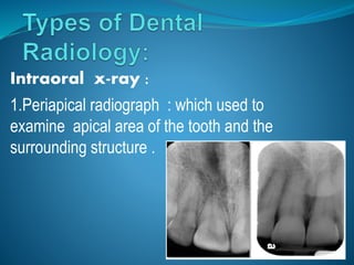
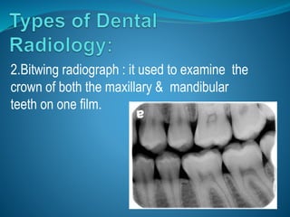
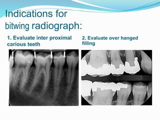
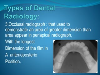
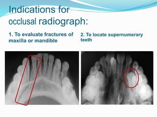
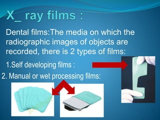
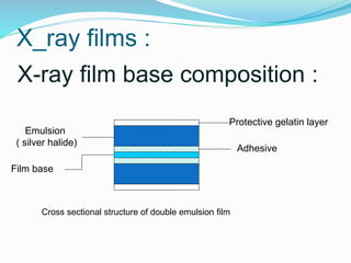
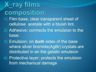
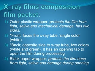
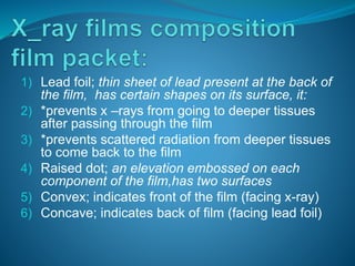
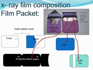
Radiation: is energy that comes from a
source and travels through space and
may be able to penetrate various
materials. It’s may occur in two forms :
1. particulate radiation ,
2.electromagnetic radiation .
Dental radiograph: a photographic image
produced on film by passage of x –ray
through teeth & related structures.
Intraoral x-ray :
1.Periapical radiograph : which used to
examine apical area of the tooth and the
surrounding structure .
Indications for
Periapical radiograph:
1.Detection of apical cyst 2.Evaluation of implants
2.Bitwing radiograph : it used to examine the
crown of both the maxillary & mandibular
teeth on one film.
Indications for
bitwing radiograph:
1. Evaluate inter proximal
carious teeth
2. Evaluate over hanged
filling
3.Occlusal radiograph : that used to
demonstrate an area of greater dimension than
area appear in periapical radiograph,
With the longest
Dimension of the film in
A anterioposterio
Position.
Indications for
occlusal radiograph:
1. To evaluate fractures of
maxilla or mandible
2. To locate supernumerary
teeth
Dental films:The media on which the
radiographic images of objects are
recorded, there is 2 types of films:
1.Self developing films :
2. Manual or wet processing films:
X_ray films :
X-ray film base composition :
Protective gelatin layer
Adhesive
Emulsion
( silver halide)
Film base
Cross sectional structure of double emulsion film
1) Film base; clear transparent sheet of
cellulose acetate with a bluish tint.
2) Adhesive; connects the emulsion to the
base.
3) Emulsion; on both sides of the base
where silver bromide(AgBr) crystals are
distributed in an the gelatin emulsion
4) Protective layer; protects the emulsion
from mechanical damage
1) Outer plastic wrapper; protects the film from
light, saliva and mechanical damage, has two
sides:
2) *Front; faces the x-ray tube, single color
(white)
3) *Back; opposite side to x-ray tube, two colors
(white and green). It has an opening tab to
open the film during processibg
4) Black paper wrapper; protects the film base
from light, saliva and damage during opening
1) Lead foil; thin sheet of lead present at the back of
the film, has certain shapes on its surface, it:
2) *prevents x –rays from going to deeper tissues
after passing through the film
3) *prevents scattered radiation from deeper tissues
to come back to the film
4) Raised dot; an elevation embossed on each
component of the film,has two surfaces
5) Convex; indicates front of the film (facing x-ray)
6) Concave; indicates back of film (facing lead foil)
Outer plastic cover
Front Back
Film
Lead
foil
Protective black paper
Dental radiology ppt

Dental radiology ppt

  • 2.
    Radiation: is energythat comes from a source and travels through space and may be able to penetrate various materials. It’s may occur in two forms : 1. particulate radiation , 2.electromagnetic radiation . Dental radiograph: a photographic image produced on film by passage of x –ray through teeth & related structures.
  • 3.
    Intraoral x-ray : 1.Periapicalradiograph : which used to examine apical area of the tooth and the surrounding structure .
  • 4.
    Indications for Periapical radiograph: 1.Detectionof apical cyst 2.Evaluation of implants
  • 5.
    2.Bitwing radiograph :it used to examine the crown of both the maxillary & mandibular teeth on one film.
  • 6.
    Indications for bitwing radiograph: 1.Evaluate inter proximal carious teeth 2. Evaluate over hanged filling
  • 7.
    3.Occlusal radiograph :that used to demonstrate an area of greater dimension than area appear in periapical radiograph, With the longest Dimension of the film in A anterioposterio Position.
  • 8.
    Indications for occlusal radiograph: 1.To evaluate fractures of maxilla or mandible 2. To locate supernumerary teeth
  • 9.
    Dental films:The mediaon which the radiographic images of objects are recorded, there is 2 types of films: 1.Self developing films : 2. Manual or wet processing films:
  • 10.
    X_ray films : X-rayfilm base composition : Protective gelatin layer Adhesive Emulsion ( silver halide) Film base Cross sectional structure of double emulsion film
  • 11.
    1) Film base;clear transparent sheet of cellulose acetate with a bluish tint. 2) Adhesive; connects the emulsion to the base. 3) Emulsion; on both sides of the base where silver bromide(AgBr) crystals are distributed in an the gelatin emulsion 4) Protective layer; protects the emulsion from mechanical damage
  • 12.
    1) Outer plasticwrapper; protects the film from light, saliva and mechanical damage, has two sides: 2) *Front; faces the x-ray tube, single color (white) 3) *Back; opposite side to x-ray tube, two colors (white and green). It has an opening tab to open the film during processibg 4) Black paper wrapper; protects the film base from light, saliva and damage during opening
  • 13.
    1) Lead foil;thin sheet of lead present at the back of the film, has certain shapes on its surface, it: 2) *prevents x –rays from going to deeper tissues after passing through the film 3) *prevents scattered radiation from deeper tissues to come back to the film 4) Raised dot; an elevation embossed on each component of the film,has two surfaces 5) Convex; indicates front of the film (facing x-ray) 6) Concave; indicates back of film (facing lead foil)
  • 14.
    Outer plastic cover FrontBack Film Lead foil Protective black paper