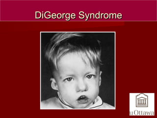
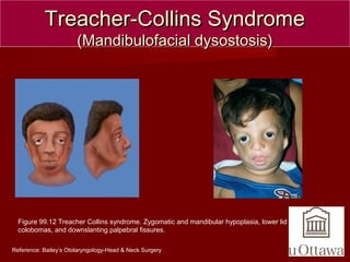
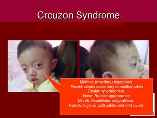
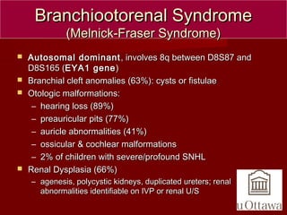
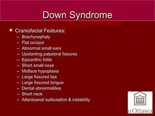
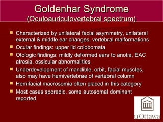
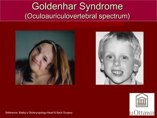
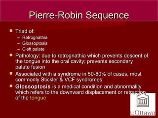
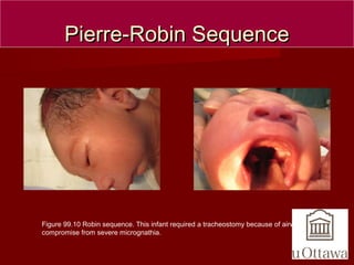
CRANIOFACIALCRANIOFACIAL
ANOMALIESANOMALIES
BY:DR IMTIAZ AHMEDBY:DR IMTIAZ AHMED
BDS, FCPSBDS, FCPS
ORTHODONTICSORTHODONTICS
DiGeorge SyndromeDiGeorge Syndrome
 Genetic disorder due to microdeletion ofGenetic disorder due to microdeletion of
ChromosomeChromosome 22q11.222q11.2 ((tbx-1tbx-1 genegene))
– The same genetic defect as VCF with differentThe same genetic defect as VCF with different
phenotypic expressionphenotypic expression
 Characterized by:Characterized by:
– Hypocalcemia (due to hypoplastic parathyroids)Hypocalcemia (due to hypoplastic parathyroids)
– Immunodeficiency due to hypoplastic thymusImmunodeficiency due to hypoplastic thymus
– Congenital heart defects of the outflow tracts (aortaCongenital heart defects of the outflow tracts (aorta
and pulmonary artery).and pulmonary artery).
•Reference: http://medicine.net
DiGeorge SyndromeDiGeorge Syndrome
Treacher-Collins SyndromeTreacher-Collins Syndrome
(Mandibulofacial dysostosis)(Mandibulofacial dysostosis)
 Autosomal dominant, 40% will have family history, otherAutosomal dominant, 40% will have family history, other
60% new mutations60% new mutations
 TCOF1TCOF1 gene found on chromosome 5q (gene found on chromosome 5q (TREACLETREACLE
genegene))
 Malformation of 1st (& 2nd) branchial archesMalformation of 1st (& 2nd) branchial arches
 Otologic: Malformed ossicles, auricular deformity, auralOtologic: Malformed ossicles, auricular deformity, aural
atresia, CHL present 30% of time, occasional SNHLatresia, CHL present 30% of time, occasional SNHL
– 50% will have hearing impairment from EAC and/or middle ear50% will have hearing impairment from EAC and/or middle ear
malformationsmalformations
 Preauricular fistulas, mandibular and malar hypoplasia,Preauricular fistulas, mandibular and malar hypoplasia,
antimongoloid palpebral fissures, coloboma of the lowerantimongoloid palpebral fissures, coloboma of the lower
eyelids, may have cleft lip and palate, normal IQeyelids, may have cleft lip and palate, normal IQ
Treacher-Collins SyndromeTreacher-Collins Syndrome
(Mandibulofacial dysostosis)(Mandibulofacial dysostosis)
Figure 99.12 Treacher Collins syndrome. Zygomatic and mandibular hypoplasia, lower lid
colobomas, and downslanting palpebral fissures.
Reference: Bailey’s Otolaryngology-Head & Neck Surgery
Apert (acrocephalosyndactyly)Apert (acrocephalosyndactyly)
 Autosomal dominant, most cases due to spontaneousAutosomal dominant, most cases due to spontaneous
mutationmutation
 Due to a mutation ofDue to a mutation of FGFR-2FGFR-2 (Fibroblast Growth Factor(Fibroblast Growth Factor
Receptor) gene (Receptor) gene (10q2610q26))
 Common findings:Common findings:
– Craniosynostosis (pre-mature fusion of the cranial sutures)Craniosynostosis (pre-mature fusion of the cranial sutures)
– Severe symmetrical syndactylySevere symmetrical syndactyly
– Low-set earsLow-set ears
– Cognitive function normal to severe mental retardationCognitive function normal to severe mental retardation
– EyesEyes: down-slanting palpebrael fissures, Hypertelorism, Exophthalmos: down-slanting palpebrael fissures, Hypertelorism, Exophthalmos
– Midface hypoplasiaMidface hypoplasia
– Mandibular prognathismMandibular prognathism
– Possible cleft palatePossible cleft palate
– NoseNose: Parrot-beaked nose, possible Choanal Atresia: Parrot-beaked nose, possible Choanal Atresia
– Syndactyly and cervical fusionSyndactyly and cervical fusion
Figure 99.4 Apert syndrome has the additional feature of
syndactyly.
Reference: Bailey’s Otolaryngology-Head & Neck Surgery
Apert (acrocephalosyndactyly)Apert (acrocephalosyndactyly)
Crouzon SyndromeCrouzon Syndrome
(Craniofacial Dysostosis)(Craniofacial Dysostosis)
 Autosomal dominant, 50% due to spontaneousAutosomal dominant, 50% due to spontaneous
mutations, complete penetrance, variable expresivitymutations, complete penetrance, variable expresivity
 Due to mutation ofDue to mutation of FGFR-2FGFR-2 (Fibroblast Growth Factor(Fibroblast Growth Factor
Receptor) gene (Receptor) gene (10q2610q26))
 Common findings:Common findings:
– Craniosynostosis (pre-mature fusion of the cranial sutures)Craniosynostosis (pre-mature fusion of the cranial sutures)
– HypertelorismHypertelorism
– ExophthalmosExophthalmos
– Midface hypoplasiaMidface hypoplasia
– Mandibular prognathismMandibular prognathism
– Parrot-beaked noseParrot-beaked nose
– No Syndactyly or cervical fusionNo Syndactyly or cervical fusion
– Cognitive function normal to severe mental retardationCognitive function normal to severe mental retardation
Crouzon SyndromeCrouzon Syndrome
 Coronal and sagittalCoronal and sagittal
sutures are mostsutures are most
commonly involvedcommonly involved
 Cloverleaf skull is rare andCloverleaf skull is rare and
occurs in the mostoccurs in the most
severely affectedseverely affected
individuals.individuals.
 HydrocephalusHydrocephalus
(progressive in 30%)(progressive in 30%)
Crouzon SyndromeCrouzon Syndrome
Midface (maxillary) hypoplasia
Exophthalmos secondary to shallow orbits
Ocular hypertelorism
Nose: Beaked appearance
Mouth: Mandibular prognathism
Narrow, high, or cleft palate and bifid uvula
Branchiootorenal SyndromeBranchiootorenal Syndrome
(Melnick-Fraser Syndrome)(Melnick-Fraser Syndrome)
 Autosomal dominantAutosomal dominant , involves 8q between D8S87 and, involves 8q between D8S87 and
D8S165 (D8S165 (EYA1 geneEYA1 gene))
 Branchial cleft anomalies (63%): cysts or fistulaeBranchial cleft anomalies (63%): cysts or fistulae
 Otologic malformations:Otologic malformations:
– hearing loss (89%)hearing loss (89%)
– preauricular pits (77%)preauricular pits (77%)
– auricle abnormalities (41%)auricle abnormalities (41%)
– ossicular & cochlear malformationsossicular & cochlear malformations
– 2% of children with severe/profound SNHL2% of children with severe/profound SNHL
 Renal Dysplasia (66%)Renal Dysplasia (66%)
– agenesis, polycystic kidneys, duplicated ureters; renalagenesis, polycystic kidneys, duplicated ureters; renal
abnormalities identifiable on IVP or renal U/Sabnormalities identifiable on IVP or renal U/S
Branchiootorenal SyndromeBranchiootorenal Syndrome
(Melnick-Fraser Syndrome)(Melnick-Fraser Syndrome)
Figure 99.6 Branchio-oto-renal syndrome. This 3-year-old boy has visible
cup-ear deformities. He also has branchial cleft fistulae and only one kidney.
Reference: Bailey’s Otolaryngology-Head & Neck Surgery
Down SyndromeDown Syndrome
 Craniofacial Features:Craniofacial Features:
– BrachycephalyBrachycephaly
– Flat occiputFlat occiput
– Abnormal small earsAbnormal small ears
– Upslanting palpebral fissuresUpslanting palpebral fissures
– Epicanthic foldsEpicanthic folds
– Short small noseShort small nose
– Midface hypoplasiaMidface hypoplasia
– Large fissured lipsLarge fissured lips
– Large fissured tongueLarge fissured tongue
– Dental abnormalitiesDental abnormalities
– Short neckShort neck
– Atlantoaxial subluxation & instabilityAtlantoaxial subluxation & instability
Down SyndromeDown Syndrome
Goldenhar SyndromeGoldenhar Syndrome
(Oculoauriculovertebral spectrum)(Oculoauriculovertebral spectrum)
 Characterized by unilateral facial asymmetry, unilateralCharacterized by unilateral facial asymmetry, unilateral
external & middle ear changes, vertebral malformationsexternal & middle ear changes, vertebral malformations
 Ocular findings: upper lid colobomataOcular findings: upper lid colobomata
 Otologic findings: mildly deformed ears to anotia, EACOtologic findings: mildly deformed ears to anotia, EAC
atresia, ossicular abnormalitiesatresia, ossicular abnormalities
 Underdevelopment of mandible, orbit, facial muscles,Underdevelopment of mandible, orbit, facial muscles,
also may have hemivertebrae of vertebral columnalso may have hemivertebrae of vertebral column
 Hemifacial macrosomia often placed in this categoryHemifacial macrosomia often placed in this category
 Most cases sporadic, some autosomal dominantMost cases sporadic, some autosomal dominant
reportedreported
Goldenhar SyndromeGoldenhar Syndrome
(Oculoauriculovertebral spectrum)(Oculoauriculovertebral spectrum)
Reference: Bailey’s Otolaryngology-Head & Neck Surgery
Pierre-Robin SequencePierre-Robin Sequence
 Triad of:Triad of:
– RetrognathiaRetrognathia
– GlossoptosisGlossoptosis
– Cleft palateCleft palate
 Pathology: due to retrognathia which prevents descent ofPathology: due to retrognathia which prevents descent of
the tongue into the oral cavity; prevents secondarythe tongue into the oral cavity; prevents secondary
palate fusionpalate fusion
 Associated with a syndrome in 50-80% of cases, mostAssociated with a syndrome in 50-80% of cases, most
commonly Stickler & VCF syndromescommonly Stickler & VCF syndromes
 GlossoptosisGlossoptosis is a medical condition and abnormalityis a medical condition and abnormality
which refers to the downward displacement or retractionwhich refers to the downward displacement or retraction
of theof the tonguetongue
Pierre-Robin SequencePierre-Robin Sequence
Figure 99.10 Robin sequence. This infant required a tracheostomy because of airway
compromise from severe micrognathia.
DiscussionDiscussion
1.1. CraniosynostosisCraniosynostosis
2.2. Cloverleaf skull syndromeCloverleaf skull syndrome
CraniosynostosisCraniosynostosis
 Primary craniosynostosisPrimary craniosynostosis: a primary defect of: a primary defect of
ossificationossification
 Secondary craniosynostosisSecondary craniosynostosis: a failure of brain: a failure of brain
growth, more commonlygrowth, more commonly
 Syndromic craniosynostosisSyndromic craniosynostosis: display other body: display other body
deformitiesdeformities
CraniosynostosisCraniosynostosis
 The coronal suture separates the 2The coronal suture separates the 2
frontal bones from the parietal bones.frontal bones from the parietal bones.
 The metopic suture separates theThe metopic suture separates the
frontal bones.frontal bones.
 The sagittal suture separates the 2The sagittal suture separates the 2
parietal bones.parietal bones.
 The lambdoid suture separates theThe lambdoid suture separates the
occipital bone from the 2 parietaloccipital bone from the 2 parietal
bones.bones.
 The primary factor that keeps suturesThe primary factor that keeps sutures
open is ongoing brain growth.open is ongoing brain growth.
 Normal skull growth occursNormal skull growth occurs
perpendicular to each suture.perpendicular to each suture.
Primary craniosynostosisPrimary craniosynostosis
 When 1 or more sutures fuse prematurely, skull growth canWhen 1 or more sutures fuse prematurely, skull growth can
be restricted perpendicular to the suture. If multiple suturesbe restricted perpendicular to the suture. If multiple sutures
fuse while the brain is still increasing in size,fuse while the brain is still increasing in size, intracranialintracranial
pressurepressure can increase.can increase.
 Cause: a primary defect in the mesenchymal layerCause: a primary defect in the mesenchymal layer
ossification in the cranial bones.ossification in the cranial bones.
 A gene locus for single suture craniosynostosis has notA gene locus for single suture craniosynostosis has not
been identified.been identified.
ScaphocephalyScaphocephaly - Early fusion of the- Early fusion of the sagittalsagittal suturesuture
Ant. plagiocephaly - Early fusion of 1 coronal sutureAnt. plagiocephaly - Early fusion of 1 coronal suture
Post. plagiocephaly - Early closure of 1 lambdoid suturePost. plagiocephaly - Early closure of 1 lambdoid suture
Brachycephaly - Early bilateral coronal suture fusionBrachycephaly - Early bilateral coronal suture fusion
Trigonocephaly - Early fusion of the metopic sutureTrigonocephaly - Early fusion of the metopic suture
Secondary craniosynostosisSecondary craniosynostosis
 More frequentMore frequent
 Early fusion of sutures due toEarly fusion of sutures due to primary failure of brain growthprimary failure of brain growth
 Intracranial pressure usually is normal, and surgery seldomIntracranial pressure usually is normal, and surgery seldom
is neededis needed
 Intrauterine space constraints may play a role in theIntrauterine space constraints may play a role in the
premature fusion of sutures in the fetal skull. This has beenpremature fusion of sutures in the fetal skull. This has been
demonstrated in coronal craniosynostosisdemonstrated in coronal craniosynostosis
 MicrocephalyMicrocephaly usually suggests a secondaryusually suggests a secondary
craniosynostosiscraniosynostosis
Secondary craniosynostosisSecondary craniosynostosis
 EndocrineEndocrine
Hyperthyroidism, hypophosphatemia, vitamin D deficiency,Hyperthyroidism, hypophosphatemia, vitamin D deficiency,
renal osteodystrophy, hypercalcemia, and ricketsrenal osteodystrophy, hypercalcemia, and rickets
 Hematologic disordersHematologic disorders
Which cause bone marrow hyperplasia (eg, sickle cellWhich cause bone marrow hyperplasia (eg, sickle cell
disease, thalassemia)disease, thalassemia)
 Inadequate brain growthInadequate brain growth
Microcephaly and its causes and shunted hydrocephalusMicrocephaly and its causes and shunted hydrocephalus
Treatment of CraniosynostosisTreatment of Craniosynostosis
 Do not operate in patients without IICP until theDo not operate in patients without IICP until the
shape of the head does not improve byshape of the head does not improve by age 2-4age 2-4
monthsmonths, then the abnormality is unlikely to resolve, then the abnormality is unlikely to resolve
with agewith age
 Cosmetic surgery is performed in infantsCosmetic surgery is performed in infants aged 3-6aged 3-6
monthsmonths in the author's practicein the author's practice

Syndromes

  • 1.
    CRANIOFACIALCRANIOFACIAL ANOMALIESANOMALIES BY:DR IMTIAZ AHMEDBY:DRIMTIAZ AHMED BDS, FCPSBDS, FCPS ORTHODONTICSORTHODONTICS
  • 2.
    DiGeorge SyndromeDiGeorge Syndrome Genetic disorder due to microdeletion ofGenetic disorder due to microdeletion of ChromosomeChromosome 22q11.222q11.2 ((tbx-1tbx-1 genegene)) – The same genetic defect as VCF with differentThe same genetic defect as VCF with different phenotypic expressionphenotypic expression  Characterized by:Characterized by: – Hypocalcemia (due to hypoplastic parathyroids)Hypocalcemia (due to hypoplastic parathyroids) – Immunodeficiency due to hypoplastic thymusImmunodeficiency due to hypoplastic thymus – Congenital heart defects of the outflow tracts (aortaCongenital heart defects of the outflow tracts (aorta and pulmonary artery).and pulmonary artery). •Reference: http://medicine.net
  • 3.
  • 4.
    Treacher-Collins SyndromeTreacher-Collins Syndrome (Mandibulofacialdysostosis)(Mandibulofacial dysostosis)  Autosomal dominant, 40% will have family history, otherAutosomal dominant, 40% will have family history, other 60% new mutations60% new mutations  TCOF1TCOF1 gene found on chromosome 5q (gene found on chromosome 5q (TREACLETREACLE genegene))  Malformation of 1st (& 2nd) branchial archesMalformation of 1st (& 2nd) branchial arches  Otologic: Malformed ossicles, auricular deformity, auralOtologic: Malformed ossicles, auricular deformity, aural atresia, CHL present 30% of time, occasional SNHLatresia, CHL present 30% of time, occasional SNHL – 50% will have hearing impairment from EAC and/or middle ear50% will have hearing impairment from EAC and/or middle ear malformationsmalformations  Preauricular fistulas, mandibular and malar hypoplasia,Preauricular fistulas, mandibular and malar hypoplasia, antimongoloid palpebral fissures, coloboma of the lowerantimongoloid palpebral fissures, coloboma of the lower eyelids, may have cleft lip and palate, normal IQeyelids, may have cleft lip and palate, normal IQ
  • 5.
    Treacher-Collins SyndromeTreacher-Collins Syndrome (Mandibulofacialdysostosis)(Mandibulofacial dysostosis) Figure 99.12 Treacher Collins syndrome. Zygomatic and mandibular hypoplasia, lower lid colobomas, and downslanting palpebral fissures. Reference: Bailey’s Otolaryngology-Head & Neck Surgery
  • 6.
    Apert (acrocephalosyndactyly)Apert (acrocephalosyndactyly) Autosomal dominant, most cases due to spontaneousAutosomal dominant, most cases due to spontaneous mutationmutation  Due to a mutation ofDue to a mutation of FGFR-2FGFR-2 (Fibroblast Growth Factor(Fibroblast Growth Factor Receptor) gene (Receptor) gene (10q2610q26))  Common findings:Common findings: – Craniosynostosis (pre-mature fusion of the cranial sutures)Craniosynostosis (pre-mature fusion of the cranial sutures) – Severe symmetrical syndactylySevere symmetrical syndactyly – Low-set earsLow-set ears – Cognitive function normal to severe mental retardationCognitive function normal to severe mental retardation – EyesEyes: down-slanting palpebrael fissures, Hypertelorism, Exophthalmos: down-slanting palpebrael fissures, Hypertelorism, Exophthalmos – Midface hypoplasiaMidface hypoplasia – Mandibular prognathismMandibular prognathism – Possible cleft palatePossible cleft palate – NoseNose: Parrot-beaked nose, possible Choanal Atresia: Parrot-beaked nose, possible Choanal Atresia – Syndactyly and cervical fusionSyndactyly and cervical fusion
  • 7.
    Figure 99.4 Apertsyndrome has the additional feature of syndactyly. Reference: Bailey’s Otolaryngology-Head & Neck Surgery Apert (acrocephalosyndactyly)Apert (acrocephalosyndactyly)
  • 8.
    Crouzon SyndromeCrouzon Syndrome (CraniofacialDysostosis)(Craniofacial Dysostosis)  Autosomal dominant, 50% due to spontaneousAutosomal dominant, 50% due to spontaneous mutations, complete penetrance, variable expresivitymutations, complete penetrance, variable expresivity  Due to mutation ofDue to mutation of FGFR-2FGFR-2 (Fibroblast Growth Factor(Fibroblast Growth Factor Receptor) gene (Receptor) gene (10q2610q26))  Common findings:Common findings: – Craniosynostosis (pre-mature fusion of the cranial sutures)Craniosynostosis (pre-mature fusion of the cranial sutures) – HypertelorismHypertelorism – ExophthalmosExophthalmos – Midface hypoplasiaMidface hypoplasia – Mandibular prognathismMandibular prognathism – Parrot-beaked noseParrot-beaked nose – No Syndactyly or cervical fusionNo Syndactyly or cervical fusion – Cognitive function normal to severe mental retardationCognitive function normal to severe mental retardation
  • 9.
    Crouzon SyndromeCrouzon Syndrome Coronal and sagittalCoronal and sagittal sutures are mostsutures are most commonly involvedcommonly involved  Cloverleaf skull is rare andCloverleaf skull is rare and occurs in the mostoccurs in the most severely affectedseverely affected individuals.individuals.  HydrocephalusHydrocephalus (progressive in 30%)(progressive in 30%)
  • 10.
    Crouzon SyndromeCrouzon Syndrome Midface(maxillary) hypoplasia Exophthalmos secondary to shallow orbits Ocular hypertelorism Nose: Beaked appearance Mouth: Mandibular prognathism Narrow, high, or cleft palate and bifid uvula
  • 11.
    Branchiootorenal SyndromeBranchiootorenal Syndrome (Melnick-FraserSyndrome)(Melnick-Fraser Syndrome)  Autosomal dominantAutosomal dominant , involves 8q between D8S87 and, involves 8q between D8S87 and D8S165 (D8S165 (EYA1 geneEYA1 gene))  Branchial cleft anomalies (63%): cysts or fistulaeBranchial cleft anomalies (63%): cysts or fistulae  Otologic malformations:Otologic malformations: – hearing loss (89%)hearing loss (89%) – preauricular pits (77%)preauricular pits (77%) – auricle abnormalities (41%)auricle abnormalities (41%) – ossicular & cochlear malformationsossicular & cochlear malformations – 2% of children with severe/profound SNHL2% of children with severe/profound SNHL  Renal Dysplasia (66%)Renal Dysplasia (66%) – agenesis, polycystic kidneys, duplicated ureters; renalagenesis, polycystic kidneys, duplicated ureters; renal abnormalities identifiable on IVP or renal U/Sabnormalities identifiable on IVP or renal U/S
  • 12.
    Branchiootorenal SyndromeBranchiootorenal Syndrome (Melnick-FraserSyndrome)(Melnick-Fraser Syndrome) Figure 99.6 Branchio-oto-renal syndrome. This 3-year-old boy has visible cup-ear deformities. He also has branchial cleft fistulae and only one kidney. Reference: Bailey’s Otolaryngology-Head & Neck Surgery
  • 13.
    Down SyndromeDown Syndrome Craniofacial Features:Craniofacial Features: – BrachycephalyBrachycephaly – Flat occiputFlat occiput – Abnormal small earsAbnormal small ears – Upslanting palpebral fissuresUpslanting palpebral fissures – Epicanthic foldsEpicanthic folds – Short small noseShort small nose – Midface hypoplasiaMidface hypoplasia – Large fissured lipsLarge fissured lips – Large fissured tongueLarge fissured tongue – Dental abnormalitiesDental abnormalities – Short neckShort neck – Atlantoaxial subluxation & instabilityAtlantoaxial subluxation & instability
  • 14.
  • 15.
    Goldenhar SyndromeGoldenhar Syndrome (Oculoauriculovertebralspectrum)(Oculoauriculovertebral spectrum)  Characterized by unilateral facial asymmetry, unilateralCharacterized by unilateral facial asymmetry, unilateral external & middle ear changes, vertebral malformationsexternal & middle ear changes, vertebral malformations  Ocular findings: upper lid colobomataOcular findings: upper lid colobomata  Otologic findings: mildly deformed ears to anotia, EACOtologic findings: mildly deformed ears to anotia, EAC atresia, ossicular abnormalitiesatresia, ossicular abnormalities  Underdevelopment of mandible, orbit, facial muscles,Underdevelopment of mandible, orbit, facial muscles, also may have hemivertebrae of vertebral columnalso may have hemivertebrae of vertebral column  Hemifacial macrosomia often placed in this categoryHemifacial macrosomia often placed in this category  Most cases sporadic, some autosomal dominantMost cases sporadic, some autosomal dominant reportedreported
  • 16.
    Goldenhar SyndromeGoldenhar Syndrome (Oculoauriculovertebralspectrum)(Oculoauriculovertebral spectrum) Reference: Bailey’s Otolaryngology-Head & Neck Surgery
  • 17.
    Pierre-Robin SequencePierre-Robin Sequence Triad of:Triad of: – RetrognathiaRetrognathia – GlossoptosisGlossoptosis – Cleft palateCleft palate  Pathology: due to retrognathia which prevents descent ofPathology: due to retrognathia which prevents descent of the tongue into the oral cavity; prevents secondarythe tongue into the oral cavity; prevents secondary palate fusionpalate fusion  Associated with a syndrome in 50-80% of cases, mostAssociated with a syndrome in 50-80% of cases, most commonly Stickler & VCF syndromescommonly Stickler & VCF syndromes  GlossoptosisGlossoptosis is a medical condition and abnormalityis a medical condition and abnormality which refers to the downward displacement or retractionwhich refers to the downward displacement or retraction of theof the tonguetongue
  • 18.
    Pierre-Robin SequencePierre-Robin Sequence Figure99.10 Robin sequence. This infant required a tracheostomy because of airway compromise from severe micrognathia.
  • 19.
  • 20.
    CraniosynostosisCraniosynostosis  Primary craniosynostosisPrimarycraniosynostosis: a primary defect of: a primary defect of ossificationossification  Secondary craniosynostosisSecondary craniosynostosis: a failure of brain: a failure of brain growth, more commonlygrowth, more commonly  Syndromic craniosynostosisSyndromic craniosynostosis: display other body: display other body deformitiesdeformities
  • 21.
    CraniosynostosisCraniosynostosis  The coronalsuture separates the 2The coronal suture separates the 2 frontal bones from the parietal bones.frontal bones from the parietal bones.  The metopic suture separates theThe metopic suture separates the frontal bones.frontal bones.  The sagittal suture separates the 2The sagittal suture separates the 2 parietal bones.parietal bones.  The lambdoid suture separates theThe lambdoid suture separates the occipital bone from the 2 parietaloccipital bone from the 2 parietal bones.bones.  The primary factor that keeps suturesThe primary factor that keeps sutures open is ongoing brain growth.open is ongoing brain growth.  Normal skull growth occursNormal skull growth occurs perpendicular to each suture.perpendicular to each suture.
  • 22.
    Primary craniosynostosisPrimary craniosynostosis When 1 or more sutures fuse prematurely, skull growth canWhen 1 or more sutures fuse prematurely, skull growth can be restricted perpendicular to the suture. If multiple suturesbe restricted perpendicular to the suture. If multiple sutures fuse while the brain is still increasing in size,fuse while the brain is still increasing in size, intracranialintracranial pressurepressure can increase.can increase.  Cause: a primary defect in the mesenchymal layerCause: a primary defect in the mesenchymal layer ossification in the cranial bones.ossification in the cranial bones.  A gene locus for single suture craniosynostosis has notA gene locus for single suture craniosynostosis has not been identified.been identified.
  • 23.
    ScaphocephalyScaphocephaly - Earlyfusion of the- Early fusion of the sagittalsagittal suturesuture
  • 24.
    Ant. plagiocephaly -Early fusion of 1 coronal sutureAnt. plagiocephaly - Early fusion of 1 coronal suture Post. plagiocephaly - Early closure of 1 lambdoid suturePost. plagiocephaly - Early closure of 1 lambdoid suture
  • 25.
    Brachycephaly - Earlybilateral coronal suture fusionBrachycephaly - Early bilateral coronal suture fusion
  • 26.
    Trigonocephaly - Earlyfusion of the metopic sutureTrigonocephaly - Early fusion of the metopic suture
  • 27.
    Secondary craniosynostosisSecondary craniosynostosis More frequentMore frequent  Early fusion of sutures due toEarly fusion of sutures due to primary failure of brain growthprimary failure of brain growth  Intracranial pressure usually is normal, and surgery seldomIntracranial pressure usually is normal, and surgery seldom is neededis needed  Intrauterine space constraints may play a role in theIntrauterine space constraints may play a role in the premature fusion of sutures in the fetal skull. This has beenpremature fusion of sutures in the fetal skull. This has been demonstrated in coronal craniosynostosisdemonstrated in coronal craniosynostosis  MicrocephalyMicrocephaly usually suggests a secondaryusually suggests a secondary craniosynostosiscraniosynostosis
  • 28.
    Secondary craniosynostosisSecondary craniosynostosis EndocrineEndocrine Hyperthyroidism, hypophosphatemia, vitamin D deficiency,Hyperthyroidism, hypophosphatemia, vitamin D deficiency, renal osteodystrophy, hypercalcemia, and ricketsrenal osteodystrophy, hypercalcemia, and rickets  Hematologic disordersHematologic disorders Which cause bone marrow hyperplasia (eg, sickle cellWhich cause bone marrow hyperplasia (eg, sickle cell disease, thalassemia)disease, thalassemia)  Inadequate brain growthInadequate brain growth Microcephaly and its causes and shunted hydrocephalusMicrocephaly and its causes and shunted hydrocephalus
  • 29.
    Treatment of CraniosynostosisTreatmentof Craniosynostosis  Do not operate in patients without IICP until theDo not operate in patients without IICP until the shape of the head does not improve byshape of the head does not improve by age 2-4age 2-4 monthsmonths, then the abnormality is unlikely to resolve, then the abnormality is unlikely to resolve with agewith age  Cosmetic surgery is performed in infantsCosmetic surgery is performed in infants aged 3-6aged 3-6 monthsmonths in the author's practicein the author's practice