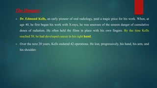
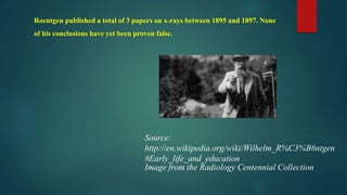
Oral Radiology
Dr.Rawand Samy Mohamed Abu Nahla
Oral Medicine,periodontology&oral Radiology
Department.
Dr. Haydar.A.Shafy Faculity Of Dentistery.
El Azhar University.
Scientific Content Of Subject Curricula:
1. Radiation history.
2. Radiation physics.
3. Dental x-ray image characteristics.
4. Image receptors.
5. Introduction to radiographic examination.
6. Exposure and technique errors.
7. Processing of x-ray films.
8. Normal anatomical landmarks.
9. Radiation protection.
10. Biological effects of radiation.
11. Extra-oral radiography.
12. Panoramic radiography.
13. Alternative and specialized techniques.
14. The periapical tissues.
15. Interpretation of dental caries.
16. Interpretation of periodontal diseases.
17. Radiological differential diagnosis describing a lesion.
18. Differential diagnosis of radiolucent lesions of the jaws.
19. Differential diagnosis of radiopaque lesions of the jaws.
20. Cysts of the jaws.
21. Benign tumors of the jaws.
22. Malignant tumors of the jaws.
23. Trauma of teeth and facial structures.
24. The temporomandibular joint.
25. Bone diseases of radiological importance.
26. Disorders of salivary glands and sialography.
Lecture 1:
The History Of Oral Radiology.
The History Of Oral
Radiology:
The Discovery
 On November 8th, 1895, Wilhelm Conrad Roentgen was working
in his laboratory in Wurzburg, Germany, using Crooke’s tubes.
 In the darkened laboratory, he noticed that a sheet of cardboard,
placed several feet away, was glowing in the shape of the letter A
that a student had painted in liquid Barium platinocyanide.
 He noticed that the effect was fluorescent, that is, the glow stopped
if the current was interrupted or the rays were blocked. Further, he
discovered that different materials attenuated the beam differently.
 Roentgen then isolated himself in his laboratory for the next
seven weeks. From his carefully crafted experiments, much of
what is known about the physics of x-radiation was derived. He
worked in secrecy, afraid that others might publish their results
before him. It has also been postulated that he worked alone
because he did not wish to be ridiculed if he were wrong.
In those seven weeks, Roentgen discovered that:
• Although the new rays were invisible, they caused fluorescence in certain
substances
• They darkened photographic plates
• They were propagated in straight lines
• Their behavior differed fundamentally from that of cathode rays
• They were neither reflected nor refracted by (then known) experimental methods
• They were not deviated by the influence of electro-magnetic fields
• Air traversed by the rays was made electrically conductive.
• He investigated the startling penetration of the rays through many materials and
observed the "hardening" of x-ray beams after penetrating several absorbers.
• He noted that different types of scattered and of secondary radiations were
produced.
Source: http://compepid.tuskegee.edu/syllabi/clinical/small/radiology/chapter2
On December 28th, 1895,Roentgen presented his findings to the Wurzburg Physical-Medical Society.
He presented several radiographs, including the one of his wife’s hand. By January 1896, words of his
discovery had spread throughout the world.
Enter Dentistry
In January 1896, a dentist in Braunschweig, Germany made the first dental
radiograph. Using a glass photographic plate wrapped in black rubber dam
and with an exposure of 25 minutes.
Dr. Otto Walkoff produced this intraoral radiograph
This radiograph was made on February 1st,
1896 by Dr. Walter Konig of Germany (Fig.1).
Dr Kells used impression compound to
stabilized the film during exposure (Fig.2).
Fig.2
Fig.1
It is thought that the first dental radiograph made in the United States was by Dr. Edmund Kells of
New Orleans, Louisiana.
Roentgen’s Apparatus
 Roentgen originally used a variation on the Hittorf-Crooke’s tube, developed by Philipp Lenard.
 Lenard placed a small window of thin aluminum foil over an opening in the tube.
 This allowed the cathode rays to escape the tube more readily, as the current was applied.
 The surprise was that the fluorescent effect occurred while using the heavier-walled Hittorf-Crooke’s
tube.
Cathode
ray tube
Ruhmkorff
Coil
Roentgen’s Apparatus
This is a German Ruhmkorff mini coil from the
beginning of the 20th Century. Image from the
Cathode Ray Tube Site
Fig.3
Fig.4
Hittorf-crookes' Tubes
Image From The Cathode Ray Tube Site
Fig.5
Roentgen’s Apparatus:
Cathode Ray:
Cathode rays are streams of electrons observed in vacuum tubes, i.e. evacuated glass tubes
that are equipped with at least two electrodes, a cathode (negative electrode) and an anode
(positive electrode) in a configuration known as a diode When the cathode is heated, it emits
some radiation which travels to the anode. If the inner glass walls behind the anode are
coated with a phosphorescent material, they glow. A metal shape placed between the
electrodes casts a shadow on the glowing coating. This suggested that the cause of the light
emission was comprised of rays emitted by the cathode and hitting the coating. They travel
towards the anode in straight lines, and continue past it for some distance.
Cathode Ray Tubes:
 The Cross Vacuum Scale demonstrates the phenomenon of discharge
at different pressures(vacuum) inside the tubes. The pressures varies
between 40 Torr (mmHg) lowest vacuum (left tube) to 0.03 Torr, the
highest vacuum (right tube.)
 In this high vacuum, used in many Crookes tubes, X-Rays are
produced.
 The glass emits here a green glow.
 On February 22nd, 1890 in Philadelphia, PA, A.W. Goodspeed and William
Jennings produced this radiogram by placing the coins on top of a glass photographic
plate.
 The coins and plate were exposed to radiation via a Hittorf – Crooke’s tube.
 They had realized the significance of their findings, they would have beaten Roentgen
to the discovery of X-rays by more than 5 years. Additionally, early radiographs might
have be called “good speedograms.” This proves that sometimes things are better off
as they are!
Contemporaries
 Lenard also experimented, with Hittorf – Crooke’s
tubes and, undoubtedly, produced X-rays, but did immediately grasp the
importance of the finding.
 He resented Roentgen throughout his life.
 In 1933 he joined the National Socialist Party(Nazis.)
 He rose to the rank of Chief of Aryan Physics.
Philipp
vonLenard
1862 – 1947
Heinrich
Rudolf Hertz
1857-1894
From 1885 to 1889 Hertz became the first person
to broadcast and receive radio waves, and to
establish the fact that light was a form of
electromagnetic radiation.
Experimented with cathode rays. (Gugliemo
Marconi didn't begin his own wireless experiments
until 1894, based on the earlier work of Hertz,
Maxwell, and others.) Hertz probably would have
gone on to make many more scientific contributions,
but he died quite young, less than a month before
his 37th birthday.
Timeline Leading to Roentgen’s Discovery:
300 Years of Discoveries Leading to Roentgen’s Work
• 1600 Gilbert's (1540-1603) De Magnete created the foundation for the sciences of magnetism and
electricity.
• 1675 Newton (1643-1727) built a more efficient electrostatic generator with a rotating glass sphere.
• 1729 Gray (1696-1736) distinguished conductors of electricity from nonconductors.
• 1750 Franklin (1706-1790) defined positive and negative electricity.
• 1785 Morgan (?-1785) in vacuum experiments, possibly produced x-rays.
• 1800 Volta (1745-1827) constructed the first electrical battery, the Voltaic pile.
• 1820 Oersted (1777-1851) discovered the link between electricity and magnetism.
• 1827 Ohm (1787-1854) formulated Ohm's law, stating the relation between electric current,
electromotive force, and resistance.
• 1821 Faraday (1791-1867) and Henry (1797-1878) discovered electromagnetic induction.
• 1850 Plucker (1801-1868) observed green-glass fluorescence opposite the negative electrode in a
vacuum tube.
• 1860 Toepler, Holtz, and Wimshurst improved electrostatic machines.
• 1873 Maxwell (1831-1879) published his famous equations in the book, "Treatise on Electricity
and Magnetism ."
• 1879 Crookes (1832-1919) found that cathode rays can be deflected by a magnet, and believed
that he was dealing with "a fourth state of matter."
• 1892 Lenard (1862-1947) built improved cathode-ray tubes and made
important observations on the properties of cathode rays.
• 1893 J. J. Thomson (1856-1940) published a supplementary volume to
Maxwell's treatise, describing in detail the passage of electricity through gases.
Source: http://compepid.tuskegee.edu/syllabi/clinical/small/radiology/chapter2.html
Niels Bohr (1885-1962) William Morton (1845-1920)
William Morton (1845-1920)
• New York dentist
• Also credited (along with Kells) with the first dental radiograph exposed in the United
States
• Wrote a book on X-rays in 1896
William H. Rollins (1852-1929)
• Boston Dentist
• Publish more than 200 articles on radiation effects and safety between 1896 and1904
• "Notes on X-Light", published in 1904 called for radiation protection for radiation
workers and patients
The Rollins Collimator
Image from the Radiology Centennial Collection
This collimator is the forerunner of our rectangular collimators!
William H. Rollins
• Alas, Rollins was ignored by his colleagues, much to their detriment. It was not until atomic bombs were exploded
over Hiroshima and Nagasaki that dentistry took
a serious look at radiation safety.
Dr. Frank Van Woert (1856-1927)
Dr. Van Woert was another pioneer of oral and maxillofacial radiology. He made a
practical demonstration of dental radiography before the NewYork Odontological
Society in1897. He was one of the first to use Kodak film (instead of glass plates).
These were wrapped in rubber dam and held in place with compound. He later
invented a metal film holder, an improved bisecting angulator, an automatic
timing switch, and the daylight processing tank.
Dr. Frank Van Woert
(1856-1927)
Howard Riley Raper (1886-1978)
Dr. Raper had one of the first oral and maxillofacial radiology offices (as opposed to a laboratory).
He was the inventor of the bitewing radiograph in 1924, the first to introduce oral and
maxillofacial radiology into the dental curriculum (in 1910 at the Indiana Dental School), and the
first to write a textbook on the subject (Elementary and Dental Radiology, 1913).
Howard Riley Raper
(1886-1978)
Early dental x-ray unit
• The G.E. CDX was the first modern type dental x-ray unit on the market in 1921.
Others soon followed.
Early dental x-ray unit
• Picker dental unit from the 1930’s
The Dangers
Although many had noted difficulties associated with "X-ray burns," it was not until
the death of Clarence Dally (1865- 1904), Edison's longtime assistant in X-ray
manufacture and testing, that observers finally agreed that the magic rays could kill as
well as cure.
Image from the Radiology Centennial Collection
The Dangers
 Dr. Edmund Kells, an early pioneer of oral radiology, paid a tragic price for his work. When, at
age 40, he first began his work with X-rays, he was unaware of the unseen danger of cumulative
doses of radiation. He often held the films in place with his own fingers. By the time Kells
reached 50, he had developed cancer in his right hand.
 Over the next 20 years, Kells endured 42 operations. He lost, progressively, his hand, his arm, and
his shoulder.
 In spite of agonizing pain, 42 operations, and numerous skin grafts, his 20-year battle against
the ill effects of the exposure from dental X-rays was about to come to an end.
 Throughout the trauma, Kells still served the profession. He lectured on preventive dentistry
and the conservation of teeth. He wrote books, and he contributed over 150 articles to
dental journals. At last, however, Kells suffering became intolerable. On May 7, 1928, at age
72, he committed suicide.
Source:http://www.ac.cc.md.us/dental/98-1newsletter/EdmundKells.html
http://clarionherald.org/20000427/
 Röntgen had family in the United States (in Iowa) and at one time he planned to emigrate. Although
he accepted an appointment at Columbia University in New York City and had actually purchased
transatlantic tickets, the outbreak of World War I changed his plans and he remained in Munich for the
rest of his career.
 Röntgen died in 1923 of carcinoma of the bowel. It is not believed his carcinoma was a result of his
work with ionizing radiation because his investigations were only for a short time and he was one of the
few pioneers in the field who used protective lead shields routinely.
Source: http://en.wikipedia.org/wiki/Wilhelm_R%C3%B6ntgen#Early_life_and_education
Roentgen published a total of 3 papers on x-rays between 1895 and 1897. None
of his conclusions have yet been proven false.
Source:
http://en.wikipedia.org/wiki/Wilhelm_R%C3%B6ntgen
#Early_life_and_education
Image from the Radiology Centennial Collection
Thankyou

Oral radiology lecture 1

  • 1.
    Oral Radiology Dr.Rawand SamyMohamed Abu Nahla Oral Medicine,periodontology&oral Radiology Department. Dr. Haydar.A.Shafy Faculity Of Dentistery. El Azhar University.
  • 2.
    Scientific Content OfSubject Curricula: 1. Radiation history. 2. Radiation physics. 3. Dental x-ray image characteristics. 4. Image receptors. 5. Introduction to radiographic examination. 6. Exposure and technique errors. 7. Processing of x-ray films. 8. Normal anatomical landmarks. 9. Radiation protection. 10. Biological effects of radiation.
  • 3.
    11. Extra-oral radiography. 12.Panoramic radiography. 13. Alternative and specialized techniques. 14. The periapical tissues. 15. Interpretation of dental caries. 16. Interpretation of periodontal diseases. 17. Radiological differential diagnosis describing a lesion. 18. Differential diagnosis of radiolucent lesions of the jaws. 19. Differential diagnosis of radiopaque lesions of the jaws. 20. Cysts of the jaws. 21. Benign tumors of the jaws. 22. Malignant tumors of the jaws. 23. Trauma of teeth and facial structures. 24. The temporomandibular joint. 25. Bone diseases of radiological importance. 26. Disorders of salivary glands and sialography.
  • 4.
    Lecture 1: The HistoryOf Oral Radiology.
  • 5.
    The History OfOral Radiology:
  • 6.
    The Discovery  OnNovember 8th, 1895, Wilhelm Conrad Roentgen was working in his laboratory in Wurzburg, Germany, using Crooke’s tubes.  In the darkened laboratory, he noticed that a sheet of cardboard, placed several feet away, was glowing in the shape of the letter A that a student had painted in liquid Barium platinocyanide.  He noticed that the effect was fluorescent, that is, the glow stopped if the current was interrupted or the rays were blocked. Further, he discovered that different materials attenuated the beam differently.
  • 7.
     Roentgen thenisolated himself in his laboratory for the next seven weeks. From his carefully crafted experiments, much of what is known about the physics of x-radiation was derived. He worked in secrecy, afraid that others might publish their results before him. It has also been postulated that he worked alone because he did not wish to be ridiculed if he were wrong.
  • 8.
    In those sevenweeks, Roentgen discovered that: • Although the new rays were invisible, they caused fluorescence in certain substances • They darkened photographic plates • They were propagated in straight lines • Their behavior differed fundamentally from that of cathode rays • They were neither reflected nor refracted by (then known) experimental methods • They were not deviated by the influence of electro-magnetic fields
  • 9.
    • Air traversedby the rays was made electrically conductive. • He investigated the startling penetration of the rays through many materials and observed the "hardening" of x-ray beams after penetrating several absorbers. • He noted that different types of scattered and of secondary radiations were produced. Source: http://compepid.tuskegee.edu/syllabi/clinical/small/radiology/chapter2
  • 10.
    On December 28th,1895,Roentgen presented his findings to the Wurzburg Physical-Medical Society. He presented several radiographs, including the one of his wife’s hand. By January 1896, words of his discovery had spread throughout the world. Enter Dentistry In January 1896, a dentist in Braunschweig, Germany made the first dental radiograph. Using a glass photographic plate wrapped in black rubber dam and with an exposure of 25 minutes. Dr. Otto Walkoff produced this intraoral radiograph
  • 11.
    This radiograph wasmade on February 1st, 1896 by Dr. Walter Konig of Germany (Fig.1). Dr Kells used impression compound to stabilized the film during exposure (Fig.2). Fig.2 Fig.1
  • 12.
    It is thoughtthat the first dental radiograph made in the United States was by Dr. Edmund Kells of New Orleans, Louisiana. Roentgen’s Apparatus  Roentgen originally used a variation on the Hittorf-Crooke’s tube, developed by Philipp Lenard.  Lenard placed a small window of thin aluminum foil over an opening in the tube.  This allowed the cathode rays to escape the tube more readily, as the current was applied.  The surprise was that the fluorescent effect occurred while using the heavier-walled Hittorf-Crooke’s tube.
  • 13.
    Cathode ray tube Ruhmkorff Coil Roentgen’s Apparatus Thisis a German Ruhmkorff mini coil from the beginning of the 20th Century. Image from the Cathode Ray Tube Site Fig.3 Fig.4
  • 14.
    Hittorf-crookes' Tubes Image FromThe Cathode Ray Tube Site Fig.5
  • 15.
    Roentgen’s Apparatus: Cathode Ray: Cathoderays are streams of electrons observed in vacuum tubes, i.e. evacuated glass tubes that are equipped with at least two electrodes, a cathode (negative electrode) and an anode (positive electrode) in a configuration known as a diode When the cathode is heated, it emits some radiation which travels to the anode. If the inner glass walls behind the anode are coated with a phosphorescent material, they glow. A metal shape placed between the electrodes casts a shadow on the glowing coating. This suggested that the cause of the light emission was comprised of rays emitted by the cathode and hitting the coating. They travel towards the anode in straight lines, and continue past it for some distance.
  • 16.
    Cathode Ray Tubes: The Cross Vacuum Scale demonstrates the phenomenon of discharge at different pressures(vacuum) inside the tubes. The pressures varies between 40 Torr (mmHg) lowest vacuum (left tube) to 0.03 Torr, the highest vacuum (right tube.)  In this high vacuum, used in many Crookes tubes, X-Rays are produced.  The glass emits here a green glow.
  • 17.
     On February22nd, 1890 in Philadelphia, PA, A.W. Goodspeed and William Jennings produced this radiogram by placing the coins on top of a glass photographic plate.  The coins and plate were exposed to radiation via a Hittorf – Crooke’s tube.  They had realized the significance of their findings, they would have beaten Roentgen to the discovery of X-rays by more than 5 years. Additionally, early radiographs might have be called “good speedograms.” This proves that sometimes things are better off as they are!
  • 18.
    Contemporaries  Lenard alsoexperimented, with Hittorf – Crooke’s tubes and, undoubtedly, produced X-rays, but did immediately grasp the importance of the finding.  He resented Roentgen throughout his life.  In 1933 he joined the National Socialist Party(Nazis.)  He rose to the rank of Chief of Aryan Physics. Philipp vonLenard 1862 – 1947
  • 19.
    Heinrich Rudolf Hertz 1857-1894 From 1885to 1889 Hertz became the first person to broadcast and receive radio waves, and to establish the fact that light was a form of electromagnetic radiation. Experimented with cathode rays. (Gugliemo Marconi didn't begin his own wireless experiments until 1894, based on the earlier work of Hertz, Maxwell, and others.) Hertz probably would have gone on to make many more scientific contributions, but he died quite young, less than a month before his 37th birthday.
  • 20.
    Timeline Leading toRoentgen’s Discovery: 300 Years of Discoveries Leading to Roentgen’s Work • 1600 Gilbert's (1540-1603) De Magnete created the foundation for the sciences of magnetism and electricity. • 1675 Newton (1643-1727) built a more efficient electrostatic generator with a rotating glass sphere. • 1729 Gray (1696-1736) distinguished conductors of electricity from nonconductors. • 1750 Franklin (1706-1790) defined positive and negative electricity. • 1785 Morgan (?-1785) in vacuum experiments, possibly produced x-rays. • 1800 Volta (1745-1827) constructed the first electrical battery, the Voltaic pile. • 1820 Oersted (1777-1851) discovered the link between electricity and magnetism. • 1827 Ohm (1787-1854) formulated Ohm's law, stating the relation between electric current, electromotive force, and resistance.
  • 21.
    • 1821 Faraday(1791-1867) and Henry (1797-1878) discovered electromagnetic induction. • 1850 Plucker (1801-1868) observed green-glass fluorescence opposite the negative electrode in a vacuum tube. • 1860 Toepler, Holtz, and Wimshurst improved electrostatic machines. • 1873 Maxwell (1831-1879) published his famous equations in the book, "Treatise on Electricity and Magnetism ." • 1879 Crookes (1832-1919) found that cathode rays can be deflected by a magnet, and believed that he was dealing with "a fourth state of matter."
  • 22.
    • 1892 Lenard(1862-1947) built improved cathode-ray tubes and made important observations on the properties of cathode rays. • 1893 J. J. Thomson (1856-1940) published a supplementary volume to Maxwell's treatise, describing in detail the passage of electricity through gases. Source: http://compepid.tuskegee.edu/syllabi/clinical/small/radiology/chapter2.html
  • 23.
    Niels Bohr (1885-1962)William Morton (1845-1920)
  • 24.
    William Morton (1845-1920) •New York dentist • Also credited (along with Kells) with the first dental radiograph exposed in the United States • Wrote a book on X-rays in 1896 William H. Rollins (1852-1929) • Boston Dentist • Publish more than 200 articles on radiation effects and safety between 1896 and1904 • "Notes on X-Light", published in 1904 called for radiation protection for radiation workers and patients
  • 25.
    The Rollins Collimator Imagefrom the Radiology Centennial Collection This collimator is the forerunner of our rectangular collimators! William H. Rollins • Alas, Rollins was ignored by his colleagues, much to their detriment. It was not until atomic bombs were exploded over Hiroshima and Nagasaki that dentistry took a serious look at radiation safety.
  • 26.
    Dr. Frank VanWoert (1856-1927) Dr. Van Woert was another pioneer of oral and maxillofacial radiology. He made a practical demonstration of dental radiography before the NewYork Odontological Society in1897. He was one of the first to use Kodak film (instead of glass plates). These were wrapped in rubber dam and held in place with compound. He later invented a metal film holder, an improved bisecting angulator, an automatic timing switch, and the daylight processing tank. Dr. Frank Van Woert (1856-1927)
  • 27.
    Howard Riley Raper(1886-1978) Dr. Raper had one of the first oral and maxillofacial radiology offices (as opposed to a laboratory). He was the inventor of the bitewing radiograph in 1924, the first to introduce oral and maxillofacial radiology into the dental curriculum (in 1910 at the Indiana Dental School), and the first to write a textbook on the subject (Elementary and Dental Radiology, 1913). Howard Riley Raper (1886-1978)
  • 28.
    Early dental x-rayunit • The G.E. CDX was the first modern type dental x-ray unit on the market in 1921. Others soon followed. Early dental x-ray unit • Picker dental unit from the 1930’s
  • 29.
    The Dangers Although manyhad noted difficulties associated with "X-ray burns," it was not until the death of Clarence Dally (1865- 1904), Edison's longtime assistant in X-ray manufacture and testing, that observers finally agreed that the magic rays could kill as well as cure. Image from the Radiology Centennial Collection
  • 30.
    The Dangers  Dr.Edmund Kells, an early pioneer of oral radiology, paid a tragic price for his work. When, at age 40, he first began his work with X-rays, he was unaware of the unseen danger of cumulative doses of radiation. He often held the films in place with his own fingers. By the time Kells reached 50, he had developed cancer in his right hand.  Over the next 20 years, Kells endured 42 operations. He lost, progressively, his hand, his arm, and his shoulder.
  • 31.
     In spiteof agonizing pain, 42 operations, and numerous skin grafts, his 20-year battle against the ill effects of the exposure from dental X-rays was about to come to an end.  Throughout the trauma, Kells still served the profession. He lectured on preventive dentistry and the conservation of teeth. He wrote books, and he contributed over 150 articles to dental journals. At last, however, Kells suffering became intolerable. On May 7, 1928, at age 72, he committed suicide. Source:http://www.ac.cc.md.us/dental/98-1newsletter/EdmundKells.html http://clarionherald.org/20000427/
  • 32.
     Röntgen hadfamily in the United States (in Iowa) and at one time he planned to emigrate. Although he accepted an appointment at Columbia University in New York City and had actually purchased transatlantic tickets, the outbreak of World War I changed his plans and he remained in Munich for the rest of his career.  Röntgen died in 1923 of carcinoma of the bowel. It is not believed his carcinoma was a result of his work with ionizing radiation because his investigations were only for a short time and he was one of the few pioneers in the field who used protective lead shields routinely. Source: http://en.wikipedia.org/wiki/Wilhelm_R%C3%B6ntgen#Early_life_and_education
  • 33.
    Roentgen published atotal of 3 papers on x-rays between 1895 and 1897. None of his conclusions have yet been proven false. Source: http://en.wikipedia.org/wiki/Wilhelm_R%C3%B6ntgen #Early_life_and_education Image from the Radiology Centennial Collection
  • 34.