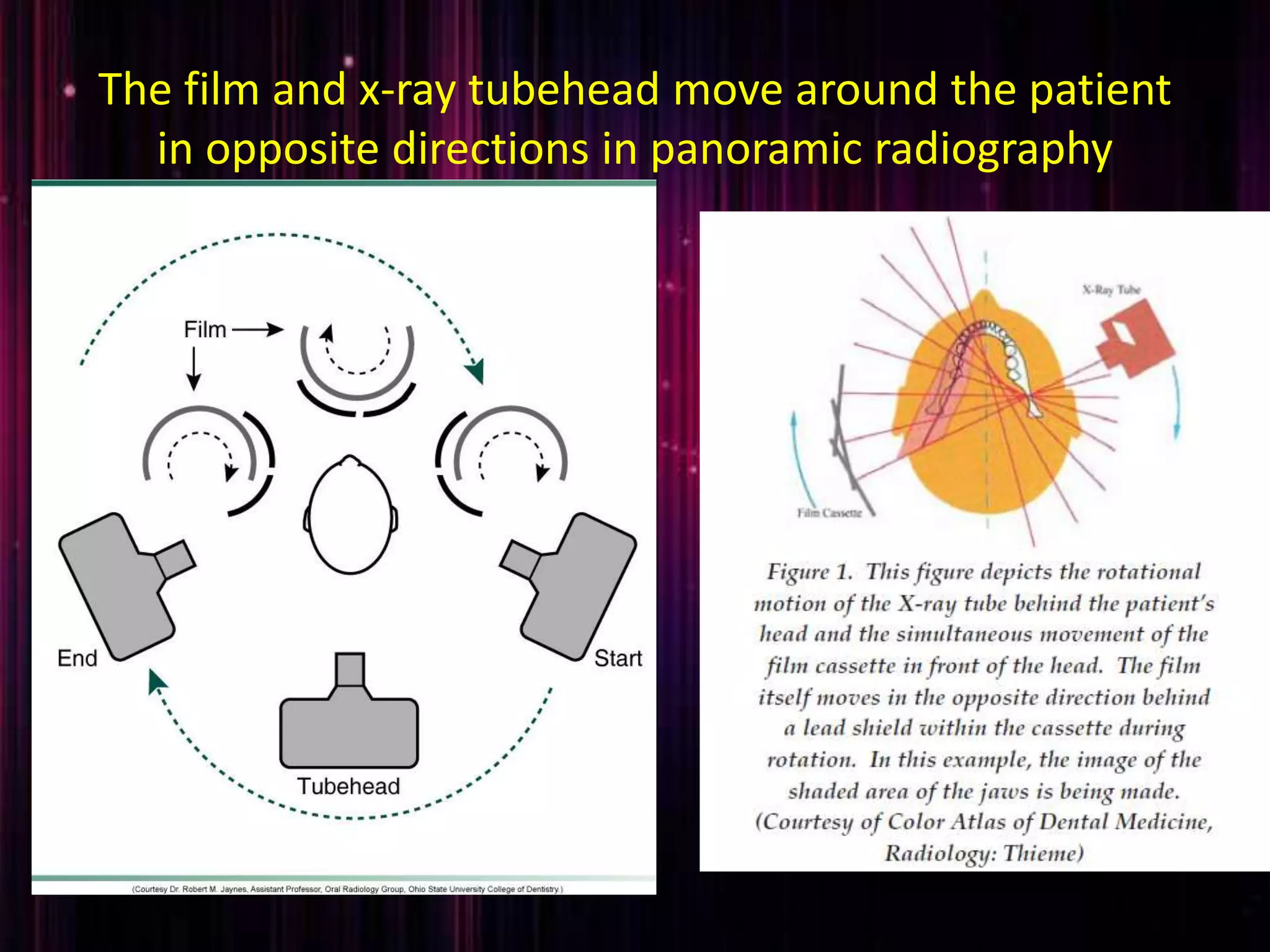
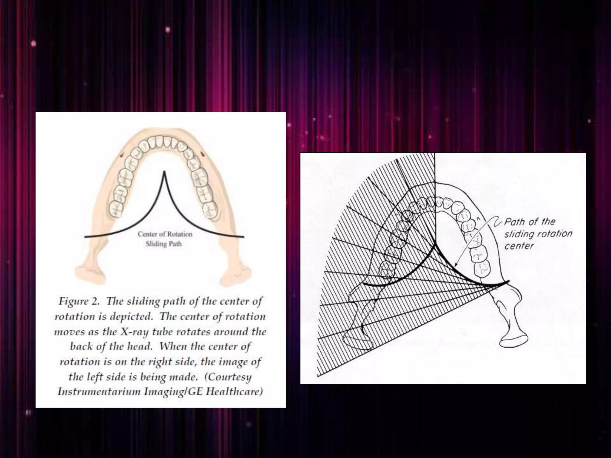
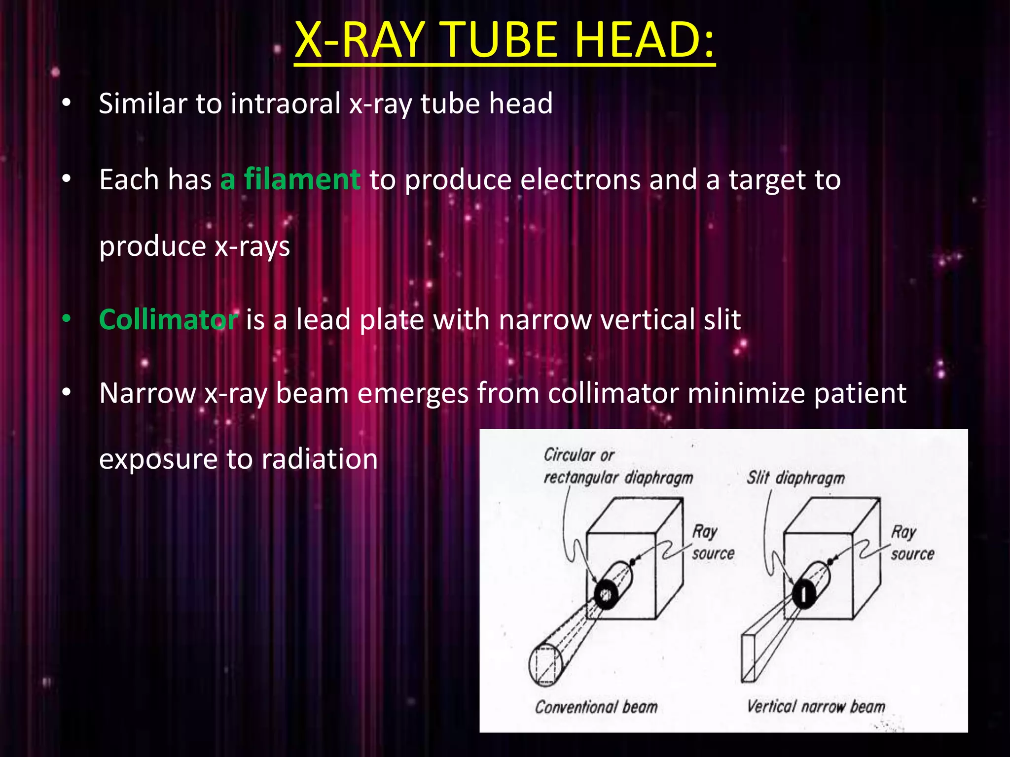
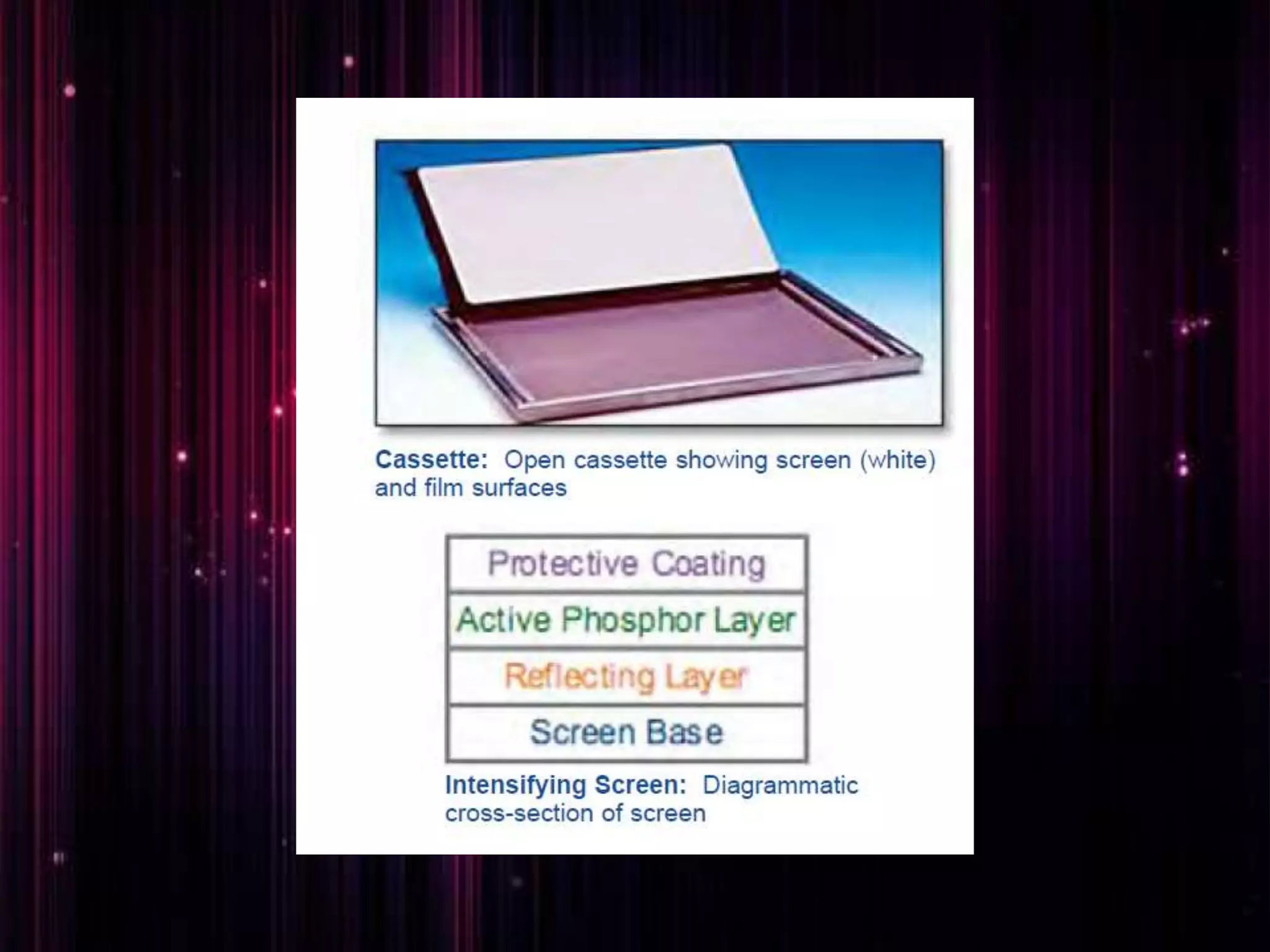
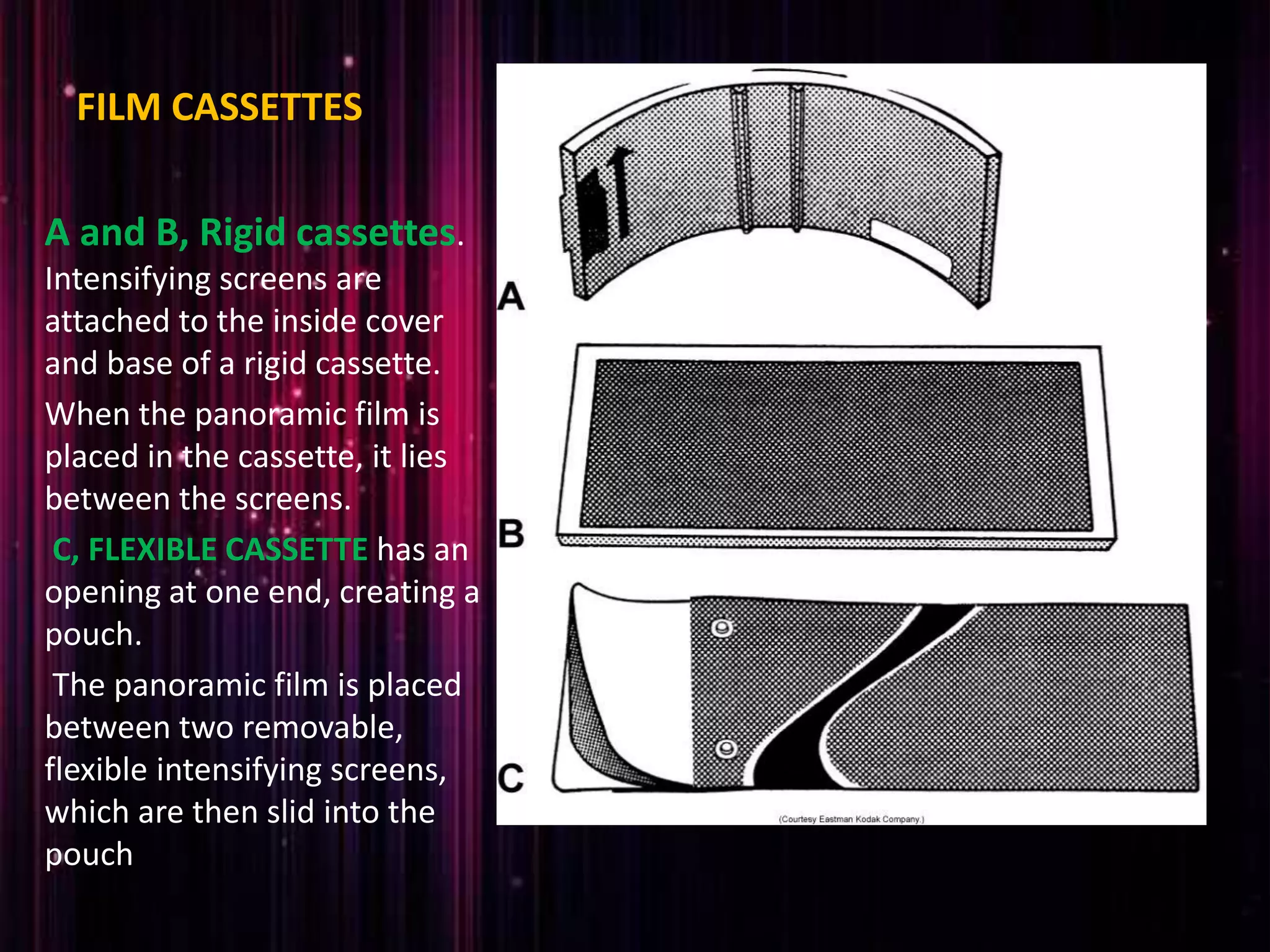
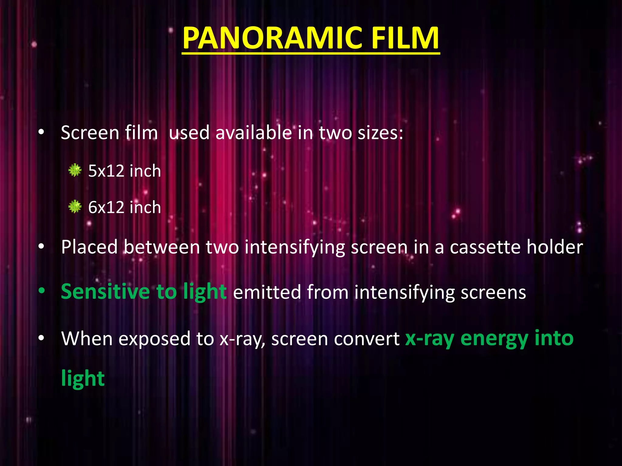
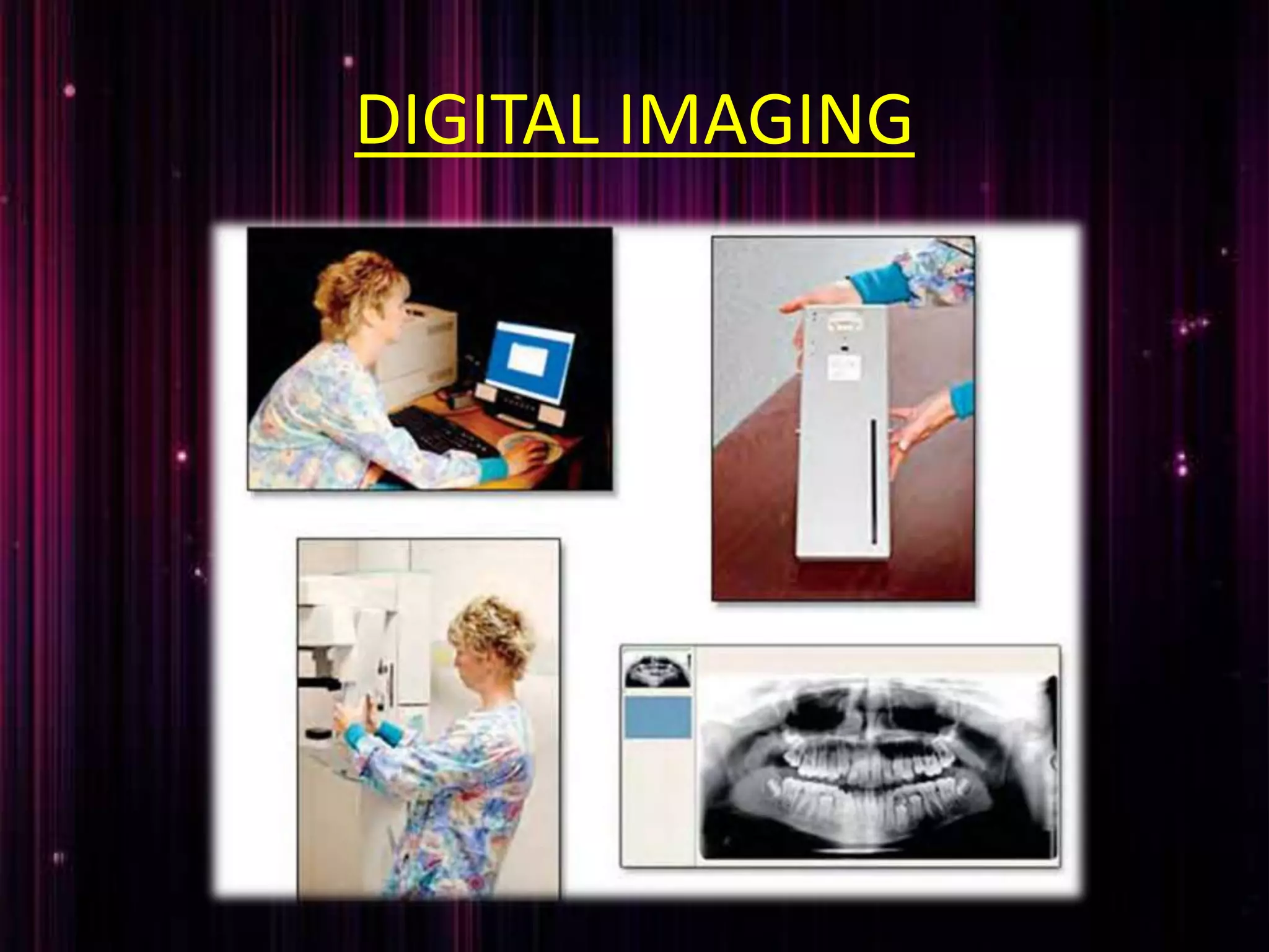
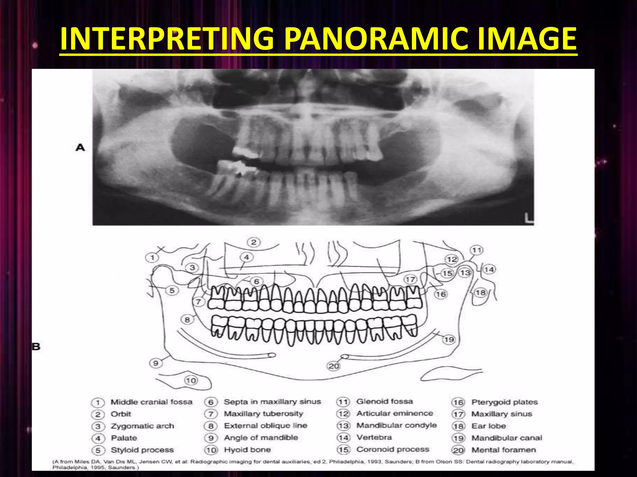
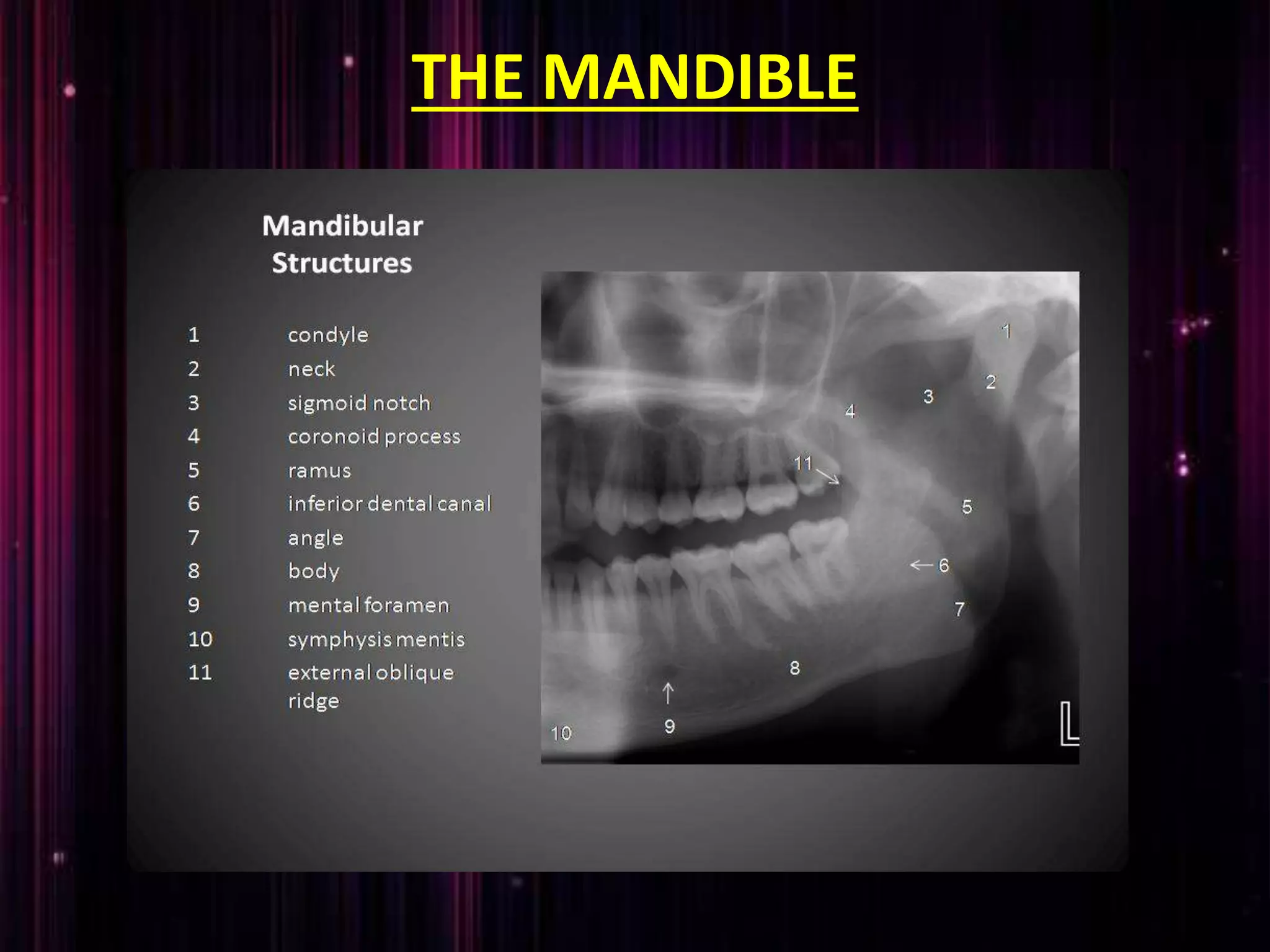
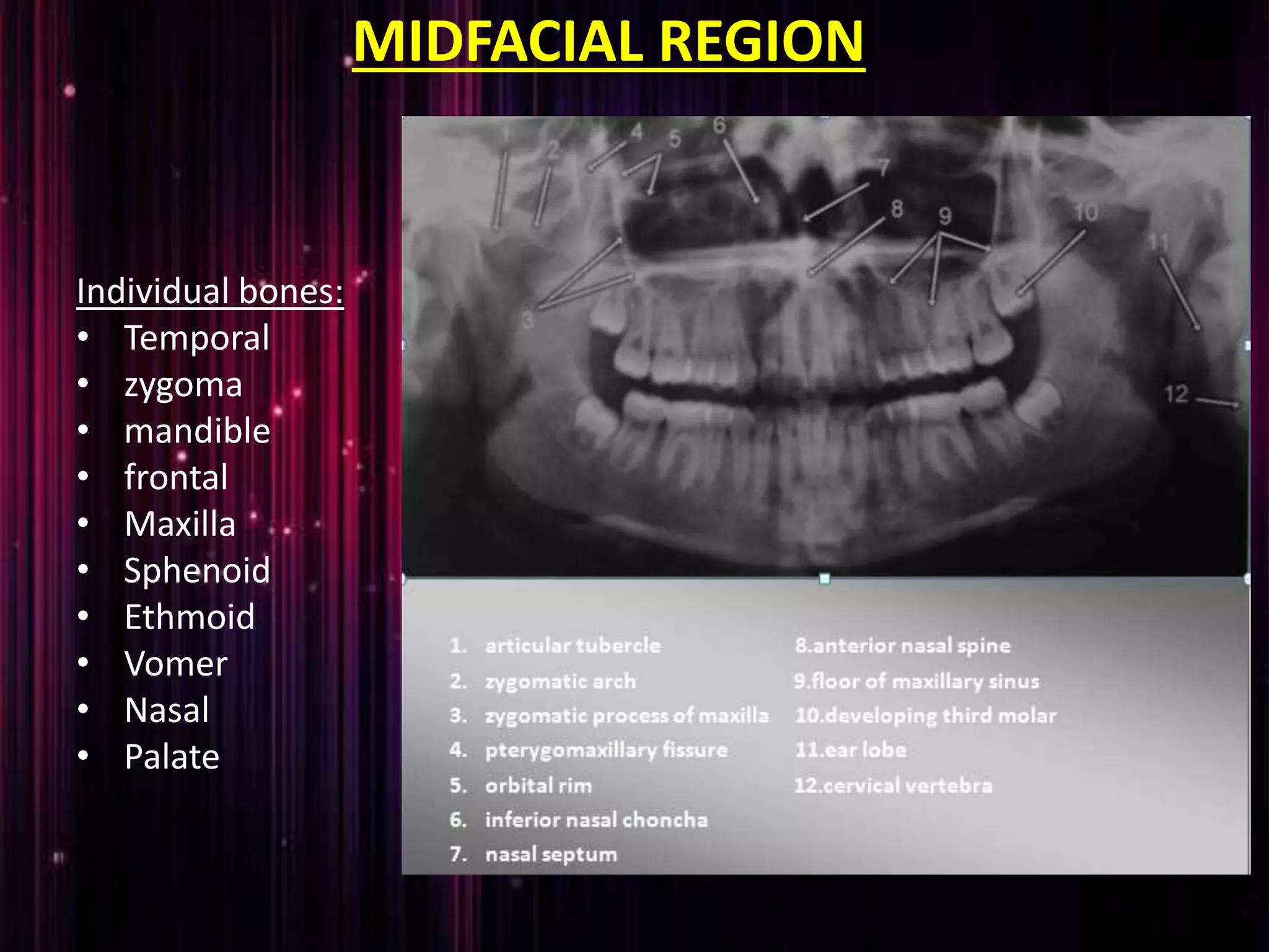
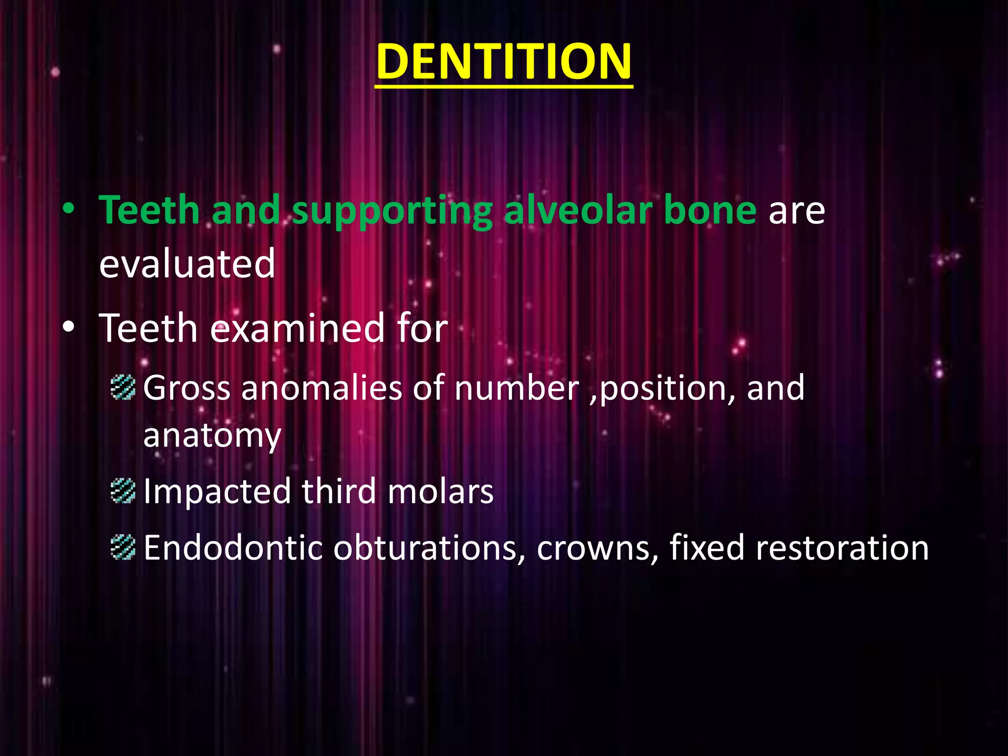
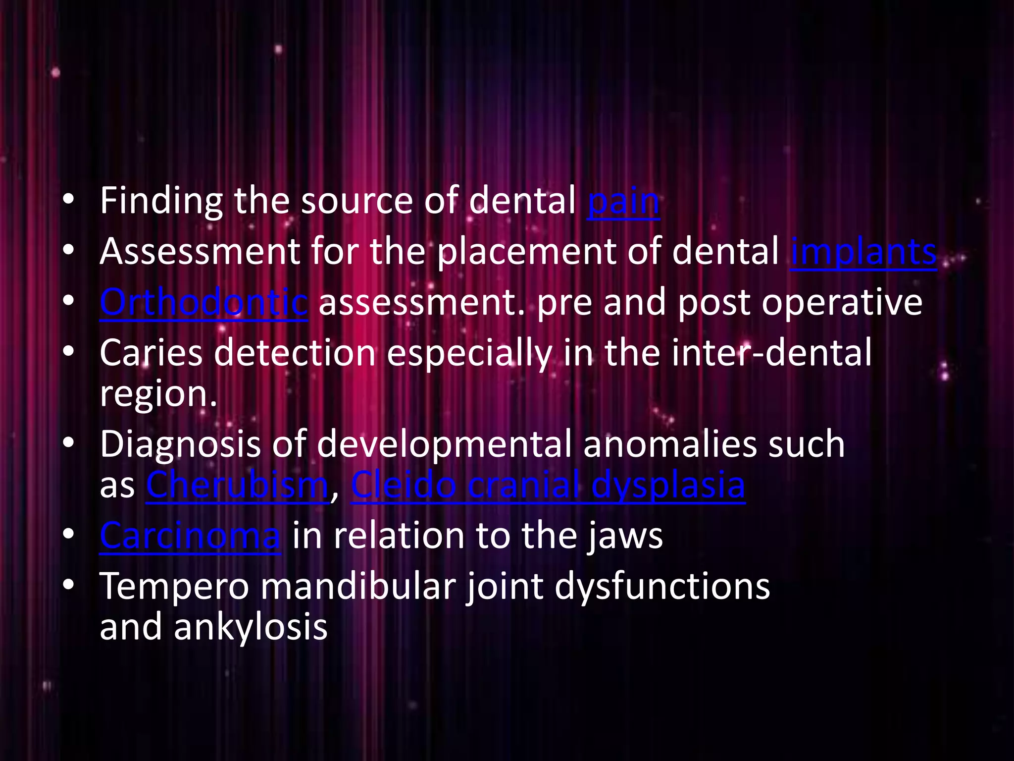
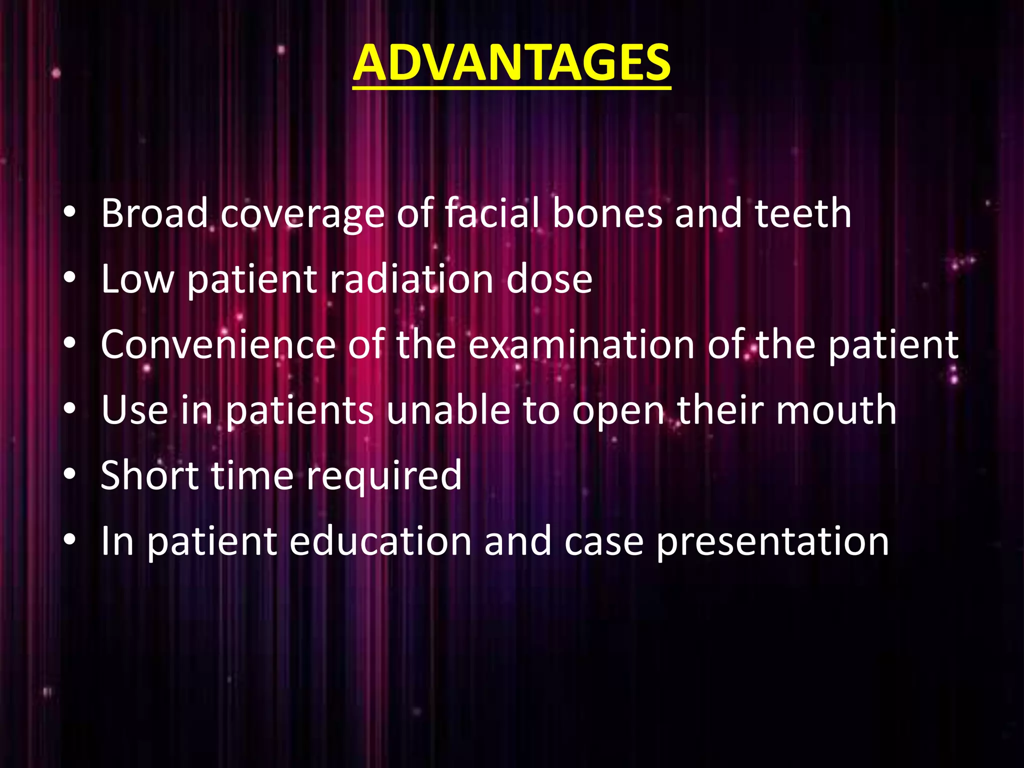
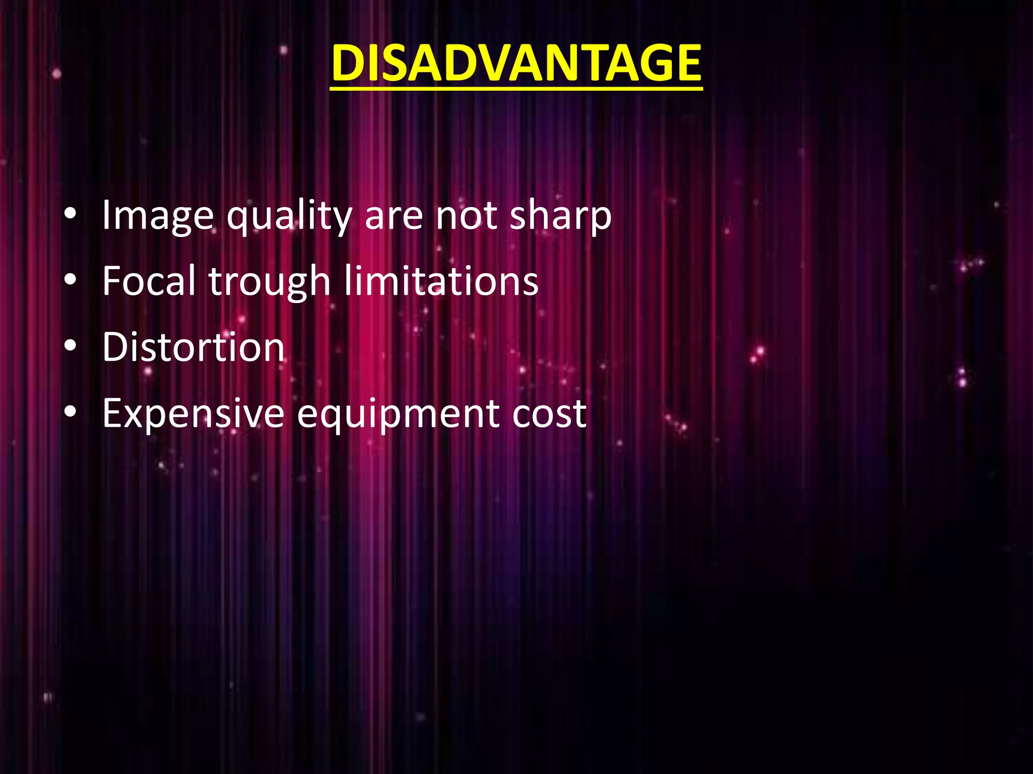
SlideShare a Scribd company logo
ORTHOPANTOMOGRAM
Dr. SHIFAYA NASRIN
CRRI(2009-2014)
CONTENTS
 Introduction
 Principles of panoramic imaging
 Image layer
 Panoramic machines
 Panoramic film
 Patient positioning
 Interpreting the panoramic imaging
 Indication
 Advantages
 Disadvantages
 Conclusion
 References
INTRODUCTION
• Panoramic imaging also called
pantomography is a technique for producing a
single tomographic image of facial structures
that includes both the maxillary and
mandibular dental arches and their supporting
structures .
• This is a curvilinear variant of conventional
tomography.
PRINCIPLES OF PANORAMIC IMAGE
FORMATION
• Patero and Numata - describe the principles of panoramic
radiography
• based on the principle of reciprocal movement of x-ray
source and an image receptor around a central point or
plane called the image layer, in which the object of image is
located.
• Object in front or behind this image are not clearly captured
because of their movement relative to the centre of rotation
of the receptor and the x-ray source.
The film and x-ray tubehead move around the patient
in opposite directions in panoramic radiography
ROTATION CENTER
 The pivotal point or axis around which the cassette carrier
and tube head rotate is termed rotation center
 Three basic rotation center used in panoramic radiography
Double centre rotation
Triple centre rotation
moving centre rotation
 The location and number of rotational centers influence
size and shape of focal trough
IMAGE LAYER
• Also known as focal trough
• It is a three dimensional curved zone where the structures lying
within this layer are reasonably well defined on final panoramic
image.
• The structures seen on a panoramic image are primarily those
located within image layer.
• Objects outside the image layer are blurred magnified are reduced
in size. Even distorted to the extent of not being recognizable.
• This shape of image layer varies with the brand of equipment used.
FOCAL TROUGH
FACTORS AFFECTING SIZE OF IMAGE LAYER:
Arc path
Velocity of receptor and X-ray tube head
Alignment of x-ray beam
Collimator width
The location of image layer change with extensive machine
used so recalibration may be necessary if consistently
suboptimal images are produced.
As a position of object is moved within the image layer size
and shape of image layer change.
PANORAMIC UNIT
A, Orthophos XG Plus extraoral x-ray machine. B,
Orthoralix 8500 extraoral x-ray machine. C, Example of
a digital panoramic system
PARTS OF PANORAMIC UNITS
a. x-ray tube head
b. head positioner:
chin rest
notched bite block
forehead rest
lateral head support
c. exposure controls
X-RAY TUBE HEAD:
• Similar to intraoral x-ray tube head
• Each has a filament to produce electrons and a target to
produce x-rays
• Collimator is a lead plate with narrow vertical slit
• Narrow x-ray beam emerges from collimator minimize patient
exposure to radiation
• Tube head is fixed in position and rotates
behind the patient head
• Film positioner is used to align the patients
teeth accurately in focal trough
Image receptor
CASSETTE
• It is a device used to hold the extra oral film
and intensifying screens
• Light tight to protect the film from exposure
• Two types
Rigid
Flexible
FILM CASSETTES
A and B, Rigid cassettes.
Intensifying screens are
attached to the inside cover
and base of a rigid cassette.
When the panoramic film is
placed in the cassette, it lies
between the screens.
C, FLEXIBLE CASSETTE has an
opening at one end, creating a
pouch.
The panoramic film is placed
between two removable,
flexible intensifying screens,
which are then slid into the
pouch
PANORAMIC FILM
• Screen film used available in two sizes:
5x12 inch
6x12 inch
• Placed between two intensifying screen in a cassette holder
• Sensitive to light emitted from intensifying screens
• When exposed to x-ray, screen convert x-ray energy into
light
INTENSIFYING SCREENS
• Calcium tungstate –emit blue light
• Rare earth –emit green light, less x-ray
exposure
• Two types
Other receptor
• PSP - photostimulable phosphor
plate
• CCD - charge-coupled device
DIGITAL IMAGING
PATIENT POSITIONING AND HEAD
ALIGNMENT
• Dental appliance earrings ,necklace, hairpins, and any other metallic
objects should be removed
• Instruct the patient to stand as tall as possible with back straight
and stand erect .
• Vertical column must be straight
• Instruct the patient to bite on the plastic bite block tooth must be
positioned in edge to edge position in the groove present in the
bite block it is used to align the teeth in the focal trough
A double-sided lead apron is recommended for
use during exposure of a panoramic film
• Midsagittal plane
perpendicular to floor
• Frankfort horizontal plane
parallel to the floor
• Tongue must be positioned on
the roof of the mouth
• Instruct the patient to remain
still while machine is rotating
INTERPRETING PANORAMIC IMAGE
THE MANDIBLE
• Condylar process and TMJ: a bony rounded
radioopaque projection extending from ramus of
mandible
• Coronoid process: triangular radio opacity posterior
to tuberosity region
• Ramus: shadow of other structure may
superimposed over the ramus such as
• Pharyngeal airway shadow
• Posterior wall of pharynx
• Cervical vertebra
• Ear lobe
• Nasal cartilage
• Soft palate and uvula
• Dorsum of tongue
• Ghost shadow
• Body and angle : radiopaque bony structure
where the ramus join the body of the
mandible
• mandibular dentition and alveolus
MIDFACIAL REGION
Individual bones:
• Temporal
• zygoma
• mandible
• frontal
• Maxilla
• Sphenoid
• Ethmoid
• Vomer
• Nasal
• Palate
• Cortical boundary of maxilla including
posterior border and alveolar ridge
• Pterygomaxillary fissure : radiolucent area
between the lateral pterygoid plate and
maxilla
• Maxillary sinuses: paired radiolucencies
located above the apices of premolars and
molars
• Zygomatic complex or buttresses of midface:
includes lateral and inferior orbital rims
zygomatic process of maxilla zygomatic arch
• Nasal cavity and conchae: radiolucent area
above the maxillary incissors
• TMJ
• Maxillary dentition and alveolus
SOFT TISSUES
• Tongue under the hard palate: radiopaque area
superimposed over the maxillary posterior teeth
• Lip line: seen in the region of anterior teeth
• Soft palate: extending posteriorly from hard
palate
• Posterior wall of pharynx
• Nasal septum
• Ear lobes
• Nose and nasolabial fold
DENTITION
• Teeth and supporting alveolar bone are
evaluated
• Teeth examined for
Gross anomalies of number ,position, and
anatomy
Impacted third molars
Endodontic obturations, crowns, fixed restoration
INDICATION
• To evaluate impacted teeth
• To evaluate eruption patterns, growth and
development
• To detect diseases ,lesions and conditions of
the jaw
• To examine extent of large lesions
• To evaluate trauma periodontal bone loss and
periapical involvement.
• Finding the source of dental pain
• Assessment for the placement of dental implants
• Orthodontic assessment. pre and post operative
• Caries detection especially in the inter-dental
region.
• Diagnosis of developmental anomalies such
as Cherubism, Cleido cranial dysplasia
• Carcinoma in relation to the jaws
• Tempero mandibular joint dysfunctions
and ankylosis
ADVANTAGES
• Broad coverage of facial bones and teeth
• Low patient radiation dose
• Convenience of the examination of the patient
• Use in patients unable to open their mouth
• Short time required
• In patient education and case presentation
DISADVANTAGE
• Image quality are not sharp
• Focal trough limitations
• Distortion
• Expensive equipment cost
CONCLUSION
• As OPG has several advantages in the field of
dentistry and its inevitable role in diagnosis
every dentist should know about it.
• Compared with the conventional radiographic
technique involving atleast 16 intraoral
exposures OPG has several advantage it takes
fairly easy; takes one minute and shows entire
oral cavity in one minute however resulting
image produce less detail than IOPA
REFERENCES
• Freny R.Karjodkar :Text book of Dental and maxillofacial
radiology 2nd edition page number :236-255
• Laura Jansen ,Joen M.Ianucci Harring :Dental radiography
Principles and techniques of Oral radiology: 3rd edition page
number:305-319
• white and pharaoh : Oral radiology principles and
interpretation 6th edition; page number: 175-189

More Related Content

What's hot

Intra oral radiographs
Intra oral radiographsIntra oral radiographs
Intra oral radiographs
melbia shine
 
Radiographic techniques
Radiographic techniquesRadiographic techniques
Radiographic techniquesanusushanth
 
Extra-oral Radiology Techniques I
Extra-oral Radiology Techniques IExtra-oral Radiology Techniques I
Extra-oral Radiology Techniques I
IAU Dent
 
Cone beam computed tomography
Cone beam computed tomographyCone beam computed tomography
Cone beam computed tomography
Vashi Narula
 
Common errors in opg
Common errors in opgCommon errors in opg
Common errors in opg
Dr.Harkaran Pandher
 
Temporomandibular joint imaging
Temporomandibular joint imagingTemporomandibular joint imaging
Temporomandibular joint imaging
Dr Jinki Singha
 
Cbct
CbctCbct
Factors controlling the beam of x-ray
Factors controlling the beam of x-rayFactors controlling the beam of x-ray
Factors controlling the beam of x-ray
Anu Jose
 
Cbct
CbctCbct
TMJ Imaging
TMJ ImagingTMJ Imaging
Quality assurance in dental radiography
Quality assurance in dental radiographyQuality assurance in dental radiography
Quality assurance in dental radiography
Mammootty Ik
 
Digital imaging IN DENTISTRY
Digital imaging IN DENTISTRYDigital imaging IN DENTISTRY
Digital imaging IN DENTISTRY
Revath Vyas Devulapalli
 
Orthopantomogram
OrthopantomogramOrthopantomogram
Orthopantomogram
Dr.shifaya nasrin
 
Radiology in dentistry
Radiology in dentistry Radiology in dentistry
Radiology in dentistry
Masuma Ryzvee
 
Waters projection
Waters projectionWaters projection
Waters projection
Amirhoseyn Sarayani
 
radio-graphic-techniques-bisecting-and-occlusal
 radio-graphic-techniques-bisecting-and-occlusal radio-graphic-techniques-bisecting-and-occlusal
radio-graphic-techniques-bisecting-and-occlusalParth Thakkar
 
RADIO VISIO GRAPHY, Dr Anirudh Singh Chauhan
RADIO VISIO GRAPHY, Dr Anirudh Singh ChauhanRADIO VISIO GRAPHY, Dr Anirudh Singh Chauhan
RADIO VISIO GRAPHY, Dr Anirudh Singh Chauhan
Anirudh Singh Chauhan
 
SIALOGRAPHY.pptx
SIALOGRAPHY.pptxSIALOGRAPHY.pptx
SIALOGRAPHY.pptx
Pooja461465
 
Faulty radiographs
Faulty     radiographsFaulty     radiographs
Faulty radiographs
melbia shine
 
Bisecting angle vs paralleling technique /orthodontic courses by Indian denta...
Bisecting angle vs paralleling technique /orthodontic courses by Indian denta...Bisecting angle vs paralleling technique /orthodontic courses by Indian denta...
Bisecting angle vs paralleling technique /orthodontic courses by Indian denta...
Indian dental academy
 

What's hot (20)

Intra oral radiographs
Intra oral radiographsIntra oral radiographs
Intra oral radiographs
 
Radiographic techniques
Radiographic techniquesRadiographic techniques
Radiographic techniques
 
Extra-oral Radiology Techniques I
Extra-oral Radiology Techniques IExtra-oral Radiology Techniques I
Extra-oral Radiology Techniques I
 
Cone beam computed tomography
Cone beam computed tomographyCone beam computed tomography
Cone beam computed tomography
 
Common errors in opg
Common errors in opgCommon errors in opg
Common errors in opg
 
Temporomandibular joint imaging
Temporomandibular joint imagingTemporomandibular joint imaging
Temporomandibular joint imaging
 
Cbct
CbctCbct
Cbct
 
Factors controlling the beam of x-ray
Factors controlling the beam of x-rayFactors controlling the beam of x-ray
Factors controlling the beam of x-ray
 
Cbct
CbctCbct
Cbct
 
TMJ Imaging
TMJ ImagingTMJ Imaging
TMJ Imaging
 
Quality assurance in dental radiography
Quality assurance in dental radiographyQuality assurance in dental radiography
Quality assurance in dental radiography
 
Digital imaging IN DENTISTRY
Digital imaging IN DENTISTRYDigital imaging IN DENTISTRY
Digital imaging IN DENTISTRY
 
Orthopantomogram
OrthopantomogramOrthopantomogram
Orthopantomogram
 
Radiology in dentistry
Radiology in dentistry Radiology in dentistry
Radiology in dentistry
 
Waters projection
Waters projectionWaters projection
Waters projection
 
radio-graphic-techniques-bisecting-and-occlusal
 radio-graphic-techniques-bisecting-and-occlusal radio-graphic-techniques-bisecting-and-occlusal
radio-graphic-techniques-bisecting-and-occlusal
 
RADIO VISIO GRAPHY, Dr Anirudh Singh Chauhan
RADIO VISIO GRAPHY, Dr Anirudh Singh ChauhanRADIO VISIO GRAPHY, Dr Anirudh Singh Chauhan
RADIO VISIO GRAPHY, Dr Anirudh Singh Chauhan
 
SIALOGRAPHY.pptx
SIALOGRAPHY.pptxSIALOGRAPHY.pptx
SIALOGRAPHY.pptx
 
Faulty radiographs
Faulty     radiographsFaulty     radiographs
Faulty radiographs
 
Bisecting angle vs paralleling technique /orthodontic courses by Indian denta...
Bisecting angle vs paralleling technique /orthodontic courses by Indian denta...Bisecting angle vs paralleling technique /orthodontic courses by Indian denta...
Bisecting angle vs paralleling technique /orthodontic courses by Indian denta...
 

Similar to Orthopantomogram.ppt

Panoramic imaging (1)
Panoramic imaging (1)Panoramic imaging (1)
Panoramic imaging (1)
fidesmuriel
 
EXTRA ORAL RADIOGRAPHY
EXTRA ORAL RADIOGRAPHYEXTRA ORAL RADIOGRAPHY
EXTRA ORAL RADIOGRAPHY
AnweshaBiswas13
 
Lecture 6 panoramic radiograph
Lecture 6 panoramic radiographLecture 6 panoramic radiograph
Lecture 6 panoramic radiograph
Lama K Banna
 
Radiographic Considerations in Implants
Radiographic Considerations in ImplantsRadiographic Considerations in Implants
Radiographic Considerations in Implants
Dr.Richa Sahai
 
Imaging Modalities in Dental Implant
Imaging Modalities in Dental ImplantImaging Modalities in Dental Implant
Imaging Modalities in Dental Implant
assem awad
 
Radiograph sem
Radiograph semRadiograph sem
Radiograph semHema Latha
 
Radiographic aids in dental implants
Radiographic aids in dental implantsRadiographic aids in dental implants
Radiographic aids in dental implants
Thaslim Fathima
 
Opg and lateral cephalometric
Opg and lateral cephalometricOpg and lateral cephalometric
Opg and lateral cephalometric
zubir shazli
 
Radiograph techniques & landmarks
Radiograph techniques & landmarksRadiograph techniques & landmarks
Radiograph techniques & landmarks
Indian dental academy
 
Radiography
RadiographyRadiography
Cephalometrics (3) /certified fixed orthodontic courses by Indian dental acad...
Cephalometrics (3) /certified fixed orthodontic courses by Indian dental acad...Cephalometrics (3) /certified fixed orthodontic courses by Indian dental acad...
Cephalometrics (3) /certified fixed orthodontic courses by Indian dental acad...
Indian dental academy
 
Recent advances in radiographic technique in orthodontics
Recent advances in radiographic technique in orthodonticsRecent advances in radiographic technique in orthodontics
Recent advances in radiographic technique in orthodontics
Indian dental academy
 
Recent advances in radiographic technique in orthodontics
Recent advances in radiographic technique in orthodonticsRecent advances in radiographic technique in orthodontics
Recent advances in radiographic technique in orthodontics
Indian dental academy
 
Imaging in implants
Imaging in implantsImaging in implants
Imaging in implants
Dr Arpita Dutta
 
Cephalometrics introdction basics
Cephalometrics introdction basicsCephalometrics introdction basics
Cephalometrics introdction basics
Indian dental academy
 
Limitations of cephalometric radiographs
Limitations of cephalometric radiographsLimitations of cephalometric radiographs
Limitations of cephalometric radiographs
Indian dental academy
 
CEPHALOMETRICS.ppt A Scientific approach to the scrutiny of human craniofacia...
CEPHALOMETRICS.ppt A Scientific approach to the scrutiny of human craniofacia...CEPHALOMETRICS.ppt A Scientific approach to the scrutiny of human craniofacia...
CEPHALOMETRICS.ppt A Scientific approach to the scrutiny of human craniofacia...
SadhuAbhijeet
 
Panoramic imaging
Panoramic imagingPanoramic imaging
Panoramic imaging
fidesmuriel
 
Introduction to Digital Dentistry
Introduction to Digital DentistryIntroduction to Digital Dentistry
Introduction to Digital Dentistry
Ibrahim Tayyan
 

Similar to Orthopantomogram.ppt (20)

Panoramic imaging (1)
Panoramic imaging (1)Panoramic imaging (1)
Panoramic imaging (1)
 
EXTRA ORAL RADIOGRAPHY
EXTRA ORAL RADIOGRAPHYEXTRA ORAL RADIOGRAPHY
EXTRA ORAL RADIOGRAPHY
 
Lecture 6 panoramic radiograph
Lecture 6 panoramic radiographLecture 6 panoramic radiograph
Lecture 6 panoramic radiograph
 
Panaromic radiography
Panaromic radiographyPanaromic radiography
Panaromic radiography
 
Radiographic Considerations in Implants
Radiographic Considerations in ImplantsRadiographic Considerations in Implants
Radiographic Considerations in Implants
 
Imaging Modalities in Dental Implant
Imaging Modalities in Dental ImplantImaging Modalities in Dental Implant
Imaging Modalities in Dental Implant
 
Radiograph sem
Radiograph semRadiograph sem
Radiograph sem
 
Radiographic aids in dental implants
Radiographic aids in dental implantsRadiographic aids in dental implants
Radiographic aids in dental implants
 
Opg and lateral cephalometric
Opg and lateral cephalometricOpg and lateral cephalometric
Opg and lateral cephalometric
 
Radiograph techniques & landmarks
Radiograph techniques & landmarksRadiograph techniques & landmarks
Radiograph techniques & landmarks
 
Radiography
RadiographyRadiography
Radiography
 
Cephalometrics (3) /certified fixed orthodontic courses by Indian dental acad...
Cephalometrics (3) /certified fixed orthodontic courses by Indian dental acad...Cephalometrics (3) /certified fixed orthodontic courses by Indian dental acad...
Cephalometrics (3) /certified fixed orthodontic courses by Indian dental acad...
 
Recent advances in radiographic technique in orthodontics
Recent advances in radiographic technique in orthodonticsRecent advances in radiographic technique in orthodontics
Recent advances in radiographic technique in orthodontics
 
Recent advances in radiographic technique in orthodontics
Recent advances in radiographic technique in orthodonticsRecent advances in radiographic technique in orthodontics
Recent advances in radiographic technique in orthodontics
 
Imaging in implants
Imaging in implantsImaging in implants
Imaging in implants
 
Cephalometrics introdction basics
Cephalometrics introdction basicsCephalometrics introdction basics
Cephalometrics introdction basics
 
Limitations of cephalometric radiographs
Limitations of cephalometric radiographsLimitations of cephalometric radiographs
Limitations of cephalometric radiographs
 
CEPHALOMETRICS.ppt A Scientific approach to the scrutiny of human craniofacia...
CEPHALOMETRICS.ppt A Scientific approach to the scrutiny of human craniofacia...CEPHALOMETRICS.ppt A Scientific approach to the scrutiny of human craniofacia...
CEPHALOMETRICS.ppt A Scientific approach to the scrutiny of human craniofacia...
 
Panoramic imaging
Panoramic imagingPanoramic imaging
Panoramic imaging
 
Introduction to Digital Dentistry
Introduction to Digital DentistryIntroduction to Digital Dentistry
Introduction to Digital Dentistry
 

More from Dr.shifaya nasrin

Osmf
OsmfOsmf
periodontal flap techniques
periodontal flap techniquesperiodontal flap techniques
periodontal flap techniques
Dr.shifaya nasrin
 
Medical emergencies
Medical emergenciesMedical emergencies
Medical emergencies
Dr.shifaya nasrin
 
Role of biomarkers and chemopreventives in oral carcinogenesis
Role of biomarkers and chemopreventives in oral carcinogenesisRole of biomarkers and chemopreventives in oral carcinogenesis
Role of biomarkers and chemopreventives in oral carcinogenesis
Dr.shifaya nasrin
 
Role of biomarkers and chemopreventives in oral carcinogenesis
Role of biomarkers and chemopreventives in oral carcinogenesisRole of biomarkers and chemopreventives in oral carcinogenesis
Role of biomarkers and chemopreventives in oral carcinogenesisDr.shifaya nasrin
 
Teledentistry
TeledentistryTeledentistry
Teledentistry
Dr.shifaya nasrin
 
Upper airway and cranial morphology
Upper airway and cranial morphologyUpper airway and cranial morphology
Upper airway and cranial morphology
Dr.shifaya nasrin
 
Pressable ceramics
Pressable ceramicsPressable ceramics
Pressable ceramics
Dr.shifaya nasrin
 

More from Dr.shifaya nasrin (8)

Osmf
OsmfOsmf
Osmf
 
periodontal flap techniques
periodontal flap techniquesperiodontal flap techniques
periodontal flap techniques
 
Medical emergencies
Medical emergenciesMedical emergencies
Medical emergencies
 
Role of biomarkers and chemopreventives in oral carcinogenesis
Role of biomarkers and chemopreventives in oral carcinogenesisRole of biomarkers and chemopreventives in oral carcinogenesis
Role of biomarkers and chemopreventives in oral carcinogenesis
 
Role of biomarkers and chemopreventives in oral carcinogenesis
Role of biomarkers and chemopreventives in oral carcinogenesisRole of biomarkers and chemopreventives in oral carcinogenesis
Role of biomarkers and chemopreventives in oral carcinogenesis
 
Teledentistry
TeledentistryTeledentistry
Teledentistry
 
Upper airway and cranial morphology
Upper airway and cranial morphologyUpper airway and cranial morphology
Upper airway and cranial morphology
 
Pressable ceramics
Pressable ceramicsPressable ceramics
Pressable ceramics
 

Recently uploaded

Factory Supply Best Quality Pmk Oil CAS 28578–16–7 PMK Powder in Stock
Factory Supply Best Quality Pmk Oil CAS 28578–16–7 PMK Powder in StockFactory Supply Best Quality Pmk Oil CAS 28578–16–7 PMK Powder in Stock
Factory Supply Best Quality Pmk Oil CAS 28578–16–7 PMK Powder in Stock
rebeccabio
 
Prix Galien International 2024 Forum Program
Prix Galien International 2024 Forum ProgramPrix Galien International 2024 Forum Program
Prix Galien International 2024 Forum Program
Levi Shapiro
 
The Normal Electrocardiogram - Part I of II
The Normal Electrocardiogram - Part I of IIThe Normal Electrocardiogram - Part I of II
The Normal Electrocardiogram - Part I of II
MedicoseAcademics
 
For Better Surat #ℂall #Girl Service ❤85270-49040❤ Surat #ℂall #Girls
For Better Surat #ℂall #Girl Service ❤85270-49040❤ Surat #ℂall #GirlsFor Better Surat #ℂall #Girl Service ❤85270-49040❤ Surat #ℂall #Girls
For Better Surat #ℂall #Girl Service ❤85270-49040❤ Surat #ℂall #Girls
Savita Shen $i11
 
Tom Selleck Health: A Comprehensive Look at the Iconic Actor’s Wellness Journey
Tom Selleck Health: A Comprehensive Look at the Iconic Actor’s Wellness JourneyTom Selleck Health: A Comprehensive Look at the Iconic Actor’s Wellness Journey
Tom Selleck Health: A Comprehensive Look at the Iconic Actor’s Wellness Journey
greendigital
 
Alcohol_Dr. Jeenal Mistry MD Pharmacology.pdf
Alcohol_Dr. Jeenal Mistry MD Pharmacology.pdfAlcohol_Dr. Jeenal Mistry MD Pharmacology.pdf
Alcohol_Dr. Jeenal Mistry MD Pharmacology.pdf
Dr Jeenal Mistry
 
Triangles of Neck and Clinical Correlation by Dr. RIG.pptx
Triangles of Neck and Clinical Correlation by Dr. RIG.pptxTriangles of Neck and Clinical Correlation by Dr. RIG.pptx
Triangles of Neck and Clinical Correlation by Dr. RIG.pptx
Dr. Rabia Inam Gandapore
 
Phone Us ❤85270-49040❤ #ℂall #gIRLS In Surat By Surat @ℂall @Girls Hotel With...
Phone Us ❤85270-49040❤ #ℂall #gIRLS In Surat By Surat @ℂall @Girls Hotel With...Phone Us ❤85270-49040❤ #ℂall #gIRLS In Surat By Surat @ℂall @Girls Hotel With...
Phone Us ❤85270-49040❤ #ℂall #gIRLS In Surat By Surat @ℂall @Girls Hotel With...
Savita Shen $i11
 
Ophthalmology Clinical Tests for OSCE exam
Ophthalmology Clinical Tests for OSCE examOphthalmology Clinical Tests for OSCE exam
Ophthalmology Clinical Tests for OSCE exam
KafrELShiekh University
 
heat stroke and heat exhaustion in children
heat stroke and heat exhaustion in childrenheat stroke and heat exhaustion in children
heat stroke and heat exhaustion in children
SumeraAhmad5
 
Hemodialysis: Chapter 3, Dialysis Water Unit - Dr.Gawad
Hemodialysis: Chapter 3, Dialysis Water Unit - Dr.GawadHemodialysis: Chapter 3, Dialysis Water Unit - Dr.Gawad
Hemodialysis: Chapter 3, Dialysis Water Unit - Dr.Gawad
NephroTube - Dr.Gawad
 
New Directions in Targeted Therapeutic Approaches for Older Adults With Mantl...
New Directions in Targeted Therapeutic Approaches for Older Adults With Mantl...New Directions in Targeted Therapeutic Approaches for Older Adults With Mantl...
New Directions in Targeted Therapeutic Approaches for Older Adults With Mantl...
i3 Health
 
KDIGO 2024 guidelines for diabetologists
KDIGO 2024 guidelines for diabetologistsKDIGO 2024 guidelines for diabetologists
KDIGO 2024 guidelines for diabetologists
د.محمود نجيب
 
Maxilla, Mandible & Hyoid Bone & Clinical Correlations by Dr. RIG.pptx
Maxilla, Mandible & Hyoid Bone & Clinical Correlations by Dr. RIG.pptxMaxilla, Mandible & Hyoid Bone & Clinical Correlations by Dr. RIG.pptx
Maxilla, Mandible & Hyoid Bone & Clinical Correlations by Dr. RIG.pptx
Dr. Rabia Inam Gandapore
 
Cervical & Brachial Plexus By Dr. RIG.pptx
Cervical & Brachial Plexus By Dr. RIG.pptxCervical & Brachial Plexus By Dr. RIG.pptx
Cervical & Brachial Plexus By Dr. RIG.pptx
Dr. Rabia Inam Gandapore
 
HOT NEW PRODUCT! BIG SALES FAST SHIPPING NOW FROM CHINA!! EU KU DB BK substit...
HOT NEW PRODUCT! BIG SALES FAST SHIPPING NOW FROM CHINA!! EU KU DB BK substit...HOT NEW PRODUCT! BIG SALES FAST SHIPPING NOW FROM CHINA!! EU KU DB BK substit...
HOT NEW PRODUCT! BIG SALES FAST SHIPPING NOW FROM CHINA!! EU KU DB BK substit...
GL Anaacs
 
basicmodesofventilation2022-220313203758.pdf
basicmodesofventilation2022-220313203758.pdfbasicmodesofventilation2022-220313203758.pdf
basicmodesofventilation2022-220313203758.pdf
aljamhori teaching hospital
 
Charaka Samhita Sutra sthana Chapter 15 Upakalpaniyaadhyaya
Charaka Samhita Sutra sthana Chapter 15 UpakalpaniyaadhyayaCharaka Samhita Sutra sthana Chapter 15 Upakalpaniyaadhyaya
Charaka Samhita Sutra sthana Chapter 15 Upakalpaniyaadhyaya
Dr KHALID B.M
 
TEST BANK for Operations Management, 14th Edition by William J. Stevenson, Ve...
TEST BANK for Operations Management, 14th Edition by William J. Stevenson, Ve...TEST BANK for Operations Management, 14th Edition by William J. Stevenson, Ve...
TEST BANK for Operations Management, 14th Edition by William J. Stevenson, Ve...
kevinkariuki227
 
Ocular injury ppt Upendra pal optometrist upums saifai etawah
Ocular injury  ppt  Upendra pal  optometrist upums saifai etawahOcular injury  ppt  Upendra pal  optometrist upums saifai etawah
Ocular injury ppt Upendra pal optometrist upums saifai etawah
pal078100
 

Recently uploaded (20)

Factory Supply Best Quality Pmk Oil CAS 28578–16–7 PMK Powder in Stock
Factory Supply Best Quality Pmk Oil CAS 28578–16–7 PMK Powder in StockFactory Supply Best Quality Pmk Oil CAS 28578–16–7 PMK Powder in Stock
Factory Supply Best Quality Pmk Oil CAS 28578–16–7 PMK Powder in Stock
 
Prix Galien International 2024 Forum Program
Prix Galien International 2024 Forum ProgramPrix Galien International 2024 Forum Program
Prix Galien International 2024 Forum Program
 
The Normal Electrocardiogram - Part I of II
The Normal Electrocardiogram - Part I of IIThe Normal Electrocardiogram - Part I of II
The Normal Electrocardiogram - Part I of II
 
For Better Surat #ℂall #Girl Service ❤85270-49040❤ Surat #ℂall #Girls
For Better Surat #ℂall #Girl Service ❤85270-49040❤ Surat #ℂall #GirlsFor Better Surat #ℂall #Girl Service ❤85270-49040❤ Surat #ℂall #Girls
For Better Surat #ℂall #Girl Service ❤85270-49040❤ Surat #ℂall #Girls
 
Tom Selleck Health: A Comprehensive Look at the Iconic Actor’s Wellness Journey
Tom Selleck Health: A Comprehensive Look at the Iconic Actor’s Wellness JourneyTom Selleck Health: A Comprehensive Look at the Iconic Actor’s Wellness Journey
Tom Selleck Health: A Comprehensive Look at the Iconic Actor’s Wellness Journey
 
Alcohol_Dr. Jeenal Mistry MD Pharmacology.pdf
Alcohol_Dr. Jeenal Mistry MD Pharmacology.pdfAlcohol_Dr. Jeenal Mistry MD Pharmacology.pdf
Alcohol_Dr. Jeenal Mistry MD Pharmacology.pdf
 
Triangles of Neck and Clinical Correlation by Dr. RIG.pptx
Triangles of Neck and Clinical Correlation by Dr. RIG.pptxTriangles of Neck and Clinical Correlation by Dr. RIG.pptx
Triangles of Neck and Clinical Correlation by Dr. RIG.pptx
 
Phone Us ❤85270-49040❤ #ℂall #gIRLS In Surat By Surat @ℂall @Girls Hotel With...
Phone Us ❤85270-49040❤ #ℂall #gIRLS In Surat By Surat @ℂall @Girls Hotel With...Phone Us ❤85270-49040❤ #ℂall #gIRLS In Surat By Surat @ℂall @Girls Hotel With...
Phone Us ❤85270-49040❤ #ℂall #gIRLS In Surat By Surat @ℂall @Girls Hotel With...
 
Ophthalmology Clinical Tests for OSCE exam
Ophthalmology Clinical Tests for OSCE examOphthalmology Clinical Tests for OSCE exam
Ophthalmology Clinical Tests for OSCE exam
 
heat stroke and heat exhaustion in children
heat stroke and heat exhaustion in childrenheat stroke and heat exhaustion in children
heat stroke and heat exhaustion in children
 
Hemodialysis: Chapter 3, Dialysis Water Unit - Dr.Gawad
Hemodialysis: Chapter 3, Dialysis Water Unit - Dr.GawadHemodialysis: Chapter 3, Dialysis Water Unit - Dr.Gawad
Hemodialysis: Chapter 3, Dialysis Water Unit - Dr.Gawad
 
New Directions in Targeted Therapeutic Approaches for Older Adults With Mantl...
New Directions in Targeted Therapeutic Approaches for Older Adults With Mantl...New Directions in Targeted Therapeutic Approaches for Older Adults With Mantl...
New Directions in Targeted Therapeutic Approaches for Older Adults With Mantl...
 
KDIGO 2024 guidelines for diabetologists
KDIGO 2024 guidelines for diabetologistsKDIGO 2024 guidelines for diabetologists
KDIGO 2024 guidelines for diabetologists
 
Maxilla, Mandible & Hyoid Bone & Clinical Correlations by Dr. RIG.pptx
Maxilla, Mandible & Hyoid Bone & Clinical Correlations by Dr. RIG.pptxMaxilla, Mandible & Hyoid Bone & Clinical Correlations by Dr. RIG.pptx
Maxilla, Mandible & Hyoid Bone & Clinical Correlations by Dr. RIG.pptx
 
Cervical & Brachial Plexus By Dr. RIG.pptx
Cervical & Brachial Plexus By Dr. RIG.pptxCervical & Brachial Plexus By Dr. RIG.pptx
Cervical & Brachial Plexus By Dr. RIG.pptx
 
HOT NEW PRODUCT! BIG SALES FAST SHIPPING NOW FROM CHINA!! EU KU DB BK substit...
HOT NEW PRODUCT! BIG SALES FAST SHIPPING NOW FROM CHINA!! EU KU DB BK substit...HOT NEW PRODUCT! BIG SALES FAST SHIPPING NOW FROM CHINA!! EU KU DB BK substit...
HOT NEW PRODUCT! BIG SALES FAST SHIPPING NOW FROM CHINA!! EU KU DB BK substit...
 
basicmodesofventilation2022-220313203758.pdf
basicmodesofventilation2022-220313203758.pdfbasicmodesofventilation2022-220313203758.pdf
basicmodesofventilation2022-220313203758.pdf
 
Charaka Samhita Sutra sthana Chapter 15 Upakalpaniyaadhyaya
Charaka Samhita Sutra sthana Chapter 15 UpakalpaniyaadhyayaCharaka Samhita Sutra sthana Chapter 15 Upakalpaniyaadhyaya
Charaka Samhita Sutra sthana Chapter 15 Upakalpaniyaadhyaya
 
TEST BANK for Operations Management, 14th Edition by William J. Stevenson, Ve...
TEST BANK for Operations Management, 14th Edition by William J. Stevenson, Ve...TEST BANK for Operations Management, 14th Edition by William J. Stevenson, Ve...
TEST BANK for Operations Management, 14th Edition by William J. Stevenson, Ve...
 
Ocular injury ppt Upendra pal optometrist upums saifai etawah
Ocular injury  ppt  Upendra pal  optometrist upums saifai etawahOcular injury  ppt  Upendra pal  optometrist upums saifai etawah
Ocular injury ppt Upendra pal optometrist upums saifai etawah
 

Orthopantomogram.ppt

  • 2. CONTENTS  Introduction  Principles of panoramic imaging  Image layer  Panoramic machines  Panoramic film  Patient positioning  Interpreting the panoramic imaging  Indication  Advantages  Disadvantages  Conclusion  References
  • 3. INTRODUCTION • Panoramic imaging also called pantomography is a technique for producing a single tomographic image of facial structures that includes both the maxillary and mandibular dental arches and their supporting structures . • This is a curvilinear variant of conventional tomography.
  • 4. PRINCIPLES OF PANORAMIC IMAGE FORMATION • Patero and Numata - describe the principles of panoramic radiography • based on the principle of reciprocal movement of x-ray source and an image receptor around a central point or plane called the image layer, in which the object of image is located. • Object in front or behind this image are not clearly captured because of their movement relative to the centre of rotation of the receptor and the x-ray source.
  • 5. The film and x-ray tubehead move around the patient in opposite directions in panoramic radiography
  • 6. ROTATION CENTER  The pivotal point or axis around which the cassette carrier and tube head rotate is termed rotation center  Three basic rotation center used in panoramic radiography Double centre rotation Triple centre rotation moving centre rotation  The location and number of rotational centers influence size and shape of focal trough
  • 7.
  • 8. IMAGE LAYER • Also known as focal trough • It is a three dimensional curved zone where the structures lying within this layer are reasonably well defined on final panoramic image. • The structures seen on a panoramic image are primarily those located within image layer. • Objects outside the image layer are blurred magnified are reduced in size. Even distorted to the extent of not being recognizable. • This shape of image layer varies with the brand of equipment used.
  • 10. FACTORS AFFECTING SIZE OF IMAGE LAYER: Arc path Velocity of receptor and X-ray tube head Alignment of x-ray beam Collimator width The location of image layer change with extensive machine used so recalibration may be necessary if consistently suboptimal images are produced. As a position of object is moved within the image layer size and shape of image layer change.
  • 12. A, Orthophos XG Plus extraoral x-ray machine. B, Orthoralix 8500 extraoral x-ray machine. C, Example of a digital panoramic system
  • 13. PARTS OF PANORAMIC UNITS a. x-ray tube head b. head positioner: chin rest notched bite block forehead rest lateral head support c. exposure controls
  • 14. X-RAY TUBE HEAD: • Similar to intraoral x-ray tube head • Each has a filament to produce electrons and a target to produce x-rays • Collimator is a lead plate with narrow vertical slit • Narrow x-ray beam emerges from collimator minimize patient exposure to radiation
  • 15. • Tube head is fixed in position and rotates behind the patient head • Film positioner is used to align the patients teeth accurately in focal trough
  • 17. CASSETTE • It is a device used to hold the extra oral film and intensifying screens • Light tight to protect the film from exposure • Two types Rigid Flexible
  • 18.
  • 19. FILM CASSETTES A and B, Rigid cassettes. Intensifying screens are attached to the inside cover and base of a rigid cassette. When the panoramic film is placed in the cassette, it lies between the screens. C, FLEXIBLE CASSETTE has an opening at one end, creating a pouch. The panoramic film is placed between two removable, flexible intensifying screens, which are then slid into the pouch
  • 20. PANORAMIC FILM • Screen film used available in two sizes: 5x12 inch 6x12 inch • Placed between two intensifying screen in a cassette holder • Sensitive to light emitted from intensifying screens • When exposed to x-ray, screen convert x-ray energy into light
  • 21. INTENSIFYING SCREENS • Calcium tungstate –emit blue light • Rare earth –emit green light, less x-ray exposure • Two types
  • 22. Other receptor • PSP - photostimulable phosphor plate • CCD - charge-coupled device
  • 24. PATIENT POSITIONING AND HEAD ALIGNMENT • Dental appliance earrings ,necklace, hairpins, and any other metallic objects should be removed • Instruct the patient to stand as tall as possible with back straight and stand erect . • Vertical column must be straight • Instruct the patient to bite on the plastic bite block tooth must be positioned in edge to edge position in the groove present in the bite block it is used to align the teeth in the focal trough
  • 25.
  • 26. A double-sided lead apron is recommended for use during exposure of a panoramic film
  • 27. • Midsagittal plane perpendicular to floor • Frankfort horizontal plane parallel to the floor • Tongue must be positioned on the roof of the mouth • Instruct the patient to remain still while machine is rotating
  • 30. • Condylar process and TMJ: a bony rounded radioopaque projection extending from ramus of mandible • Coronoid process: triangular radio opacity posterior to tuberosity region • Ramus: shadow of other structure may superimposed over the ramus such as • Pharyngeal airway shadow • Posterior wall of pharynx • Cervical vertebra • Ear lobe • Nasal cartilage • Soft palate and uvula • Dorsum of tongue • Ghost shadow
  • 31. • Body and angle : radiopaque bony structure where the ramus join the body of the mandible • mandibular dentition and alveolus
  • 32. MIDFACIAL REGION Individual bones: • Temporal • zygoma • mandible • frontal • Maxilla • Sphenoid • Ethmoid • Vomer • Nasal • Palate
  • 33. • Cortical boundary of maxilla including posterior border and alveolar ridge • Pterygomaxillary fissure : radiolucent area between the lateral pterygoid plate and maxilla • Maxillary sinuses: paired radiolucencies located above the apices of premolars and molars
  • 34. • Zygomatic complex or buttresses of midface: includes lateral and inferior orbital rims zygomatic process of maxilla zygomatic arch • Nasal cavity and conchae: radiolucent area above the maxillary incissors • TMJ • Maxillary dentition and alveolus
  • 36. • Tongue under the hard palate: radiopaque area superimposed over the maxillary posterior teeth • Lip line: seen in the region of anterior teeth • Soft palate: extending posteriorly from hard palate • Posterior wall of pharynx • Nasal septum • Ear lobes • Nose and nasolabial fold
  • 37. DENTITION • Teeth and supporting alveolar bone are evaluated • Teeth examined for Gross anomalies of number ,position, and anatomy Impacted third molars Endodontic obturations, crowns, fixed restoration
  • 38. INDICATION • To evaluate impacted teeth • To evaluate eruption patterns, growth and development • To detect diseases ,lesions and conditions of the jaw • To examine extent of large lesions • To evaluate trauma periodontal bone loss and periapical involvement.
  • 39. • Finding the source of dental pain • Assessment for the placement of dental implants • Orthodontic assessment. pre and post operative • Caries detection especially in the inter-dental region. • Diagnosis of developmental anomalies such as Cherubism, Cleido cranial dysplasia • Carcinoma in relation to the jaws • Tempero mandibular joint dysfunctions and ankylosis
  • 40. ADVANTAGES • Broad coverage of facial bones and teeth • Low patient radiation dose • Convenience of the examination of the patient • Use in patients unable to open their mouth • Short time required • In patient education and case presentation
  • 41. DISADVANTAGE • Image quality are not sharp • Focal trough limitations • Distortion • Expensive equipment cost
  • 42. CONCLUSION • As OPG has several advantages in the field of dentistry and its inevitable role in diagnosis every dentist should know about it. • Compared with the conventional radiographic technique involving atleast 16 intraoral exposures OPG has several advantage it takes fairly easy; takes one minute and shows entire oral cavity in one minute however resulting image produce less detail than IOPA
  • 43. REFERENCES • Freny R.Karjodkar :Text book of Dental and maxillofacial radiology 2nd edition page number :236-255 • Laura Jansen ,Joen M.Ianucci Harring :Dental radiography Principles and techniques of Oral radiology: 3rd edition page number:305-319 • white and pharaoh : Oral radiology principles and interpretation 6th edition; page number: 175-189