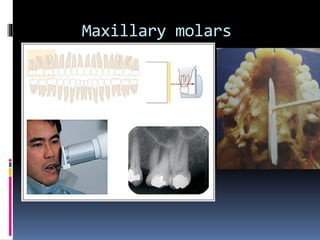
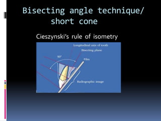
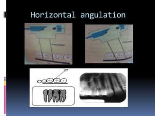
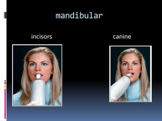
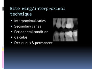
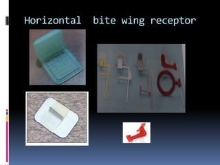
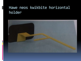
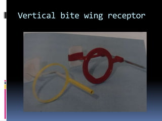
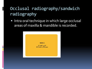
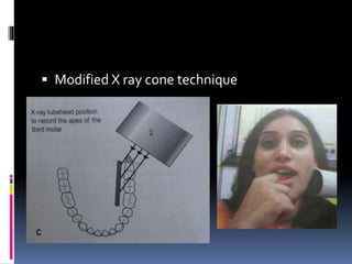
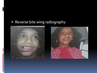
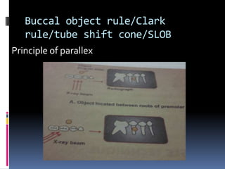
INTRA ORAL RADIOGRAPHIC
TECHNIQUES
MELBIA SHINY
FIRST yr MDS
contents
 Periapical
 Bite wing
 Occlusal
 specialised
Basic principles
Types
 Periapical
 Bite wing
 occlusal
Full mouth set
Periapical radiography
 Radiographic technique of oral structure
taken by placing film in oral cavity while X ray
source is outside oral cavity.
 Paralleling technique
 Bisecting angle technique
Indication
 Apical lesion ,cyst & apical surgery
 Dental caries
 Mixed dentition analysis
 Pulp pathology
 Periodontal status
 Trauma
 Unerupted tooth
 Endodontic treatment & implant prosthesis
Limitation
 Trismus
 Gagging
 Large cyst & tumours
 Two dimensional image
Image receptor
 Film
 Charge coupled device (CCD)
 Complementary metal oxide
semiconductors(CMOS)
 Sensors
 Storage phosphorous plates
Paralleling/fitzgerald
technique
Principle
Receptor holding instrument
Rinn XCP film holder Rinn XCP-DS solid
state digital sensor
holder
Digital phosphor plate holder
Masel precision all in one
metal holder
Hawe Neos holder
Patient positioning
Angulation of tube head
Receptor placement
Maxillary central incisors
Maxillary lateral incisors
Maxillary canine
Maxillary premolars
Maxillary molars
Distal oblique
Mandibular incisors
Mandibular canines
Mandibular premolars
Mandibular molars
Distal oblique
Bisecting angle technique/
short cone
Cieszynski’s rule of isometry
Indications
 Large rigid sensors
 Anatomy of patient
Receptor holding instruments
Receptor placement
Flim & its placement
Position of patient
Angulation of tube head
Vertical : max mad
 Incisors +40 -15
 Canines +45 -20
 Premolars +30 -10
 Molars +20 -5
Horizontal angulation
Tube head position
Le master technique/modified
bisecting angle technique
Maxillary
central incisors
lateral incisor
Maxillary canine
Maxillary
Premolar
Molar
Third molar
mandibular
incisors canine
Mandibular
Premolars molars
Third molar
comparison
Bisecting Paralleling technique
1)Distortion of image <
2)Superimposition of no
zygomatic bone
3)Cone cut not
4)Alveolar bone fills
Interproximal space true relation
5)Easier & < space
6)Long teeth seen by
Vertical angulation
Bite wing/interproximal
technique
 Interproximal caries
 Secondary caries
 Periodontal condition
 Calculus
 Deciduous & permanent
Principle
Films
Angulation
Horizontal bite wing receptor
Hawe neos kwikbite horizontal
holder
Horizontal bite wing
Rinn XCP DS solid planmeca solid state
statedigital sensor digital sensor
holder
Vertical bite wing receptor
Premolar molar
Anterior bite wing
Occlusal radiography/sandwich
radiography
 Intra oral technique in which large occlusal
areas of maxilla & mandible is recorded.
Indication
 Trismus
 Precise location of roots, supernumerary,
unerupted, & impacted teeth.
 Calculi in salivary gland duct
 Maxillary sinus
 Trauma
 Size & extend of cyst & tumours
 Position of unerupted canine
Maxillary anterior occlusal
Anterior Mandibular occlusal
Maxillary cross section
Mandibular crossection
Lateral Maxillary occlusal
Lateral Mandibular occlusal
Specialized intra oral
techniques
1)Mandibular third molar
Surgical needle holder technique/ &
using film holder
A - emmenix /snap
-A-ray
B – worth film
holder
C – artery forceps
 Two radiograph technique
 Modified X ray cone technique
Gagging /retching
 Area sensitive to gagging
management
 Attitude
 Time limit <
 Exposure sequence – anterior
 Divert attention
 Application of LA spray, undiluted mouth
wash,placebo medicines,ice cold water,table
salts
 Use Bite block & film holding device
 Place film packet flat on occlusal plane
Endodontics
 Taping film packet
 Special endodontic film holder
 Endo ray film holder
 Two different radiographs for separation of
root canal
 Thin apex
Small X ray beam & low KV
 Rubber dam & saliva ejector
Root length assesment
Edentulous ridge
Paralleling technique with cotton rolls
Paediatric patient
 Film size – 0
 < 10 yrs – 50% reduce mA ,10 to 15 yrs – 25%
reduce mA
 Show & tell approach
 Iead apron & thyroid
collar.
Modified bisected angle
technique
 Reverse bite wing radiography
Object localization
Foreign objects
 Salivary gland stones
 Canal position
 Impacted teeth
 Root piece
 Broken needle
Buccal object rule/Clark
rule/tube shift cone/SLOB
Principle of parallex
Right angle technique
stereoscopy
Radiopaque wire technique
 Edentulous patient
Shallow palate
Bony growth
Lingual frenum
Premolar area
Supine position radiography
Intra oral radiographs

Intra oral radiographs