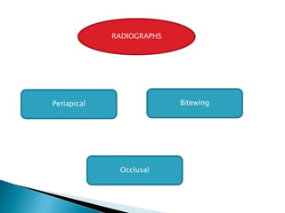
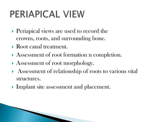
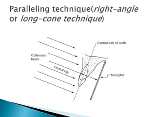
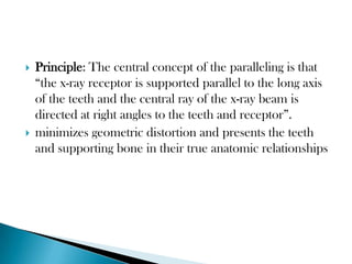
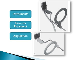
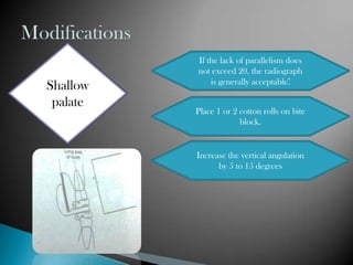
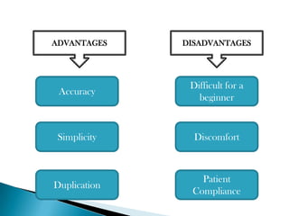
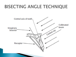
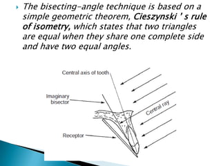
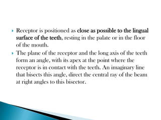
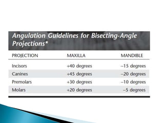
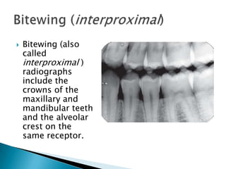
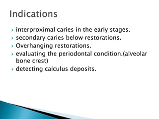
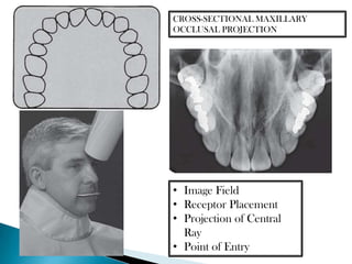
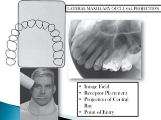
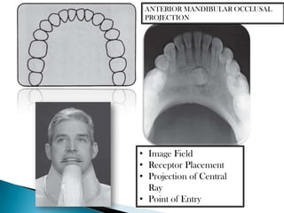
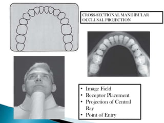
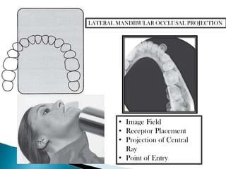
RADIOGRAPHS


Periapical                 Bitewing


              Occlusal
   Periapical views are used to record the
    crowns, roots, and surrounding bone.
   Root canal treatment.
   Assessment of root formation n completion.
   Assessment of root morphology.
    Assessment of relationship of roots to various vital
    structures.
   Implant site assessment and placement.
   Principle: The central concept of the paralleling is that
    “the x-ray receptor is supported parallel to the long axis
    of the teeth and the central ray of the x-ray beam is
    directed at right angles to the teeth and receptor”.
   minimizes geometric distortion and presents the teeth
    and supporting bone in their true anatomic relationships
Instruments


 Receptor
Placement


Angulation
If the lack of parallelism does
          not exceed 20, the radiograph
              is generally acceptable.̊
Shallow
 palate
          Place 1 or 2 cotton rolls on bite
                       block.


          Increase the vertical angulation
                by 5 to 15 degrees
For maxilla, place the
          film on far side of the
 Bony              film.
growths    For mandible, place
           film between the tori
                and tongue
ADVANTAGES    DISADVANTAGES


               Difficult for a
 Accuracy
                 beginner


 Simplicity     Discomfort


                 Patient
Duplication
               Compliance
   The bisecting-angle technique is based on a
    simple geometric theorem, Cieszynski ’ s rule
    of isometry, which states that two triangles
    are equal when they share one complete side
    and have two equal angles.
   Receptor is positioned as close as possible to the lingual
    surface of the teeth, resting in the palate or in the floor
    of the mouth.
   The plane of the receptor and the long axis of the teeth
    form an angle, with its apex at the point where the
    receptor is in contact with the teeth. An imaginary line
    that bisects this angle, direct the central ray of the beam
    at right angles to this bisector.
   No film holder required.
   Better technique when anatomical variations
    hinder paralleling tech.
   Decreased exposure time.
   To reproduce the length of each root of a
    multi-rooted tooth accurately, the central
    beam must be angled differently for each
    root. (Inaccurate)
   Another limitation of this technique is that
    the alveolar ridge often projects more
    coronally than its true position, thus
    distorting the apparent height of the alveolar
    bone around the teeth.
   to obtain three-dimensional information of location
    of an object.


       The right-angle (or
                                    The tube shift
         cross-section)
           technique
                                      technique

                              buccal object rule and Clark ’ s rule
   to obtain three-dimensional information of location
    of an object.


       The right-angle (or
                                    The tube shift
         cross-section)
           technique                  technique

                              buccal object rule and Clark ’ s rule
   Bitewing (also
    called
    interproximal )
    radiographs
    include the
    crowns of the
    maxillary and
    mandibular teeth
    and the alveolar
    crest on the
    same receptor.
   interproximal caries in the early stages.
   secondary caries below restorations.
   Overhanging restorations.
   evaluating the periodontal condition.(alveolar
    bone crest)
   detecting calculus deposits.
   Parallel with the occlusal plane.
   The aiming cylinder is positioned about + 10
    degrees to project the beam parallel with the
    occlusal plane.


    Vertical     Horizontal
    Bitewing      Bitewing
   An occlusal radiograph displays a relatively
    large segment of a dental arch.
   when patients are unable to open the mouth.
   localization of objects.
   To localize foreign bodies in the jaws and
    stones in the ducts.
   To demonstrate and evaluate the integrity of
    the outlines of the maxillary sinus
   To obtain information about the
    location, nature, extent, and displacement of
    fractures of the mandible and maxilla.
   To determine the medial and lateral extent of
    disease (e.g., cysts, osteomyelitis, tumors)
    and to detect disease in the palate or floor of
    the mouth.
ANTERIOR MAXILLARY OCCLUSAL PROJECTION


               • Image Field
               • Receptor Placement
               • Projection of Central
                 Ray
               • Point of Entry
CROSS-SECTIONAL MAXILLARY
OCCLUSAL PROJECTION


• Image Field
• Receptor Placement
• Projection of Central
  Ray
• Point of Entry
LATERAL MAXILLARY OCCLUSAL PROJECTION


    • Image Field
    • Receptor Placement
    • Projection of Central
      Ray
    • Point of Entry
ANTERIOR MANDIBULAR OCCLUSAL
 PROJECTION


• Image Field
• Receptor Placement
• Projection of Central
  Ray
• Point of Entry
CROSS-SECTIONAL MANDIBULAR
OCCLUSAL PROJECTION


• Image Field
• Receptor Placement
• Projection of Central
  Ray
• Point of Entry
LATERAL MANDIBULAR OCCLUSAL PROJECTION


        • Image Field
        • Receptor Placement
        • Projection of Central
          Ray
        • Point of Entry
INFECTION


                  trismus


             Pain and Difficulty


Extra oral                         Occlusal
TRAUMA
may have a         Use
                             periapical or
 dental or    2radiographs
                               occlusal
   facial        at right
                             radiographs
 fracture.        angle.
   lack of coordination or inability to comprehend is
    expected.
   Speedily
   Sedation
   Relax and reassure the patient
   Describe and explain the procedures
   Perform the procedure in the morning,
   Sliding the film along the palate or tongue is likely to
    stimulate the gag reflex.
   The longer the receptor stays in the mouth, the greater
    the possibility that the patient will start to gag.
   The patient should be advised to breathe rapidly
    through the nose because mouth breathing usually
    aggravates this condition.
   Shift the patient ’ s attention

Radiographic techniques

  • 2.
    RADIOGRAPHS Periapical Bitewing Occlusal
  • 3.
    Periapical views are used to record the crowns, roots, and surrounding bone.  Root canal treatment.  Assessment of root formation n completion.  Assessment of root morphology.  Assessment of relationship of roots to various vital structures.  Implant site assessment and placement.
  • 5.
    Principle: The central concept of the paralleling is that “the x-ray receptor is supported parallel to the long axis of the teeth and the central ray of the x-ray beam is directed at right angles to the teeth and receptor”.  minimizes geometric distortion and presents the teeth and supporting bone in their true anatomic relationships
  • 6.
  • 7.
    If the lackof parallelism does not exceed 20, the radiograph is generally acceptable.̊ Shallow palate Place 1 or 2 cotton rolls on bite block. Increase the vertical angulation by 5 to 15 degrees
  • 8.
    For maxilla, placethe film on far side of the Bony film. growths For mandible, place film between the tori and tongue
  • 9.
    ADVANTAGES DISADVANTAGES Difficult for a Accuracy beginner Simplicity Discomfort Patient Duplication Compliance
  • 11.
    The bisecting-angle technique is based on a simple geometric theorem, Cieszynski ’ s rule of isometry, which states that two triangles are equal when they share one complete side and have two equal angles.
  • 12.
    Receptor is positioned as close as possible to the lingual surface of the teeth, resting in the palate or in the floor of the mouth.  The plane of the receptor and the long axis of the teeth form an angle, with its apex at the point where the receptor is in contact with the teeth. An imaginary line that bisects this angle, direct the central ray of the beam at right angles to this bisector.
  • 14.
    No film holder required.  Better technique when anatomical variations hinder paralleling tech.  Decreased exposure time.
  • 15.
    To reproduce the length of each root of a multi-rooted tooth accurately, the central beam must be angled differently for each root. (Inaccurate)  Another limitation of this technique is that the alveolar ridge often projects more coronally than its true position, thus distorting the apparent height of the alveolar bone around the teeth.
  • 16.
    to obtain three-dimensional information of location of an object. The right-angle (or The tube shift cross-section) technique technique buccal object rule and Clark ’ s rule
  • 18.
    to obtain three-dimensional information of location of an object. The right-angle (or The tube shift cross-section) technique technique buccal object rule and Clark ’ s rule
  • 20.
    Bitewing (also called interproximal ) radiographs include the crowns of the maxillary and mandibular teeth and the alveolar crest on the same receptor.
  • 21.
    interproximal caries in the early stages.  secondary caries below restorations.  Overhanging restorations.  evaluating the periodontal condition.(alveolar bone crest)  detecting calculus deposits.
  • 22.
    Parallel with the occlusal plane.  The aiming cylinder is positioned about + 10 degrees to project the beam parallel with the occlusal plane. Vertical Horizontal Bitewing Bitewing
  • 23.
    An occlusal radiograph displays a relatively large segment of a dental arch.  when patients are unable to open the mouth.  localization of objects.  To localize foreign bodies in the jaws and stones in the ducts.  To demonstrate and evaluate the integrity of the outlines of the maxillary sinus
  • 24.
    To obtain information about the location, nature, extent, and displacement of fractures of the mandible and maxilla.  To determine the medial and lateral extent of disease (e.g., cysts, osteomyelitis, tumors) and to detect disease in the palate or floor of the mouth.
  • 25.
    ANTERIOR MAXILLARY OCCLUSALPROJECTION • Image Field • Receptor Placement • Projection of Central Ray • Point of Entry
  • 26.
    CROSS-SECTIONAL MAXILLARY OCCLUSAL PROJECTION •Image Field • Receptor Placement • Projection of Central Ray • Point of Entry
  • 27.
    LATERAL MAXILLARY OCCLUSALPROJECTION • Image Field • Receptor Placement • Projection of Central Ray • Point of Entry
  • 28.
    ANTERIOR MANDIBULAR OCCLUSAL PROJECTION • Image Field • Receptor Placement • Projection of Central Ray • Point of Entry
  • 29.
    CROSS-SECTIONAL MANDIBULAR OCCLUSAL PROJECTION •Image Field • Receptor Placement • Projection of Central Ray • Point of Entry
  • 30.
    LATERAL MANDIBULAR OCCLUSALPROJECTION • Image Field • Receptor Placement • Projection of Central Ray • Point of Entry
  • 31.
    INFECTION trismus Pain and Difficulty Extra oral Occlusal
  • 32.
    TRAUMA may have a Use periapical or dental or 2radiographs occlusal facial at right radiographs fracture. angle.
  • 33.
    lack of coordination or inability to comprehend is expected.  Speedily  Sedation
  • 34.
    Relax and reassure the patient  Describe and explain the procedures  Perform the procedure in the morning,  Sliding the film along the palate or tongue is likely to stimulate the gag reflex.  The longer the receptor stays in the mouth, the greater the possibility that the patient will start to gag.  The patient should be advised to breathe rapidly through the nose because mouth breathing usually aggravates this condition.  Shift the patient ’ s attention