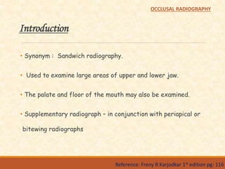
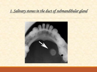
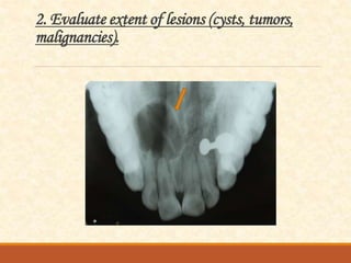
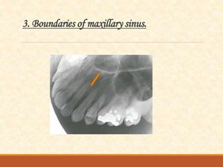
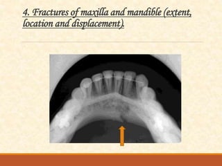
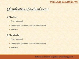
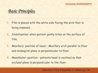
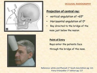
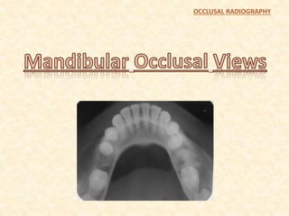
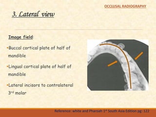
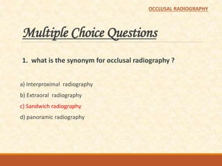
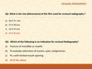
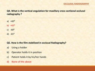
OCCLUSAL
RADIOGRAPHY
Presented By:
Parikshit Harnoor
14D0959
Final Year B.D.S.
Guide:
Dr. Pramod G. V.
Professor,
Dept. of Oral Medicine and Radiology
Objectives
To know…
 Indiactions of occlusal radiography,
 Basic principles of occlusal radiography,
 Different types of occlusal radiography,
 Methods of obtaining the different types of radiographs.
Contents:
Introduction
Film
Indications
Classification of occlusal views
Basic principles
Maxillary occlusal views
Mandibular occlusal views
Conclusion
Bibliography
OCCLUSAL RADIOGRAPHY
Introduction
• Synonym : Sandwich radiography.
• Used to examine large areas of upper and lower jaw.
• The palate and floor of the mouth may also be examined.
• Supplementary radiograph – in conjunction with periapical or
bitewing radiographs
Reference: Freny R Karjodkar 1st edition pg: 116
OCCLUSAL RADIOGRAPHY
Film
•Special films are marketed
•Bigger than IOPA films
57mm
76mm
Reference: Freny R Karjodkar 1st edition pg: 116
OCCLUSAL RADIOGRAPHY
1. Salivary stones in the duct of submandibular gland
2. Evaluate extent of lesions (cysts, tumors,
malignancies).
3. Boundaries of maxillary sinus.
4. Fractures of maxilla and mandible (extent,
location and displacement).
5. Foreign bodies in maxilla or mandible
6. Examine cleft palate.
7. Retained roots of extracted teeth.
8. Supernumerary, unerupted or impacted teeth.
Classification of occlusal views
1. Maxillary
• Cross sectional
• Topographic (anterior and posterior/lateral)
• Pediatric
2. Mandibular
• Cross sectional
• Topographic (anterior and posterior/lateral)
• Pediatric
Reference: Freny R Karjodkar 1st edition pg: 116
OCCLUSAL RADIOGRAPHY
Basic Principles.
1. Film is placed with the white side facing the arch that is
being exposed.
2. Stabilization: when patient gently bites on the surface of
film.
3. Maxillary: position of head – Maxillary arch parallel to floor
and midsagital plane is perpendicular to floor.
4. Mandibular: position – patients head is reclined so that
occlusal plane is perpendicular to the floor.
Reference: Freny R Karjodkar 1st edition pg: 116
OCCLUSAL RADIOGRAPHY
1. Cross sectional view
Image field:
Hard Palate.
Zygomatic process of maxilla.
Nasolacrimal canals.
Teeth from second molar to
second molar.
Nasal septum.
Reference: white and Pharoah 1st South Asia Edition pg: 121
OCCLUSAL RADIOGRAPHY
Projection of central ray:
• vertical angulation of +65⁰
• Horizaontal angulation of 0⁰
• Ray directed to the bridge of the
nose just below the nasion
Reference: white and Pharoah 1st South Asia Edition pg: 121
Freny R Karjodkar 1st edition pg: 117
OCCLUSAL RADIOGRAPHY
Point of Entry
Rays enter the patients face
through the bridge of the nose.
2. Topographic view - anterior
Image field:
Anterior maxilla.
Anterior floor of the nasal fossa
Teeth from canine to canine
Reference: white and Pharoah 1st South Asia Edition pg: 120
OCCLUSAL RADIOGRAPHY
Projection of central ray:
• Orient the central ray through the
tip of the nose.
• Vertical angulation of +45⁰
• Horizontal angulation of 0 ⁰
Reference: white and Pharoah 1st South Asia Edition pg: 121
Freny R Karjodkar 1st edition pg: 117
OCCLUSAL RADIOGRAPHY
Point of entry
The central ray enters the patients
face approximately through the tip
of nose.
3. Topographic view - lateral
Image field:
Quadrant of the alveolar ridge of
the maxilla
Inferolateral aspect of antrum
Tuberosity
Teeth from lateral incisor to the
contralateral third molar
Zygomatic process of the maxilla
superimposes over the roots of
molar
Reference: white and Pharoah 1st South Asia Edition pg: 122
OCCLUSAL RADIOGRAPHY
Projection of central ray:
• Orient the central ray with
• Vertical angulation of +60⁰ to a
point 2cm below the lateral
canthus of the eye
OCCLUSAL RADIOGRAPHY
Reference: white and Pharoah 1st South Asia Edition pg: 122
Freny R Karjodkar 1st edition pg: 117
Point of entry
The central ray enters at a point
approximately 2cm below the lateral
canthus of the eye.
OCCLUSAL RADIOGRAPHY
1. Cross sectional view
Image field:
Buccal cortical plate of the
mandible
Lingual cortical plate of the
mandible
Teeth from 37 – 47
Reference: white and Pharoah 1st South Asia Edition pg: 124
OCCLUSAL RADIOGRAPHY
Projection of central ray:
• At the midline through the floor of
the mouth.
• Approximately 3cm below the chin, at
90⁰ to the receptor
Reference: white and Pharoah 1st South Asia Edition pg: 124
Freny R Karjodkar 1st edition pg: 118
OCCLUSAL RADIOGRAPHY
Point of entry
Midline. Through the floor of the mouth
approximately 3cm below the chin
2. Anterior view
Image field:
Anterior portion of the mandible
Dentition from canine to canine
Inferior cortical border of
mandible
Reference: white and Pharoah 1st South Asia Edition pg: 123
OCCLUSAL RADIOGRAPHY
Receptor Placement
• Seat the patient tilted – occlusal
plane is 45⁰ above horizontal
Projection of central ray:
• -10⁰ through point of chin, giving -
45⁰angulation to receptor.
Reference: white and Pharoah 1st South Asia Edition pg: 123
Freny R Karjodkar 1st edition pg: 118
OCCLUSAL RADIOGRAPHY
Point of entry
Midline – tip of the chin
45⁰
3. Lateral view
Image field:
Buccal cortical plate of half of
mandible
Lingual cortical plate of half of
mandible
Lateral incisors to contralateral
3rd molar
Reference: white and Pharoah 1st South Asia Edition pg: 122
OCCLUSAL RADIOGRAPHY
Projection of central ray:
Perpendicular to center of receptor,
through a point beneath the chin, approx.
3cm posterior to point of chin and 3cm
lateral to midline.
Reference: white and Pharoah 1st South Asia Edition pg: 125
Freny R Karjodkar 1st edition pg: 118
OCCLUSAL RADIOGRAPHY
Point of entry
Beneath the chin, 3cm posterior to chin
and 3cm lateral to midline.
Conclusion :-
OCCLUSAL RADIOGRAPHY
Bibliography
White and Pharoah – textbook of Oral Radiology – Principles
and Interpretation – 1st South Asia Edition.
Freny R Karjodkar – Essentials Of Oral and Maxillofacial
Radiology – 1st Edition
OCCLUSAL RADIOGRAPHY
Multiple Choice Questions
1. what is the synonym for occlusal radiography ?
a) Interproximal radiography
b) Extraoral radiography
c) Sandwich radiography
d) panoramic radiography
OCCLUSAL RADIOGRAPHY
Q2. What is the size (dimensions) of the film used for occlusal radiography ?
a) 50 X 72 mm
b) 57 X 78 mm
c) 52 X 74 mm
d) 57 X 76 mm
Q3. Which of the following is an indication for occlusal Radiography?
a) Fracture of mandible or maxilla
b) To evaluate extensions of tumors, cysts, malignancies
c) Pt.s with limited mouth opening
d) All of the above
OCCLUSAL RADIOGRAPHY
Q4. What is the vertical angulation for maxillary cross sectional occlusal
radiography ?
a) +60⁰
b) +65⁰
c) -60⁰
d) -65⁰
Q5. How is the film stabilized in occlusal Radiography?
a) Using a holder
b) Operator holds it in position
c) Patient holds it by his/her hands
d) None of the above
OCCLUSAL RADIOGRAPHY
Occlusal radiography

Occlusal radiography

  • 2.
    OCCLUSAL RADIOGRAPHY Presented By: Parikshit Harnoor 14D0959 FinalYear B.D.S. Guide: Dr. Pramod G. V. Professor, Dept. of Oral Medicine and Radiology
  • 3.
    Objectives To know…  Indiactionsof occlusal radiography,  Basic principles of occlusal radiography,  Different types of occlusal radiography,  Methods of obtaining the different types of radiographs.
  • 4.
    Contents: Introduction Film Indications Classification of occlusalviews Basic principles Maxillary occlusal views Mandibular occlusal views Conclusion Bibliography OCCLUSAL RADIOGRAPHY
  • 5.
    Introduction • Synonym :Sandwich radiography. • Used to examine large areas of upper and lower jaw. • The palate and floor of the mouth may also be examined. • Supplementary radiograph – in conjunction with periapical or bitewing radiographs Reference: Freny R Karjodkar 1st edition pg: 116 OCCLUSAL RADIOGRAPHY
  • 6.
    Film •Special films aremarketed •Bigger than IOPA films 57mm 76mm Reference: Freny R Karjodkar 1st edition pg: 116 OCCLUSAL RADIOGRAPHY
  • 9.
    1. Salivary stonesin the duct of submandibular gland
  • 10.
    2. Evaluate extentof lesions (cysts, tumors, malignancies).
  • 11.
    3. Boundaries ofmaxillary sinus.
  • 12.
    4. Fractures ofmaxilla and mandible (extent, location and displacement).
  • 13.
    5. Foreign bodiesin maxilla or mandible
  • 14.
  • 15.
    7. Retained rootsof extracted teeth.
  • 16.
    8. Supernumerary, uneruptedor impacted teeth.
  • 17.
    Classification of occlusalviews 1. Maxillary • Cross sectional • Topographic (anterior and posterior/lateral) • Pediatric 2. Mandibular • Cross sectional • Topographic (anterior and posterior/lateral) • Pediatric Reference: Freny R Karjodkar 1st edition pg: 116 OCCLUSAL RADIOGRAPHY
  • 18.
    Basic Principles. 1. Filmis placed with the white side facing the arch that is being exposed. 2. Stabilization: when patient gently bites on the surface of film. 3. Maxillary: position of head – Maxillary arch parallel to floor and midsagital plane is perpendicular to floor. 4. Mandibular: position – patients head is reclined so that occlusal plane is perpendicular to the floor. Reference: Freny R Karjodkar 1st edition pg: 116 OCCLUSAL RADIOGRAPHY
  • 19.
  • 20.
    1. Cross sectionalview Image field: Hard Palate. Zygomatic process of maxilla. Nasolacrimal canals. Teeth from second molar to second molar. Nasal septum. Reference: white and Pharoah 1st South Asia Edition pg: 121 OCCLUSAL RADIOGRAPHY
  • 21.
    Projection of centralray: • vertical angulation of +65⁰ • Horizaontal angulation of 0⁰ • Ray directed to the bridge of the nose just below the nasion Reference: white and Pharoah 1st South Asia Edition pg: 121 Freny R Karjodkar 1st edition pg: 117 OCCLUSAL RADIOGRAPHY Point of Entry Rays enter the patients face through the bridge of the nose.
  • 22.
    2. Topographic view- anterior Image field: Anterior maxilla. Anterior floor of the nasal fossa Teeth from canine to canine Reference: white and Pharoah 1st South Asia Edition pg: 120 OCCLUSAL RADIOGRAPHY
  • 23.
    Projection of centralray: • Orient the central ray through the tip of the nose. • Vertical angulation of +45⁰ • Horizontal angulation of 0 ⁰ Reference: white and Pharoah 1st South Asia Edition pg: 121 Freny R Karjodkar 1st edition pg: 117 OCCLUSAL RADIOGRAPHY Point of entry The central ray enters the patients face approximately through the tip of nose.
  • 24.
    3. Topographic view- lateral Image field: Quadrant of the alveolar ridge of the maxilla Inferolateral aspect of antrum Tuberosity Teeth from lateral incisor to the contralateral third molar Zygomatic process of the maxilla superimposes over the roots of molar Reference: white and Pharoah 1st South Asia Edition pg: 122 OCCLUSAL RADIOGRAPHY
  • 25.
    Projection of centralray: • Orient the central ray with • Vertical angulation of +60⁰ to a point 2cm below the lateral canthus of the eye OCCLUSAL RADIOGRAPHY Reference: white and Pharoah 1st South Asia Edition pg: 122 Freny R Karjodkar 1st edition pg: 117 Point of entry The central ray enters at a point approximately 2cm below the lateral canthus of the eye.
  • 26.
  • 27.
    1. Cross sectionalview Image field: Buccal cortical plate of the mandible Lingual cortical plate of the mandible Teeth from 37 – 47 Reference: white and Pharoah 1st South Asia Edition pg: 124 OCCLUSAL RADIOGRAPHY
  • 28.
    Projection of centralray: • At the midline through the floor of the mouth. • Approximately 3cm below the chin, at 90⁰ to the receptor Reference: white and Pharoah 1st South Asia Edition pg: 124 Freny R Karjodkar 1st edition pg: 118 OCCLUSAL RADIOGRAPHY Point of entry Midline. Through the floor of the mouth approximately 3cm below the chin
  • 29.
    2. Anterior view Imagefield: Anterior portion of the mandible Dentition from canine to canine Inferior cortical border of mandible Reference: white and Pharoah 1st South Asia Edition pg: 123 OCCLUSAL RADIOGRAPHY
  • 30.
    Receptor Placement • Seatthe patient tilted – occlusal plane is 45⁰ above horizontal Projection of central ray: • -10⁰ through point of chin, giving - 45⁰angulation to receptor. Reference: white and Pharoah 1st South Asia Edition pg: 123 Freny R Karjodkar 1st edition pg: 118 OCCLUSAL RADIOGRAPHY Point of entry Midline – tip of the chin 45⁰
  • 31.
    3. Lateral view Imagefield: Buccal cortical plate of half of mandible Lingual cortical plate of half of mandible Lateral incisors to contralateral 3rd molar Reference: white and Pharoah 1st South Asia Edition pg: 122 OCCLUSAL RADIOGRAPHY
  • 32.
    Projection of centralray: Perpendicular to center of receptor, through a point beneath the chin, approx. 3cm posterior to point of chin and 3cm lateral to midline. Reference: white and Pharoah 1st South Asia Edition pg: 125 Freny R Karjodkar 1st edition pg: 118 OCCLUSAL RADIOGRAPHY Point of entry Beneath the chin, 3cm posterior to chin and 3cm lateral to midline.
  • 33.
  • 34.
    Bibliography White and Pharoah– textbook of Oral Radiology – Principles and Interpretation – 1st South Asia Edition. Freny R Karjodkar – Essentials Of Oral and Maxillofacial Radiology – 1st Edition OCCLUSAL RADIOGRAPHY
  • 35.
  • 36.
    Multiple Choice Questions 1.what is the synonym for occlusal radiography ? a) Interproximal radiography b) Extraoral radiography c) Sandwich radiography d) panoramic radiography OCCLUSAL RADIOGRAPHY
  • 37.
    Q2. What isthe size (dimensions) of the film used for occlusal radiography ? a) 50 X 72 mm b) 57 X 78 mm c) 52 X 74 mm d) 57 X 76 mm Q3. Which of the following is an indication for occlusal Radiography? a) Fracture of mandible or maxilla b) To evaluate extensions of tumors, cysts, malignancies c) Pt.s with limited mouth opening d) All of the above OCCLUSAL RADIOGRAPHY
  • 38.
    Q4. What isthe vertical angulation for maxillary cross sectional occlusal radiography ? a) +60⁰ b) +65⁰ c) -60⁰ d) -65⁰ Q5. How is the film stabilized in occlusal Radiography? a) Using a holder b) Operator holds it in position c) Patient holds it by his/her hands d) None of the above OCCLUSAL RADIOGRAPHY
  • 39.
    Multiple Choice Questions 1.what is the synonym for occlusal radiography ? a) Interproximal radiography b) Extraoral radiography c) Sandwich radiography d) panoramic radiography OCCLUSAL RADIOGRAPHY
  • 40.
    Q2. What isthe size (dimensions) of the film used for occlusal radiography ? a) 50 X 72 mm b) 57 X 78 mm c) 52 X 74 mm d) 57 X 76 mm Q3. Which of the following is an indication for occlusal Radiography? a) Fracture of mandible or maxilla b) To evaluate extensions of tumors, cysts, malignancies c) Pt.s with limited mouth opening d) All of the above OCCLUSAL RADIOGRAPHY
  • 41.
    Q4. What isthe vertical angulation for maxillary cross sectional occlusal radiography ? a) +60⁰ b) +65⁰ c) -60⁰ d) -65⁰ Q5. How is the film stabilized in occlusal Radiography? a) Using a holder b) Operator holds it in position c) Patient holds it by his/her hands d) None of the above OCCLUSAL RADIOGRAPHY

Editor's Notes

  • #17 And also in patient who cant open their mouth wider than few milimeters