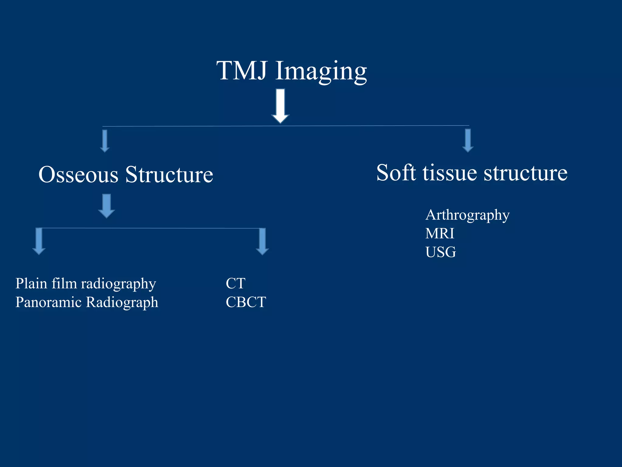
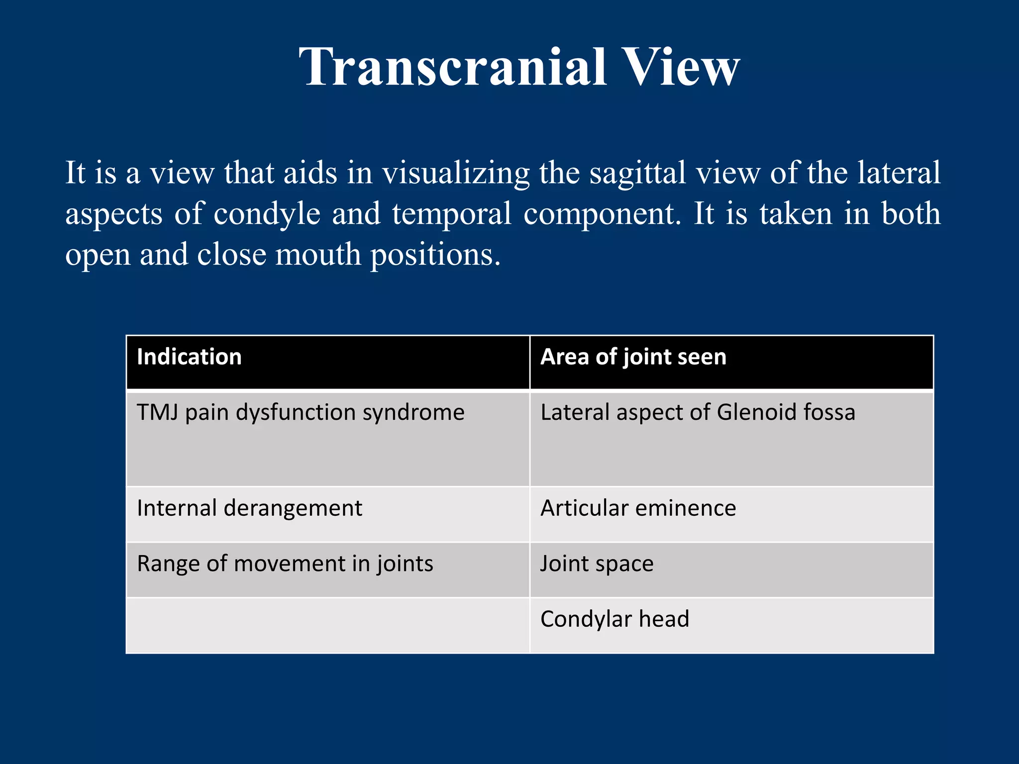
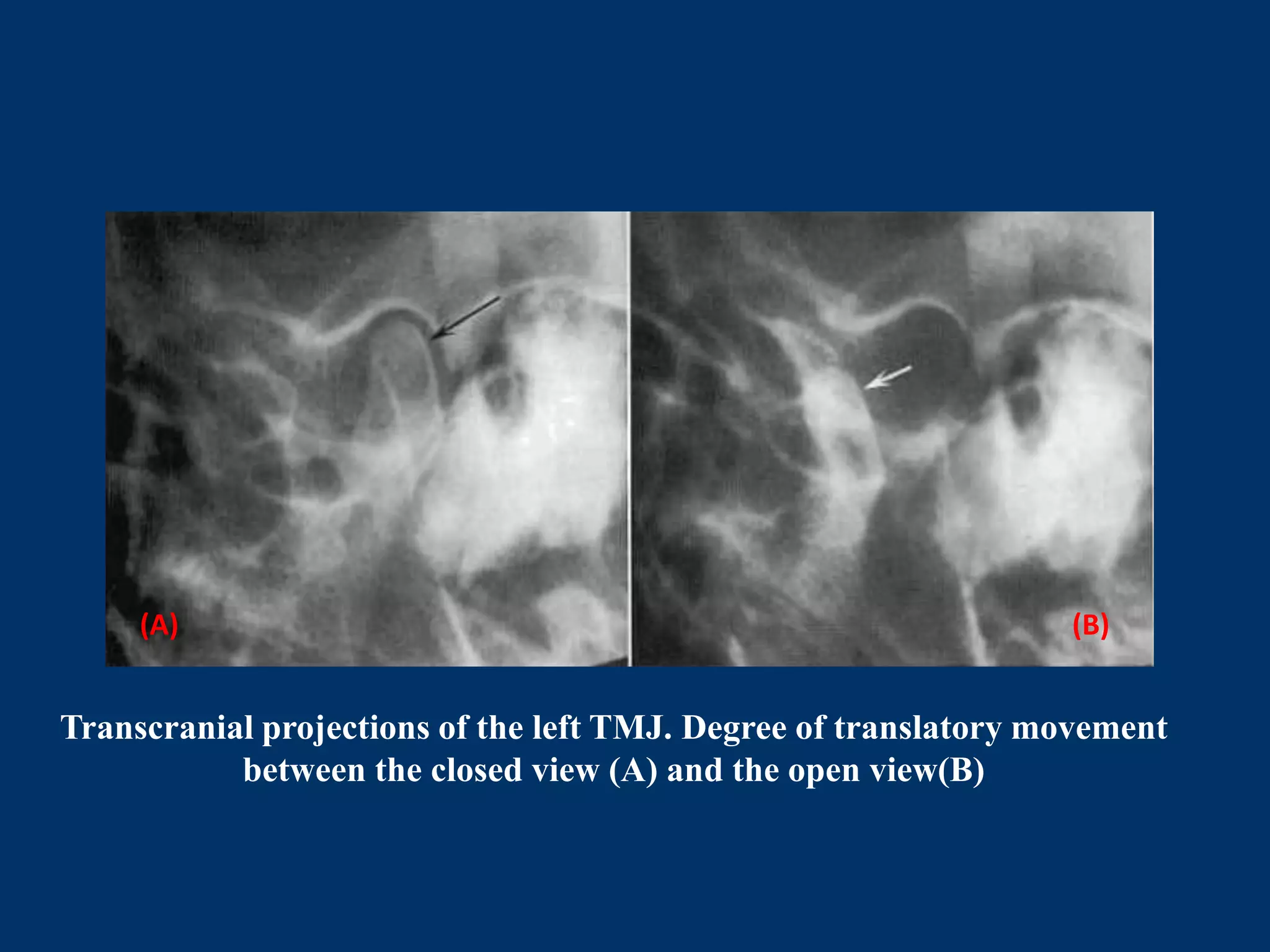
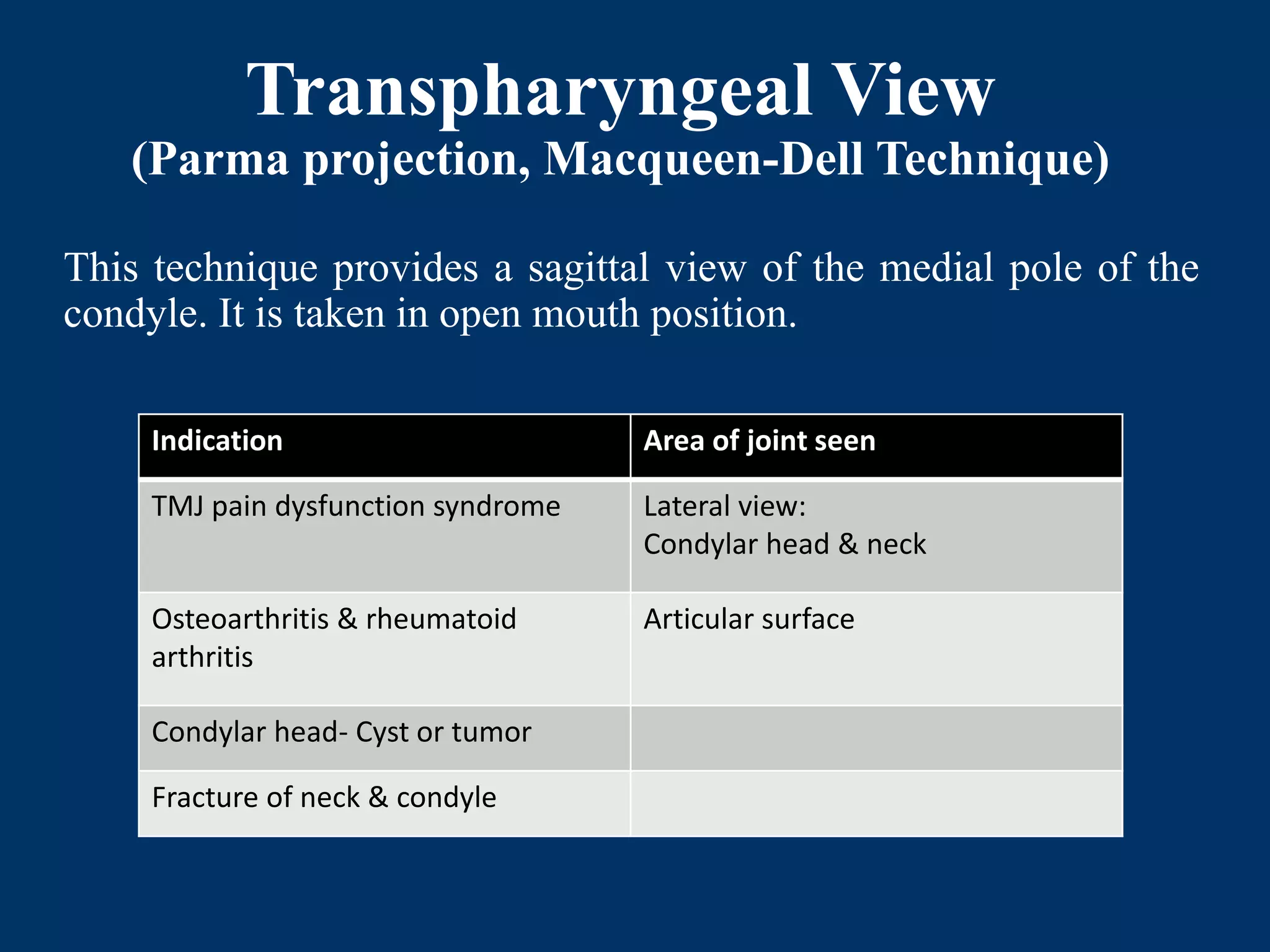
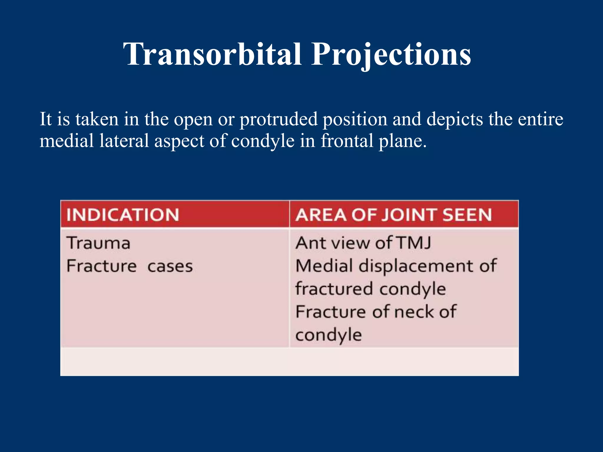
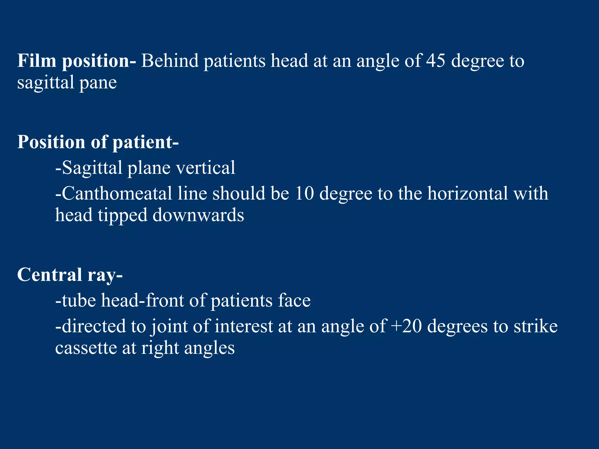
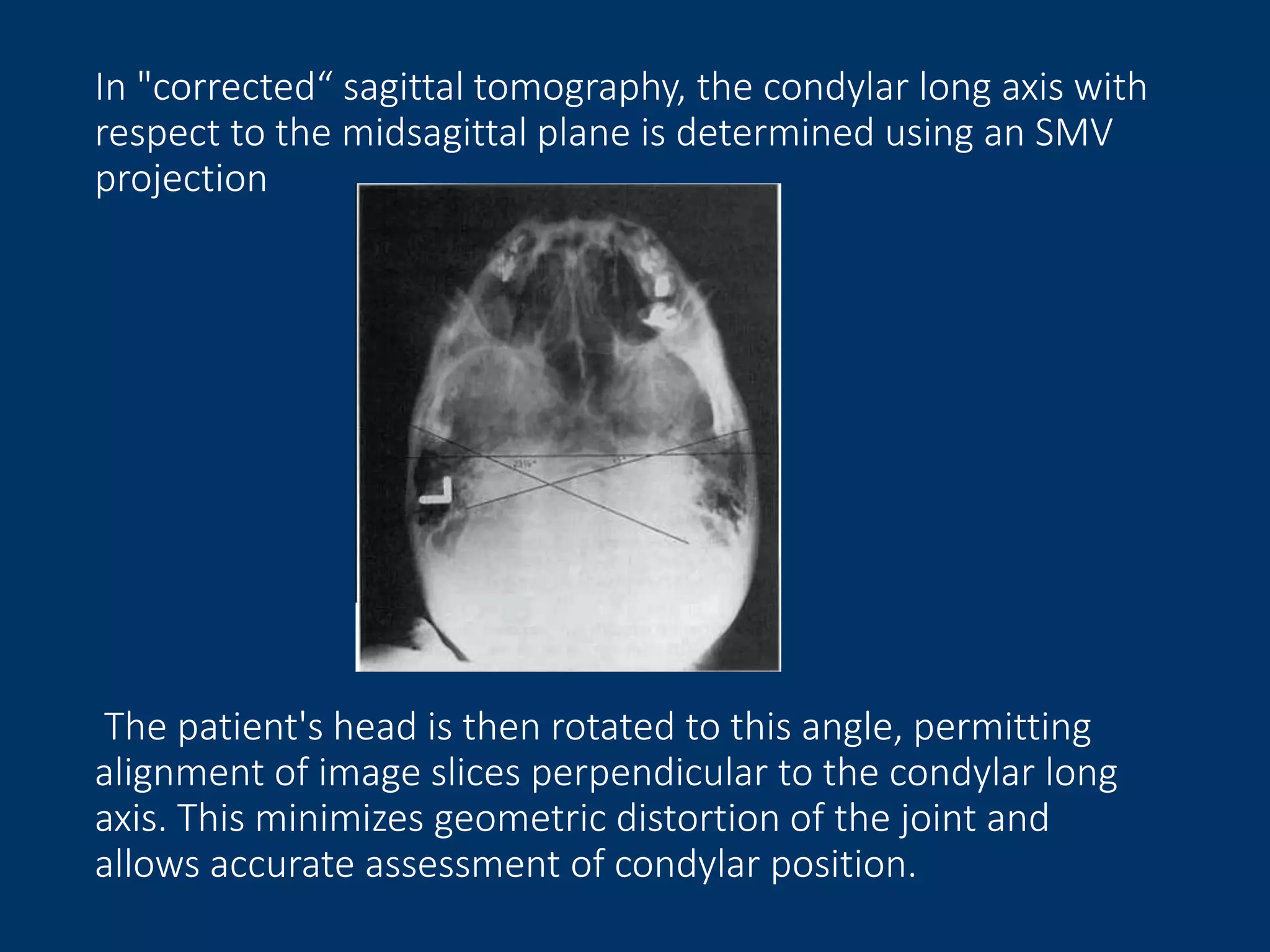
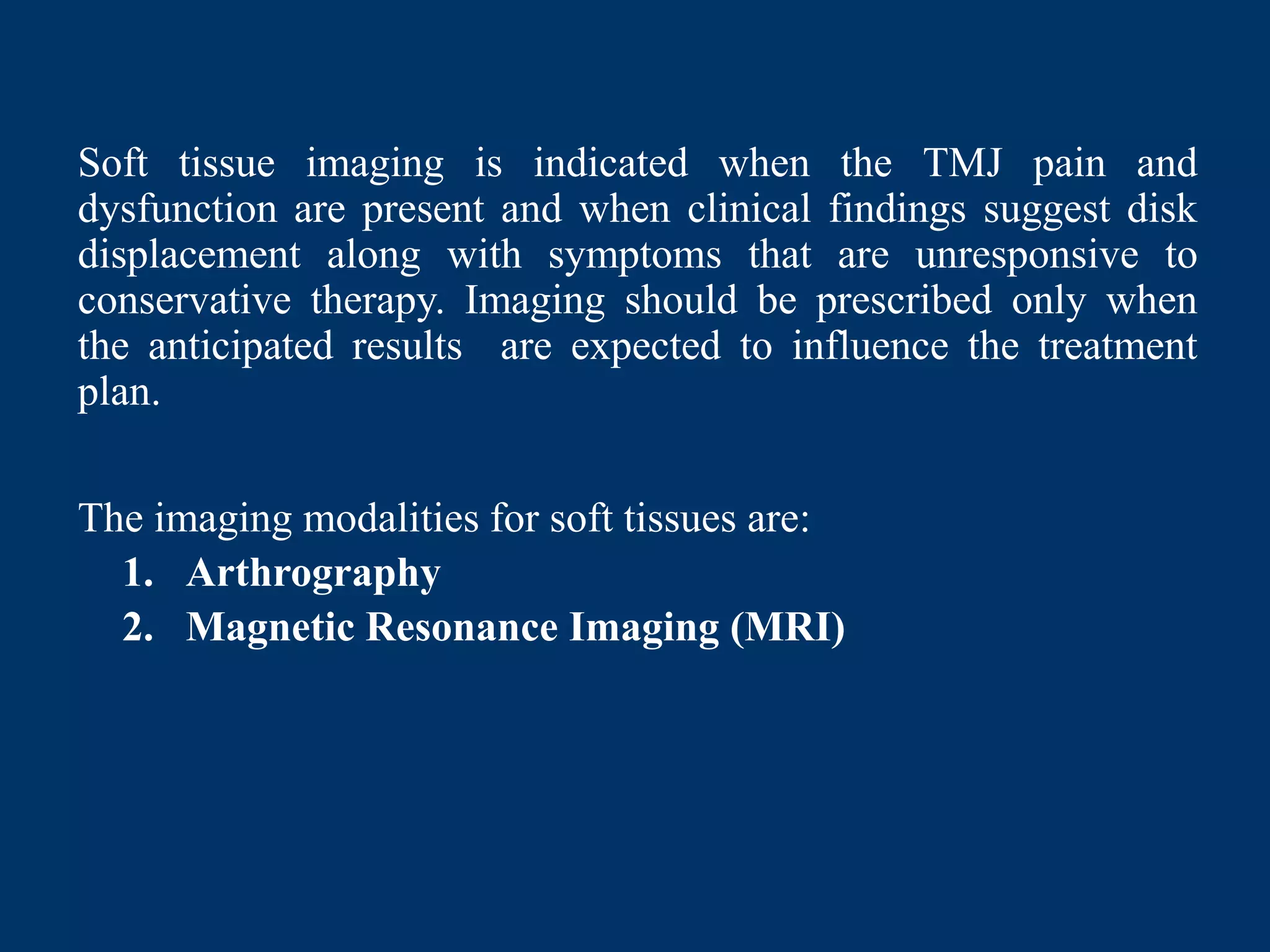
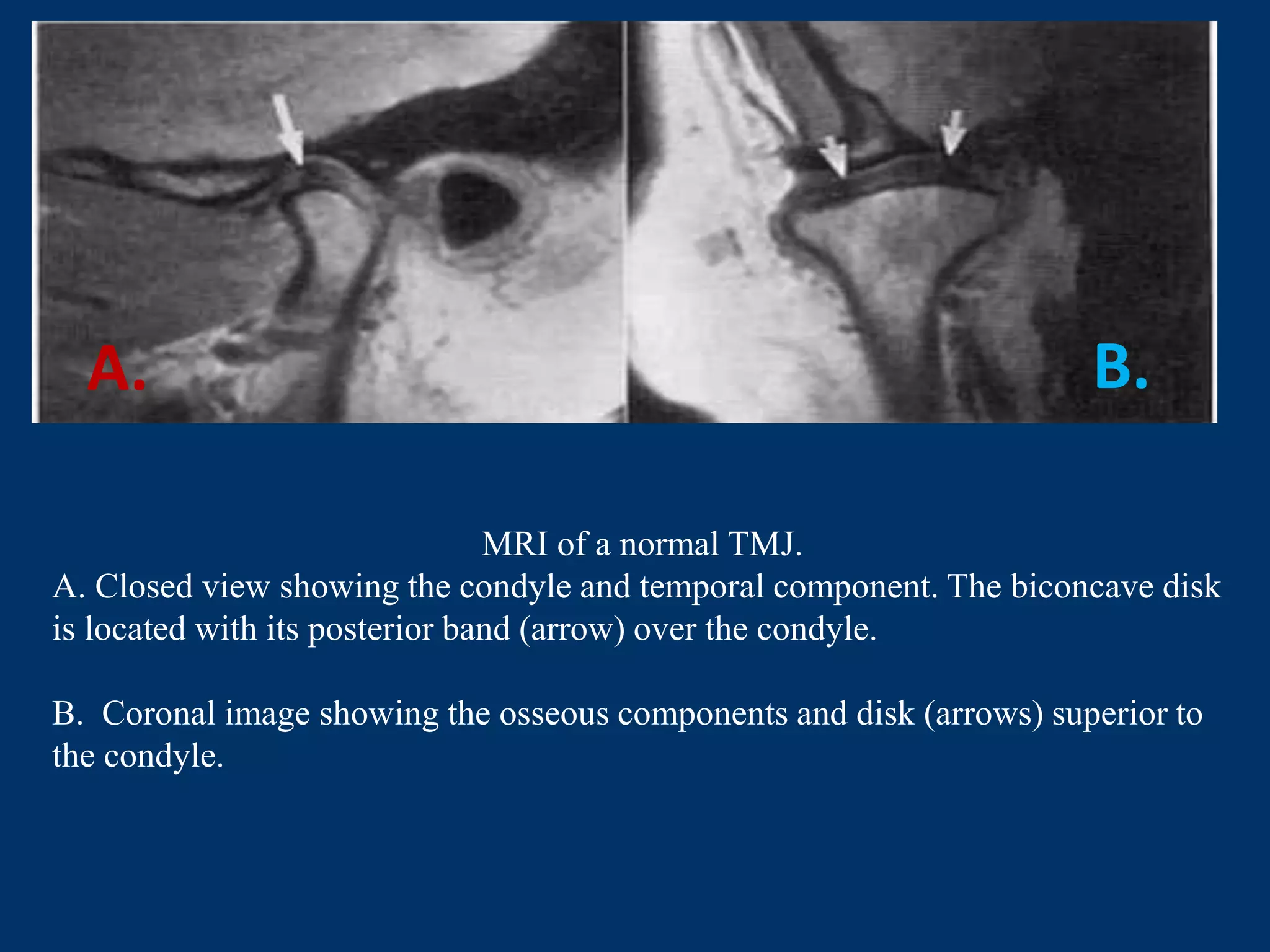
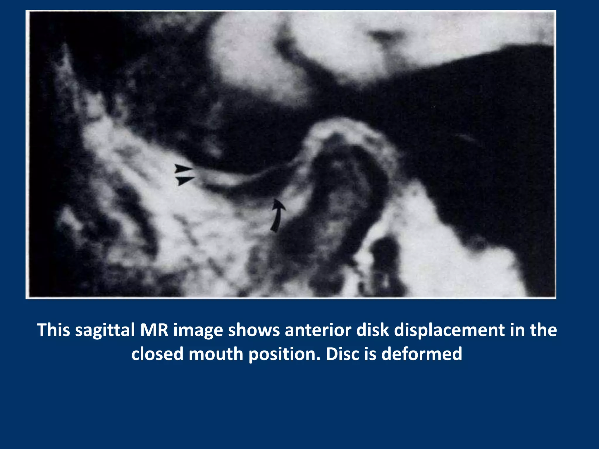
This document discusses imaging modalities for the temporomandibular joint (TMJ). It begins by introducing the anatomy and components of the TMJ. For osseous structures, imaging options include panoramic radiography, plain film radiography, computed tomography (CT), and cone beam CT. Panoramic radiography is useful for detecting gross bony changes but does not show detail or joint positions. CT and cone beam CT provide three-dimensional bone images but not of soft tissues. For soft tissues like the articular disc, magnetic resonance imaging (MRI) is the best option, as it clearly depicts disc position and abnormalities. The document reviews the techniques and indications for various imaging modalities of both osseous