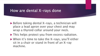
DENTAL X-RAYS
DR KR PETER
What are dental X-rays?
 Dental X-rays (radiographs) are internal images of your teeth
and jaws.
 Dentists use X-rays to examine structures they can’t see
during a routine checkup, like:
 Jawbone ,
 Nerves ,
 Sinuses
 Teeth decay (cavities)
 and teeth roots.
How do dental X-rays work?
 Like X-rays taken in other parts of your body, dental
X-rays use electromagnetic radiation to capture
images of your mouth.
 The radiation beam passes through your soft tissues
and creates images of your teeth and bones.
 Dental X-rays may be traditional (taken with film) or
digital (taken with digital sensors and a computer).
 Digital dental X-rays use 80% to 90% less radiation
compared to traditional dental X-ray machines.
What can dental X-rays detect?
Dental X-rays help your dentist
diagnose a wide range of oral
health issues.
Dental X-rays show:
Cavities, especially small areas of
decay between teeth.
Decay beneath existing fillings.
• Bone loss in your jaw.
• Areas of infection.
• The position of unerupted or impacted
teeth.
• Abscessed teeth (infection at the root of
your tooth or between your gums and
your tooth).
• Cysts and some types of tumors.
 Dentists also use X-rays to help determine your
eligibility for treatments like
 Dental implants
 Braces or
 Dentures .
 X-rays help your dentist check healing after certain
procedures, too, such as dental bone grafts and root
canal therapy.
How are dental X-rays done
 Before taking dental X-rays, a technician will
place a lead apron over your chest and may
wrap a thyroid collar around your neck.
 This helps protect you from excess radiation.
 When it’s time to take the X-rays, you’ll either
sit in a chair or stand in front of an X-ray
machine.
A technician will place the film or
sensor, and then press a button to
take the X-ray image.
It’s important to hold as still as
possible during this process.
Types of x-rays
What are the different types of dental
X-rays?
There are two main types of dental X-
rays:
1.Intraoral: The film or sensor is inside
your mouth.
2.Extraoral: The film or sensor is
outside your mouth.
INTRAORAL X-RAYS
There are different types of
intraoral X-rays:
Periapical X-rays
Bitewing X-rays.
Occlusal X-rays.
Periapical X-rays
 A periapical X-ray shows your entire tooth, from
the crown to the root tip.
 This type of X-ray helps your dentist detect:
 Decay
 Gum disease
 Bone loss
 and any other abnormalities of your tooth or
surrounding bone.
 Periapical radiography describes intraoral
techniques designed to show individual teeth and
the tissues around the apices.
 Each image usually shows two to four teeth and
provides detailed information about the teeth and
the surrounding alveolar bone.
Main indications
 The main clinical indications for periapical radiography
include:
 Detection of apical infection/inflammation
 Assessment of the periodontal status
 After trauma to the teeth and associated alveolar bone
 Assessment of the presence and position of unerupted
teeth
 Assessment of root morphology before extractions
 During endodontics
 Preoperative assessment and postoperative
appraisal of apical surgery
 Detailed evaluation of apical cysts and other lesions
within the alveolar bone
 Evaluation of implants postoperatively
Ideal positioning requirements
Diagram illustrating the ideal geometrical relationship between image receptor, tooth and X-
ray beam.
 The ideal requirements for the position of the image receptor
(film packet or digital sensor) and the X-ray beam, relative to
a tooth, are shown in the previous picture.
 They include:
 The tooth under investigation and the image receptor should
be in contact or, if not feasible, as close together as possible.
 The tooth and the image receptor should be parallel to one
another.
 The image receptor should be positioned with its long
axis vertically for incisors and canines, and horizontally
for premolars and molars with sufficient receptor beyond
the apices to record the apical tissues.
 The X-ray tubehead should be positioned so that the
beam meets the tooth and the image receptor at right
angles in both the vertical and the horizontal planes.
 The positioning should be reproducible.
A. Patient positioning (maxillary central incisor). B Diagram of the positioning. C Plan view of the positioning. D Resultant
radiograph with the main radiographic features indicated.
Bitewing X-rays
Bitewings show the upper and lower
teeth in one area of your mouth.
These dental X-rays help your dentist
detect decay between your teeth or any
changes that occur just below your gum
line.
Bitewing X-rays don’t usually show the
roots of your teeth.
 Bitewing radiographs take their name from the original technique which
required the patient to bite on a small wing attached to an intraoral film
packet
 Modern techniques use holders, as shown later, which have eliminated
the need for the wing (now termed a tab), and digital image receptors
(solid-state or phosphor plate) can be used instead of film, but the
terminology and clinical indications have remained the same.
 An individual image is designed to show the crowns of the premolar and
molar teeth on one side of the jaws.
An intraoral barrier-wrapped film packet with a wing or tab attached.
Main indications
 These include:
 • Detection of lesions of caries
 • Monitoring the progression of dental caries
 • Assessment of existing restorations
 • Assessment of the periodontal status.
Diagrams showing the ideal image receptor positions for different types of
bitewings
Ideal technique requirements
 These include:
 The image receptor should be positioned centrally within
the holder with the upper and lower edges of the image
receptor parallel to the bite-platform.
 The image receptor should be positioned with its long
axis horizontally for a horizontal bitewing or vertically for
a vertical bitewing
 The posterior teeth and the image receptor should be in
contact or as close together as possible.
 The posterior teeth and the image receptor should
be in contact or as close together as possible.
 The posterior teeth and the image receptor should
be parallel – the shape of the dental arch may
necessitate two separate image receptor positions
to achieve this requirement for both the premolar
and the molar teeth
 The positioning should be reproducible.
 It is sometimes not possible to use an image receptor
holder (with beam-aiming device) and achieve these
ideal technical requirements – particularly in children.
 Clinicians therefore still need to be aware of the original
technique of using a tab attached to the film packet or
phosphor plate and aligning the X-ray tubehead by eye.
Positioning techniques
 Using image receptor holders with beam-aiming devices
 Several image receptor holders with different beam-aiming devices have
been produced for use with film packets or digital phosphor plates and
with digital solid-state sensors, held either horizontally or vertically.
 As in periapical radiography, the choice of holder is a matter of personal
preference and dependent upon the type of image receptors being
employed.
 The various holders vary in cost and design but essentially consist of the
same three basic components that make up periapical holders.
 A mechanism for holding the image receptor parallel
to the teeth
 • A bite-platform that replaces the original wing
 • An X-ray beam-aiming device.
 The radiographic technique can be summarized as
follows:
 1. The desired holder is selected together with an appropriate-sized image
receptor – typically a 31 × 41 mm film packet or phosphor plate or the equivalent-
sized solid-state sensor.
 2. The patient is positioned with the head supported and with the occlusal plane
horizontal.
 3. The holder is inserted carefully into the lingual sulcus opposite the posterior
teeth.
 4. The anterior edge of the image receptor should be positioned opposite the
distal aspect of the lower canine – in this position the image receptor extends
usually just beyond the mesial aspect of the lower third molar.
 5. The patient is asked to close the teeth firmly together onto
the bite platform. (Note: Extra care needs to be taken of
solid-state sensor cables.)
 6. The X-ray tubehead is aligned accurately using the beam-
aiming device to achieve optimal horizontal and vertical
angulations.
Occlusal X-rays
 Occlusal radiography is defined as those intraoral
radiographic techniques taken using a dental X-ray
set where the image receptor (film packet or digital
phosphor plate – 5.7 × 7.6 cm) is placed in the
occlusal plane.
 Suitable-sized solid-state digital sensors are not
currently available.
Occlusal X-rays
 Occlusal X-rays help your dentist detect any issues
in the floor or roof of your mouth.
 These images are helpful when diagnosing
fractured or impacted teeth or evaluating the roots
of your front teeth.
 Occlusal images can also help identify cysts,
abscesses and jaw fractures.
 Pediatric dentists may use occlusal X-rays to
evaluate developing teeth.
Maxillary occlusal projections
 • Upper standard (or anterior) occlusal (standard
occlusal)
 • Upper oblique occlusal (oblique occlusal)
 • Vertex occlusal (vertex occlusal) – no longer used.
Mandibular occlusal projections
 • Lower 90° occlusal (true occlusal)
 • Lower 45° (or anterior) occlusal (standard
occlusal)
 • Lower oblique occlusal (oblique occlusal).
Upper standard (or anterior) occlusal
 This projection shows the anterior part of the maxilla and the upper anterior teeth.
Main clinical indications
 • Periapical assessment of the upper anterior teeth, especially in children but also
in adults unable to tolerate periapical holders
 • Detecting the presence of unerupted canines, supernumeraries and odontomes
 • As the midline view, when using the parallax method for determining the
bucco/palatal position of unerupted canines.
 • Evaluation of the size and extent of lesions such as cysts or tumours in the
anterior maxilla
 • Assessment of fractures of the anterior teeth and alveolar bone.
Technique and positioning
 1. The patient is seated with the head supported and with the
occlusal plane horizontal and parallel to the floor and is
asked to support a protective thyroid shield.
 2. The image receptor, suitably barrier wrapped, is placed flat
into the mouth on to the occlusal surfaces of the lower teeth.
 The patient is asked to bite together gently.
 The image receptor is placed centrally in the mouth with its
long axis crossways in adults and anteroposteriorly in
children.
An example of an upper standard occlusal radiograph with the main anatomical
features indicated
 3. The X-ray tubehead is positioned above the
patient in the midline, aiming downwards through
the bridge of the nose at an angle of 65–70° to the
image receptor
 Upper oblique occlusal
 This projection shows the posterior part of the
maxilla and the upper posterior teeth on one
side.
 Main clinical indications
 • Periapical assessment of the upper posterior teeth,
especially in adults unable to tolerate periapical
image receptor holders
 • Evaluation of the size and extent of lesions such as
cysts, tumours or other bone lesions affecting the
posterior maxilla
Extraoral X-rays
There are several types of
extraoral X-rays:
• Panoramic X-rays.
• Cephalometric X-rays.
• Cone beam CT scan.
Panoramic x-rays
 Panoramic X-rays show a broad view of the jaws,
teeth, sinuses, nose and the jaw joints.
 These X-rays do not find cavities but do show problems such as
:
Impacted teeth,
Bone abnormalities,
Cysts ,
Tumours ,
Infections and fractures.
It is a 2D view.
Cephalometric X-rays
A cephalometric X-ray shows your
entire head from the side.
It shows your dentist the location of
your teeth in relation to your jaw.
Orthodontists (dentists who specialize
in correcting bites) often use
cephalometric X-rays to plan treatment.
Lateral x-ray
PA- (Posterior –anterior)-SKULL
Cone beam CT scan
 What is a Cone Beam CT Scan?
 Dental cone beam computed tomography (CBCT) is
x-ray used when regular dental x-rays are not
 We may use this technology to produce three
images of your teeth, soft tissues, nerve pathways
single scan.
 Images obtained with cone beam CT allow for more
treatment planning.
 CBCT is not the same as conventional CT.
 However, dental cone beam CT can be used to produce images that
are similar to those produced by conventional CT imaging.
 With cone beam CT, an x-ray beam in the shape of a cone is moved
around the patient to produce a large number of images, also called
views.
 CT scans and cone beam CT both produce high-quality images.
 Dental cone beam CT was developed as a means of producing
similar types of images but with a much smaller and less expensive
machine that could be placed in the dentist’s office.
 Cone beam CT provides detailed images of the bone and is
performed to evaluate diseases of the jaw, dentition, bone structure,
nasal cavity and sinuses.
 The cone beam CT has the advantage of lower radiation exposure
compared to conventional CT.
THANK YOU

DENTAL X-RAYS-UNIT 4.pptx

  • 1.
  • 2.
    What are dentalX-rays?  Dental X-rays (radiographs) are internal images of your teeth and jaws.  Dentists use X-rays to examine structures they can’t see during a routine checkup, like:  Jawbone ,  Nerves ,  Sinuses  Teeth decay (cavities)  and teeth roots.
  • 3.
    How do dentalX-rays work?  Like X-rays taken in other parts of your body, dental X-rays use electromagnetic radiation to capture images of your mouth.  The radiation beam passes through your soft tissues and creates images of your teeth and bones.  Dental X-rays may be traditional (taken with film) or digital (taken with digital sensors and a computer).  Digital dental X-rays use 80% to 90% less radiation compared to traditional dental X-ray machines.
  • 4.
    What can dentalX-rays detect? Dental X-rays help your dentist diagnose a wide range of oral health issues. Dental X-rays show: Cavities, especially small areas of decay between teeth. Decay beneath existing fillings.
  • 5.
    • Bone lossin your jaw. • Areas of infection. • The position of unerupted or impacted teeth. • Abscessed teeth (infection at the root of your tooth or between your gums and your tooth). • Cysts and some types of tumors.
  • 6.
     Dentists alsouse X-rays to help determine your eligibility for treatments like  Dental implants  Braces or  Dentures .  X-rays help your dentist check healing after certain procedures, too, such as dental bone grafts and root canal therapy.
  • 7.
    How are dentalX-rays done  Before taking dental X-rays, a technician will place a lead apron over your chest and may wrap a thyroid collar around your neck.  This helps protect you from excess radiation.  When it’s time to take the X-rays, you’ll either sit in a chair or stand in front of an X-ray machine.
  • 8.
    A technician willplace the film or sensor, and then press a button to take the X-ray image. It’s important to hold as still as possible during this process.
  • 9.
    Types of x-rays Whatare the different types of dental X-rays? There are two main types of dental X- rays: 1.Intraoral: The film or sensor is inside your mouth. 2.Extraoral: The film or sensor is outside your mouth.
  • 10.
    INTRAORAL X-RAYS There aredifferent types of intraoral X-rays: Periapical X-rays Bitewing X-rays. Occlusal X-rays.
  • 11.
    Periapical X-rays  Aperiapical X-ray shows your entire tooth, from the crown to the root tip.  This type of X-ray helps your dentist detect:  Decay  Gum disease  Bone loss  and any other abnormalities of your tooth or surrounding bone.
  • 12.
     Periapical radiographydescribes intraoral techniques designed to show individual teeth and the tissues around the apices.  Each image usually shows two to four teeth and provides detailed information about the teeth and the surrounding alveolar bone.
  • 13.
    Main indications  Themain clinical indications for periapical radiography include:  Detection of apical infection/inflammation  Assessment of the periodontal status  After trauma to the teeth and associated alveolar bone  Assessment of the presence and position of unerupted teeth
  • 14.
     Assessment ofroot morphology before extractions  During endodontics  Preoperative assessment and postoperative appraisal of apical surgery  Detailed evaluation of apical cysts and other lesions within the alveolar bone  Evaluation of implants postoperatively
  • 16.
    Ideal positioning requirements Diagramillustrating the ideal geometrical relationship between image receptor, tooth and X- ray beam.
  • 17.
     The idealrequirements for the position of the image receptor (film packet or digital sensor) and the X-ray beam, relative to a tooth, are shown in the previous picture.  They include:  The tooth under investigation and the image receptor should be in contact or, if not feasible, as close together as possible.  The tooth and the image receptor should be parallel to one another.
  • 18.
     The imagereceptor should be positioned with its long axis vertically for incisors and canines, and horizontally for premolars and molars with sufficient receptor beyond the apices to record the apical tissues.  The X-ray tubehead should be positioned so that the beam meets the tooth and the image receptor at right angles in both the vertical and the horizontal planes.  The positioning should be reproducible.
  • 19.
    A. Patient positioning(maxillary central incisor). B Diagram of the positioning. C Plan view of the positioning. D Resultant radiograph with the main radiographic features indicated.
  • 25.
    Bitewing X-rays Bitewings showthe upper and lower teeth in one area of your mouth. These dental X-rays help your dentist detect decay between your teeth or any changes that occur just below your gum line. Bitewing X-rays don’t usually show the roots of your teeth.
  • 26.
     Bitewing radiographstake their name from the original technique which required the patient to bite on a small wing attached to an intraoral film packet  Modern techniques use holders, as shown later, which have eliminated the need for the wing (now termed a tab), and digital image receptors (solid-state or phosphor plate) can be used instead of film, but the terminology and clinical indications have remained the same.  An individual image is designed to show the crowns of the premolar and molar teeth on one side of the jaws.
  • 27.
    An intraoral barrier-wrappedfilm packet with a wing or tab attached.
  • 29.
    Main indications  Theseinclude:  • Detection of lesions of caries  • Monitoring the progression of dental caries  • Assessment of existing restorations  • Assessment of the periodontal status.
  • 30.
    Diagrams showing theideal image receptor positions for different types of bitewings
  • 32.
    Ideal technique requirements These include:  The image receptor should be positioned centrally within the holder with the upper and lower edges of the image receptor parallel to the bite-platform.  The image receptor should be positioned with its long axis horizontally for a horizontal bitewing or vertically for a vertical bitewing  The posterior teeth and the image receptor should be in contact or as close together as possible.
  • 33.
     The posteriorteeth and the image receptor should be in contact or as close together as possible.  The posterior teeth and the image receptor should be parallel – the shape of the dental arch may necessitate two separate image receptor positions to achieve this requirement for both the premolar and the molar teeth
  • 36.
     The positioningshould be reproducible.  It is sometimes not possible to use an image receptor holder (with beam-aiming device) and achieve these ideal technical requirements – particularly in children.  Clinicians therefore still need to be aware of the original technique of using a tab attached to the film packet or phosphor plate and aligning the X-ray tubehead by eye.
  • 37.
    Positioning techniques  Usingimage receptor holders with beam-aiming devices  Several image receptor holders with different beam-aiming devices have been produced for use with film packets or digital phosphor plates and with digital solid-state sensors, held either horizontally or vertically.  As in periapical radiography, the choice of holder is a matter of personal preference and dependent upon the type of image receptors being employed.  The various holders vary in cost and design but essentially consist of the same three basic components that make up periapical holders.
  • 39.
     A mechanismfor holding the image receptor parallel to the teeth  • A bite-platform that replaces the original wing  • An X-ray beam-aiming device.  The radiographic technique can be summarized as follows:
  • 40.
     1. Thedesired holder is selected together with an appropriate-sized image receptor – typically a 31 × 41 mm film packet or phosphor plate or the equivalent- sized solid-state sensor.  2. The patient is positioned with the head supported and with the occlusal plane horizontal.  3. The holder is inserted carefully into the lingual sulcus opposite the posterior teeth.  4. The anterior edge of the image receptor should be positioned opposite the distal aspect of the lower canine – in this position the image receptor extends usually just beyond the mesial aspect of the lower third molar.
  • 42.
     5. Thepatient is asked to close the teeth firmly together onto the bite platform. (Note: Extra care needs to be taken of solid-state sensor cables.)  6. The X-ray tubehead is aligned accurately using the beam- aiming device to achieve optimal horizontal and vertical angulations.
  • 43.
    Occlusal X-rays  Occlusalradiography is defined as those intraoral radiographic techniques taken using a dental X-ray set where the image receptor (film packet or digital phosphor plate – 5.7 × 7.6 cm) is placed in the occlusal plane.  Suitable-sized solid-state digital sensors are not currently available.
  • 44.
    Occlusal X-rays  OcclusalX-rays help your dentist detect any issues in the floor or roof of your mouth.  These images are helpful when diagnosing fractured or impacted teeth or evaluating the roots of your front teeth.  Occlusal images can also help identify cysts, abscesses and jaw fractures.  Pediatric dentists may use occlusal X-rays to evaluate developing teeth.
  • 45.
    Maxillary occlusal projections • Upper standard (or anterior) occlusal (standard occlusal)  • Upper oblique occlusal (oblique occlusal)  • Vertex occlusal (vertex occlusal) – no longer used.
  • 46.
    Mandibular occlusal projections • Lower 90° occlusal (true occlusal)  • Lower 45° (or anterior) occlusal (standard occlusal)  • Lower oblique occlusal (oblique occlusal).
  • 47.
    Upper standard (oranterior) occlusal  This projection shows the anterior part of the maxilla and the upper anterior teeth.
  • 48.
    Main clinical indications • Periapical assessment of the upper anterior teeth, especially in children but also in adults unable to tolerate periapical holders  • Detecting the presence of unerupted canines, supernumeraries and odontomes  • As the midline view, when using the parallax method for determining the bucco/palatal position of unerupted canines.  • Evaluation of the size and extent of lesions such as cysts or tumours in the anterior maxilla  • Assessment of fractures of the anterior teeth and alveolar bone.
  • 49.
    Technique and positioning 1. The patient is seated with the head supported and with the occlusal plane horizontal and parallel to the floor and is asked to support a protective thyroid shield.  2. The image receptor, suitably barrier wrapped, is placed flat into the mouth on to the occlusal surfaces of the lower teeth.  The patient is asked to bite together gently.  The image receptor is placed centrally in the mouth with its long axis crossways in adults and anteroposteriorly in children.
  • 51.
    An example ofan upper standard occlusal radiograph with the main anatomical features indicated
  • 52.
     3. TheX-ray tubehead is positioned above the patient in the midline, aiming downwards through the bridge of the nose at an angle of 65–70° to the image receptor
  • 53.
     Upper obliqueocclusal  This projection shows the posterior part of the maxilla and the upper posterior teeth on one side.
  • 54.
     Main clinicalindications  • Periapical assessment of the upper posterior teeth, especially in adults unable to tolerate periapical image receptor holders  • Evaluation of the size and extent of lesions such as cysts, tumours or other bone lesions affecting the posterior maxilla
  • 55.
    Extraoral X-rays There areseveral types of extraoral X-rays: • Panoramic X-rays. • Cephalometric X-rays. • Cone beam CT scan.
  • 56.
    Panoramic x-rays  PanoramicX-rays show a broad view of the jaws, teeth, sinuses, nose and the jaw joints.  These X-rays do not find cavities but do show problems such as : Impacted teeth, Bone abnormalities,
  • 57.
    Cysts , Tumours , Infectionsand fractures. It is a 2D view.
  • 59.
    Cephalometric X-rays A cephalometricX-ray shows your entire head from the side. It shows your dentist the location of your teeth in relation to your jaw. Orthodontists (dentists who specialize in correcting bites) often use cephalometric X-rays to plan treatment.
  • 60.
  • 61.
  • 62.
    Cone beam CTscan  What is a Cone Beam CT Scan?  Dental cone beam computed tomography (CBCT) is x-ray used when regular dental x-rays are not  We may use this technology to produce three images of your teeth, soft tissues, nerve pathways single scan.  Images obtained with cone beam CT allow for more treatment planning.
  • 64.
     CBCT isnot the same as conventional CT.  However, dental cone beam CT can be used to produce images that are similar to those produced by conventional CT imaging.  With cone beam CT, an x-ray beam in the shape of a cone is moved around the patient to produce a large number of images, also called views.  CT scans and cone beam CT both produce high-quality images.
  • 65.
     Dental conebeam CT was developed as a means of producing similar types of images but with a much smaller and less expensive machine that could be placed in the dentist’s office.  Cone beam CT provides detailed images of the bone and is performed to evaluate diseases of the jaw, dentition, bone structure, nasal cavity and sinuses.  The cone beam CT has the advantage of lower radiation exposure compared to conventional CT.
  • 66.