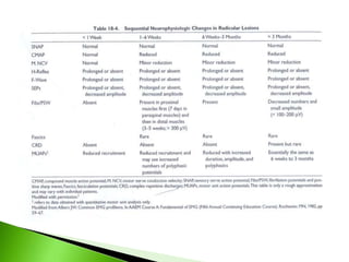
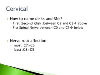
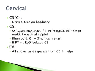
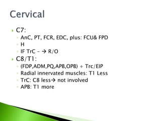
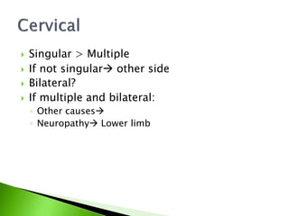
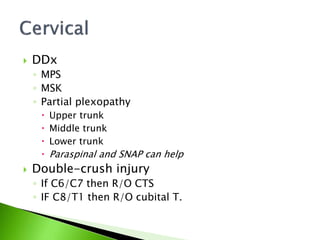
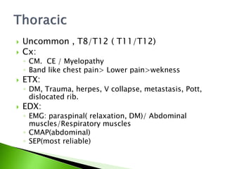
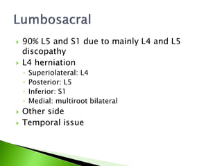
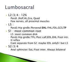
 Origins of pain
 Clinically important tip: What likes root pain?
 Pure sensory Radiculopathy?
 EDX important limitation
 Axonal loss: M&S&R loss
 Conduction Block:
◦ Non- demeylinating
◦ Demeylinating: M&S&R loss
 Conduction slowing
◦ Differential: S&R loss, NCV can be normal:
◦ Synchronized
◦ EDX= extension of neurologic examination
 SNAP:
◦ NL>>Amp>latency
◦ C6,C7,C6&C7,C8,T1
◦ L2-L3, L4,L5,S1
 When SNAP dec?
◦ Proximal DRG (severe L5)
◦ Far lateral Herniation
◦ Gangloionopathy (Dm/ herpes)
 CMAP
◦ NL>Amp>absent>latency: Why?
◦ Abnormality in 6-8 days
◦ C8/T1
◦ L5/S1
◦ S1/S2
 H reflex:( GCS)
◦ S radiculopathy 30-100% abnormality(definition)
◦ Different pathway from ankle jerk reflex
 H reflex:( FCR)
◦ C6&C7 radiculopathy :17.4, 0.7 ms
 Advantage:
◦ sensory radiculopathy
 Disadvantage:
◦ Sensitivity&Specifity
 Abnormal H(GCS)
 F wave
 Fist finding in: GBS/Vasonervoum infarct
 F-wave abnormality: MMN
 Advantage:
◦ Immediately, Not readily accessible araes
 Disadvantage:
◦ Recurrent process
◦ Motor only
◦ If one root AbNl: others can mask it
◦ Dilution
◦ Non specific
 Nerve Root Stim.
◦ C5 SPC5&C6: BB
◦ C6 SPC5-C8:TRC
◦ C7 SPC8&T1:ADM
◦ L5 SP TA
◦ S1 SP GCS(lat)
 Abnormal: amp/ latency
 Advantage:
◦ More sensitive than EMG
 Disadvantage:
◦ Doubt in stim location, only motor, dangers
 Advantage:
◦ Immediately, Sensory
 Disadvantage:
◦ Not sensitive to mild lesions
◦ Variation
◦ Dilution
◦ No perfect localization
◦ mixed: multi root assessment,
◦ segmental: really uni root, cervical?
◦ Dermatomal?
 Advantage:
◦ Probably the highest Dx yield
 Disadvantage
◦ Motor only
◦ Not immediately
◦ Sometimes no conformation to H&PE
 Myotomal charts vs. individual innervation
 Small number of fibers involved
 Cyclical presence of Fib/PSW
 Fib/PSW
◦ Advantage
 Hall mark
 The most sensitive
 Before MUAP morphology change
◦ Timeframe: 1/3/-5,6 weeks. Is it questionable?
 Inc insertional activity
◦ Run of Fib/PSW 50 ms after stopping
◦ First Abnormality after denervation
◦ Don’t base your Dx.
 CRD
◦ Chronic phase, a few muscles: Paraspinal
 Fasciculation
◦ Chronic phase, a few muscles, not paraspinal
◦ Sometimes the only finding
◦ Rare but if myotomal then highly diagnostic
 Hallmark of radiculopathy: paraspinal
denervation (post. rami innervation)
 Technique:
◦ 1-2 cm ( L: 2.5) lateral&1 cm superior to SP
◦ Angel: 45,60 then withdraw
 Special technique in cervical; interspinal
 Instability?
◦ Difficult relaxation
◦ Renervation
 Other diseases :
 MND, Inflammatory myopathy, DM
 Trauma,
 Metastasis
 iatrogenic : myelogeraphy, LP, Rhizolysis,tomy
 Dorsal rami entrapment
 Paraspinal findings after operation
◦ Denervation ,anyway Can we say on “recurrence?
 Techniques:
◦ Lateral placement:
 1 & 3 cm:+ in latter recurrence(?)
◦ Semi-quantitive:
 After 1 y: < 100, if> 300-600 recurrence
 Better in limbs as might be scar in paraS
 Avoid it
 Myotomal denervation in first 1-2 m: Axonal
loss
 The same but after >3 m:
 ongoing or progressive
◦ High Amp
◦ Proximal muscles
 Care on hematoma
 MUAP abnormalities
◦ Red.Recruitment
 Practically Difficult
 Reference data
 Not very useful in
mild lesions
◦ Variability
 In acute phase
◦ Incr.Duration
 Incr.fiber
 asynchronicity
– Polyphasic
• NL
• Needs Quantitive EMG
• Must be myotomal
_ Large, polyphasic and
high duration MUAPs
 SFEMG:
◦ Jitter : inc
◦ Block + acute
◦ Block –  chronic
 Density
 If new symptoms in 4-6 w denervation amp
matters
 Low sensitivity of EMG
◦ Sensory only, Few fibers, slowly progressive,
Temporal
◦ No FP if myotomal denervation
 Imaging: low specificity
_ 30-80% of normal individuals have
abnormal MRI
 H&P/E: not “golden standard”
 So complimentary
 How to name disks and SNs?
◦ First (Second )disk :between C2 and C3 above
◦ Fist Spinal Nerve between C0 and C1 below
 Nerve root affection:
◦ most: C7>C6
◦ least :C8>C5
 C3/C4:
◦ Nerves, tension headache
 C5:
◦ SS,IS,DeL,BB,SuP,BR if + PT,FCR,ECR then C6 or
multi, Paraspinal helpful
◦ Rhomboid: Only (findings matter)
◦ If PT + : R/O isolated C5
 C6:
◦ All above, cant separate from C5; H helps
 C7:
◦ AnC, PT, FCR, EDC, plus: FCU& FPD
◦ H
◦ IF TrC -  R/O
 C8/T1:
◦ (FDP,ADM,PQ,APB,OPB) + Trc/EIP
◦ Radial innervated muscles: T1 Less
◦ TrC: C8 less not involved
◦ APB: T1 more
 Singular > Multiple
 If not singular other side
 Bilateral?
 If multiple and bilateral:
◦ Other causes
◦ Neuropathy Lower limb
 DDx
◦ MPS
◦ MSK
◦ Partial plexopathy
 Upper trunk
 Middle trunk
 Lower trunk
 Paraspinal and SNAP can help
 Double-crush injury
◦ If C6/C7 then R/O CTS
◦ IF C8/T1 then R/O cubital T.
 Uncommon , T8/T12 ( T11/T12)
 Cx:
◦ CM. CE / Myelopathy
◦ Band like chest pain> Lower pain>wekness
 ETX:
◦ DM, Trauma, herpes, V collapse, metastasis, Pott,
dislocated rib.
 EDX:
◦ EMG: paraspinal( relaxation, DM)/ Abdominal
muscles/Respiratory muscles
◦ CMAP(abdominal)
◦ SEP(most reliable)
 90% L5 and S1 due to mainly L4 and L5
discopathy
 L4 herniation
◦ Superiolateral: L4
◦ Posterior: L5
◦ Inferior: S1
◦ Medial: multiroot bilateral
 Other side
 Temporal issue
 L2/3/4 : 12%
◦ ParaS ,IlioP,AL,Gra, Quad
◦ Few nerves, all proximal muscles
 L5 :
◦ ParaS/Hip girdle/Peroenal:EHL/FHL,FDL,GCS,TP
 S1 : most common root
◦ L5 :most common disk
◦ ParaS/Hip girdle/TFL/Post calf,EDL.EHL/Foot intr.
◦ H reflex
◦ Cant separate from S2: maybe EDL onlyS1 but L5
 S2/3/4
◦ Anal sphincter/SoL/Foot inter. Always bilateral
 L2/3/4
◦ Diabetic amyotrophy
◦ Femoral nerve injury
◦ Obturator nerve injury
◦ Lumbar plexopathy: SNAP, paraspinal
 L5:
◦ Foot drop
 Peroenal palsy: SNAP of SPN, Block, EMG
 MND
 Plexopathy
 Peripheral polyneuropathy
 L2/3/4
◦ Diabetic amyotrophy
◦ Femoral nerve injury
◦ Obturator nerve injury
◦ Lumbar plexopathy: SNAP, paraspinal
 L5:
◦ Foot drop
 Peroenal palsy: SNAP of SPN, Block, EMG
 MND
 Plexopathy
 Peripheral polyneuropathy
 S1/S2
◦ Sciatic nerve injury
◦ Sacral plexopathy
 S2/3/4
◦ Pudendal nerve injury

Radiculopathies

  • 2.
     Origins ofpain  Clinically important tip: What likes root pain?  Pure sensory Radiculopathy?
  • 3.
     EDX importantlimitation  Axonal loss: M&S&R loss  Conduction Block: ◦ Non- demeylinating ◦ Demeylinating: M&S&R loss  Conduction slowing ◦ Differential: S&R loss, NCV can be normal: ◦ Synchronized ◦ EDX= extension of neurologic examination
  • 5.
     SNAP: ◦ NL>>Amp>latency ◦C6,C7,C6&C7,C8,T1 ◦ L2-L3, L4,L5,S1  When SNAP dec? ◦ Proximal DRG (severe L5) ◦ Far lateral Herniation ◦ Gangloionopathy (Dm/ herpes)
  • 6.
     CMAP ◦ NL>Amp>absent>latency:Why? ◦ Abnormality in 6-8 days ◦ C8/T1 ◦ L5/S1 ◦ S1/S2
  • 7.
     H reflex:(GCS) ◦ S radiculopathy 30-100% abnormality(definition) ◦ Different pathway from ankle jerk reflex  H reflex:( FCR) ◦ C6&C7 radiculopathy :17.4, 0.7 ms  Advantage: ◦ sensory radiculopathy  Disadvantage: ◦ Sensitivity&Specifity  Abnormal H(GCS)
  • 8.
     F wave Fist finding in: GBS/Vasonervoum infarct  F-wave abnormality: MMN  Advantage: ◦ Immediately, Not readily accessible araes  Disadvantage: ◦ Recurrent process ◦ Motor only ◦ If one root AbNl: others can mask it ◦ Dilution ◦ Non specific
  • 9.
     Nerve RootStim. ◦ C5 SPC5&C6: BB ◦ C6 SPC5-C8:TRC ◦ C7 SPC8&T1:ADM ◦ L5 SP TA ◦ S1 SP GCS(lat)  Abnormal: amp/ latency  Advantage: ◦ More sensitive than EMG  Disadvantage: ◦ Doubt in stim location, only motor, dangers
  • 10.
     Advantage: ◦ Immediately,Sensory  Disadvantage: ◦ Not sensitive to mild lesions ◦ Variation ◦ Dilution ◦ No perfect localization ◦ mixed: multi root assessment, ◦ segmental: really uni root, cervical? ◦ Dermatomal?
  • 11.
     Advantage: ◦ Probablythe highest Dx yield  Disadvantage ◦ Motor only ◦ Not immediately ◦ Sometimes no conformation to H&PE  Myotomal charts vs. individual innervation  Small number of fibers involved  Cyclical presence of Fib/PSW
  • 12.
     Fib/PSW ◦ Advantage Hall mark  The most sensitive  Before MUAP morphology change ◦ Timeframe: 1/3/-5,6 weeks. Is it questionable?  Inc insertional activity ◦ Run of Fib/PSW 50 ms after stopping ◦ First Abnormality after denervation ◦ Don’t base your Dx.
  • 13.
     CRD ◦ Chronicphase, a few muscles: Paraspinal  Fasciculation ◦ Chronic phase, a few muscles, not paraspinal ◦ Sometimes the only finding ◦ Rare but if myotomal then highly diagnostic
  • 14.
     Hallmark ofradiculopathy: paraspinal denervation (post. rami innervation)  Technique: ◦ 1-2 cm ( L: 2.5) lateral&1 cm superior to SP ◦ Angel: 45,60 then withdraw  Special technique in cervical; interspinal  Instability? ◦ Difficult relaxation ◦ Renervation
  • 15.
     Other diseases:  MND, Inflammatory myopathy, DM  Trauma,  Metastasis  iatrogenic : myelogeraphy, LP, Rhizolysis,tomy  Dorsal rami entrapment
  • 16.
     Paraspinal findingsafter operation ◦ Denervation ,anyway Can we say on “recurrence?  Techniques: ◦ Lateral placement:  1 & 3 cm:+ in latter recurrence(?) ◦ Semi-quantitive:  After 1 y: < 100, if> 300-600 recurrence  Better in limbs as might be scar in paraS  Avoid it
  • 17.
     Myotomal denervationin first 1-2 m: Axonal loss  The same but after >3 m:  ongoing or progressive ◦ High Amp ◦ Proximal muscles  Care on hematoma
  • 18.
     MUAP abnormalities ◦Red.Recruitment  Practically Difficult  Reference data  Not very useful in mild lesions ◦ Variability  In acute phase ◦ Incr.Duration  Incr.fiber  asynchronicity – Polyphasic • NL • Needs Quantitive EMG • Must be myotomal _ Large, polyphasic and high duration MUAPs
  • 19.
     SFEMG: ◦ Jitter: inc ◦ Block + acute ◦ Block –  chronic  Density  If new symptoms in 4-6 w denervation amp matters
  • 20.
     Low sensitivityof EMG ◦ Sensory only, Few fibers, slowly progressive, Temporal ◦ No FP if myotomal denervation  Imaging: low specificity _ 30-80% of normal individuals have abnormal MRI  H&P/E: not “golden standard”  So complimentary
  • 22.
     How toname disks and SNs? ◦ First (Second )disk :between C2 and C3 above ◦ Fist Spinal Nerve between C0 and C1 below  Nerve root affection: ◦ most: C7>C6 ◦ least :C8>C5
  • 23.
     C3/C4: ◦ Nerves,tension headache  C5: ◦ SS,IS,DeL,BB,SuP,BR if + PT,FCR,ECR then C6 or multi, Paraspinal helpful ◦ Rhomboid: Only (findings matter) ◦ If PT + : R/O isolated C5  C6: ◦ All above, cant separate from C5; H helps
  • 24.
     C7: ◦ AnC,PT, FCR, EDC, plus: FCU& FPD ◦ H ◦ IF TrC -  R/O  C8/T1: ◦ (FDP,ADM,PQ,APB,OPB) + Trc/EIP ◦ Radial innervated muscles: T1 Less ◦ TrC: C8 less not involved ◦ APB: T1 more
  • 25.
     Singular >Multiple  If not singular other side  Bilateral?  If multiple and bilateral: ◦ Other causes ◦ Neuropathy Lower limb
  • 26.
     DDx ◦ MPS ◦MSK ◦ Partial plexopathy  Upper trunk  Middle trunk  Lower trunk  Paraspinal and SNAP can help  Double-crush injury ◦ If C6/C7 then R/O CTS ◦ IF C8/T1 then R/O cubital T.
  • 27.
     Uncommon ,T8/T12 ( T11/T12)  Cx: ◦ CM. CE / Myelopathy ◦ Band like chest pain> Lower pain>wekness  ETX: ◦ DM, Trauma, herpes, V collapse, metastasis, Pott, dislocated rib.  EDX: ◦ EMG: paraspinal( relaxation, DM)/ Abdominal muscles/Respiratory muscles ◦ CMAP(abdominal) ◦ SEP(most reliable)
  • 28.
     90% L5and S1 due to mainly L4 and L5 discopathy  L4 herniation ◦ Superiolateral: L4 ◦ Posterior: L5 ◦ Inferior: S1 ◦ Medial: multiroot bilateral  Other side  Temporal issue
  • 29.
     L2/3/4 :12% ◦ ParaS ,IlioP,AL,Gra, Quad ◦ Few nerves, all proximal muscles  L5 : ◦ ParaS/Hip girdle/Peroenal:EHL/FHL,FDL,GCS,TP  S1 : most common root ◦ L5 :most common disk ◦ ParaS/Hip girdle/TFL/Post calf,EDL.EHL/Foot intr. ◦ H reflex ◦ Cant separate from S2: maybe EDL onlyS1 but L5  S2/3/4 ◦ Anal sphincter/SoL/Foot inter. Always bilateral
  • 30.
     L2/3/4 ◦ Diabeticamyotrophy ◦ Femoral nerve injury ◦ Obturator nerve injury ◦ Lumbar plexopathy: SNAP, paraspinal  L5: ◦ Foot drop  Peroenal palsy: SNAP of SPN, Block, EMG  MND  Plexopathy  Peripheral polyneuropathy
  • 31.
     L2/3/4 ◦ Diabeticamyotrophy ◦ Femoral nerve injury ◦ Obturator nerve injury ◦ Lumbar plexopathy: SNAP, paraspinal  L5: ◦ Foot drop  Peroenal palsy: SNAP of SPN, Block, EMG  MND  Plexopathy  Peripheral polyneuropathy
  • 32.
     S1/S2 ◦ Sciaticnerve injury ◦ Sacral plexopathy  S2/3/4 ◦ Pudendal nerve injury