2. QUICK ANATOMY
Meningeal layers:
o Dura mater
o Arachnoid mater
o Pia mater
Meningeal spaces:
o Epidural space
o Subdural space
o Subarachnoid space
3. DEFINITION
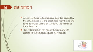
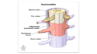
Arachnoiditis is a chronic pain disorder caused by
the inflammation of the arachnoid membrane and
subarachnoid space that surround the nerves of
the spinal cord.
The inflammation can cause the meninges to
adhere to the spinal cord and nerve roots
4. EPIDEMOLOGY
The prevalence is unknown. About 25,000 cases of
arachnoiditis occur each year, mostly in North and South
America, Asia and Europe, where spinal operations are more
prevalent.
5. ETIOLOGY
Complications from spinal surgery or multiple lumbar
punctures: Up to 90% of cases of arachnoiditis have
been linked to lumbar spine surgeries.
Direct injury to the spine: In rare cases, direct trauma
or injury to your spine, such as from a fall or vehicle
accident, can lead to arachnoiditis.
Infection from bacteria or viruses: Infections such as
viral and fungal meningitis, tuberculosis or HIV can
affect your spine and cause arachnoiditis.
6. Chemicals: Dye used in myelograms has been blamed for
some cases of arachnoiditis.
Myelograms are diagnostic tests in which a dye called radiographic contrast
media is injected into the area surrounding your spinal cord and nerves.
Chronic compression of spinal nerves: Chronic compression of
your spinal nerves due to degenerative disc disease or
advanced spinal stenosis (narrowing of your spinal column)
can cause arachnoiditis.
7. CLINICAL PRESENTATION
Severe shooting pain that can be similar to an electric
shock sensation.
Weakness in your legs.
Sensations that may feel like insects crawling on your
skin (formication) or water trickling down your leg.
Difficulty in sitting for a long time
8. SYMPTOMS
Arachnoiditis has no consistent pattern of symptoms, though the
most common symptom is pain.
The symptoms can vary based on which part of your spine
(which spinal nerve) is affected and can range from mild to
severe.
Arachnoiditis most commonly affects the nerves connecting to
your lower back and legs (lumbar spine).
9. Other symptoms, including:
o Headaches.
o Tingling, numbness or weakness in legs.
o Muscle cramps
o spasms and/or uncontrollable twitching.
o Neurogenic bladder.
o Bowel dysfunction.
o Sexual dysfunction, such as erectile dysfunction or
vaginal dryness.
10. PATHOPHYSIOLOGY
In arachnoiditis, damage to and inflammation of the
arachnoid (subarachnoid space) leads to a cascade of
events, including:
o Collagen deposits.
o Scar tissue that encloses nerve roots.
o Fibrosis (thickening or scarring of tissue).
o Decreased cerebrospinal fluid flow.
o Clumping of nerve roots.
11. o Impaired blood supply to the affected nerves.
o Nerve atrophy (wasting).
o Nerve damage.
Due to these changes in the arachnoid and nerve roots,
arachnoiditis frequently results in pain and possible
neurological deficits, such as muscle weakness and sensory
issues.
12. DIAGNOSIS
Magnetic resonance imaging (MRI):
Healthcare provider will look for certain signs of arachnoiditis, such as
nerve root thickening and clumping.
Computed tomography (CT) myelogram:
A myelogram is an imaging procedure that examines the relationship
between your vertebrae and discs, through your spinal cord, nerves and nerve
roots. Your provider will look for certain signs of arachnoiditis.
Lumbar puncture:
Examines the CSF to findout infections in the spinal fluid.
Electromyogram (EMG):
Assess the severity of the damage to the affected nerve roots by using
electrical impulses to check nerve function.
13. MEDICAL MANAGEMENT
Oral medication or medication through an intrathecal pump
such as: non-steroidal anti-inflammatory drugs (NSAIDs)
Methadon, morphine can be used to release neuropathic pain.
Antidepressants may reduce burning neuropathic pain, but in
much lower doses than for depression.
Diazepam is used for muscle relaxation
16. RECENT STUDIES
Dr. Cynthia lewis et. Al, (2006) conducted a study on “Physiotherapy
and spinal nerve root adhesion”
and concluded that The treatment of patients with spinal neuropathic
pain warrants special consideration as far as physiotherapy is
concerned: patients should only be prescribed gentle, individually
tailored exercise.
Szymon jurga et. Al, (2021) conducted case study on “Spinal
adhesive arachnoiditis”
three cases were reported, which diagnosed and confirmed using MRI
and symptoms were managed conservatively.