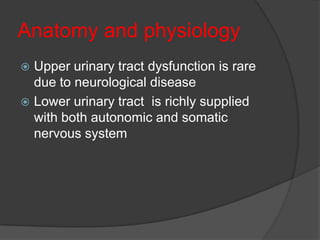
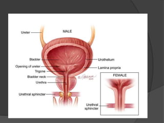
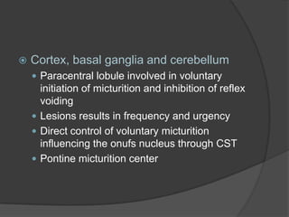
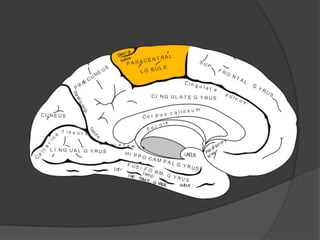
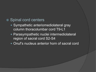
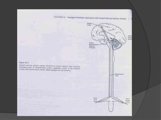
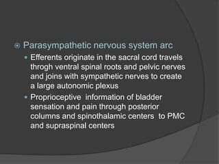
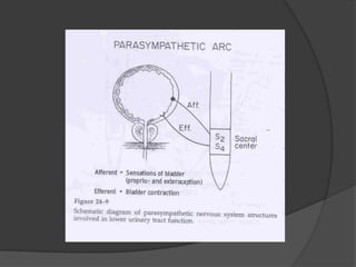
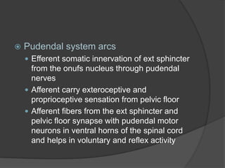
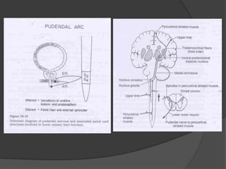
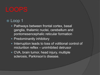
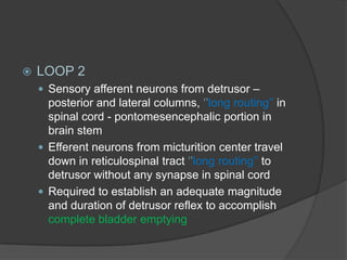
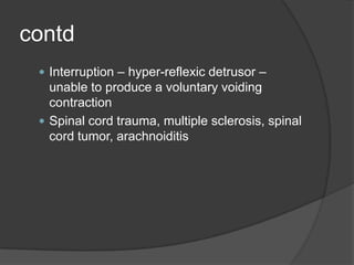
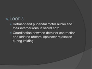
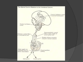
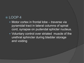
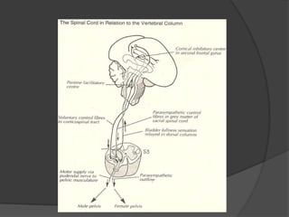
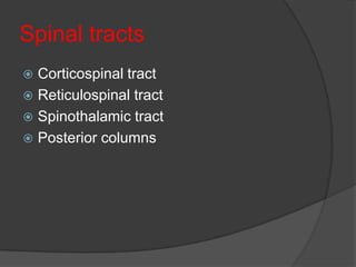
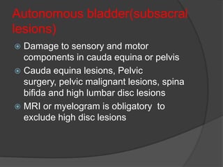
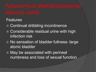
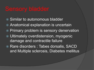
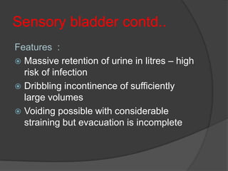
OVERVIEW
 Anatomy and physiology
 CNS centers
 Arcs and loops
 Spinal tracts
 Basic concepts of neurourological
  function
 Reflexes
 Dysfunction
 Pharmacological management
Anatomy and physiology
 Upper urinary tract dysfunction is rare
  due to neurological disease
 Lower urinary tract is richly supplied
  with both autonomic and somatic
  nervous system
Bladder anatomy
   Three anatomical layers
     Inner mucosal layer
     Muscular middle layer
      ○ Outer and inner longitudinal layer
      ○ Middle circular layer
     Outer adventitial layer
   Functinally bladder is divided in to parts
     Body
     Trigone
Receptors of the bladder
 Parasympathetic (musacrinic)
 Sympathetic
     Dual action
     Beta adrenargic
     Alpha adrenergic
CNS centers
   PONS
     Pontomesencephalic reticular formation
     Afferents from bladder receptors of
      distension
     Sphincter detrusor synergesia
     Reticulospinal tracts spincter and detrusor
      centers of the spinal cord
   Cortex, basal ganglia and cerebellum
     Paracentral lobule involved in voluntary
      initiation of micturition and inhibition of reflex
      voiding
     Lesions results in frequency and urgency
     Direct control of voluntary micturition
      influencing the onufs nucleus through CST
     Pontine micturition center
   Spinal cord centers
     Sympathetic anteriomediolateral gray
      column thoracolumbar cord T9-L1
     Parasympathetic nuclei intermediolateral
      region of sacral cord S2-S4
     Onuf’s nucleus anterior horn of sacral cord
Arcs and Loops
   Supra spinal arc
     Parasympathetic afferent input from tension
      receptor in the bladder wall to pontine
      micturition centers
     Reticulospinal tracts to centers to sacral
      cord
     3 to 4 yrs of age voiding is a reflex process
     Lesions above the brain stem manifested
      clinically by frequency and urgency with
      preserved detrusor sphincter synergesia
   Sympathetic nervous system arc
     Efferent sympathetic innervation T9-L1
      through ventral routes, sympathetic ganglia
      in the para vertebral chain preaortic and
      parvertebral chains
     Touch, pain, and temperature from bladder
      through spinothalamic tract
   Parasympathetic nervous system arc
     Efferents originate in the sacral cord travels
      throgh ventral spinal roots and pelvic nerves
      and joins with sympathetic nerves to create
      a large autonomic plexus
     Proprioceptive information of bladder
      sensation and pain through posterior
      columns and spinothalamic centers to PMC
      and supraspinal centers
   Pudendal system arcs
     Efferent somatic innervation of ext sphincter
      from the onufs nucleus through pudendal
      nerves
     Afferent carry exteroceptive and
      proprioceptive sensation from pelvic floor
     Afferent fibers from the ext sphincter and
      pelvic floor synapse with pudendal motor
      neurons in ventral horns of the spinal cord
      and helps in voluntary and reflex activity
LOOPS
   Loop 1
     Pathways between frontal cortex, basal
      ganglia, thalamic nuclei, cerebellum and
      pontomesencephalic reticular formation
     Predominantly inhibitory
     Interruption leads to loss of volitional control of
      micturition reflex – uninhibited detrusor
     CVA, brain tumor, head injury, multiple
      sclerosis, Parkinson’s disease.
   LOOP 2
     Sensory afferent neurons from detrusor –
      posterior and lateral columns, ‘’long routing’’ in
      spinal cord - pontomesencephalic portion in
      brain stem
     Efferent neurons from micturition center travel
      down in reticulospinal tract ‘’long routing’’ to
      detrusor without any synapse in spinal cord
     Required to establish an adequate magnitude
      and duration of detrusor reflex to accomplish
      complete bladder emptying
contd
  Interruption – hyper-reflexic detrusor –
   unable to produce a voluntary voiding
   contraction
  Spinal cord trauma, multiple sclerosis, spinal
   cord tumor, arachnoiditis
   LOOP 3
     Detrusor and pudendal motor nuclei and
      their interneurons in sacral cord
     Coordination between detrusor contraction
      and striated urethral sphincter relaxation
      during voiding
   LOOP 4
     Motor cortex in frontal lobe – traverse via
      pyramidal tract in lateral columns of spinal
      cord, synapse on pudendal sphicter nucleus.
     Voluntary control over striated muscle of the
      urethral sphincter during bladder storage
      and voiding
Spinal tracts
 Corticospinal tract
 Reticulospinal tract
 Spinothalamic tract
 Posterior columns
Basic concepts of
neurourological function
   Two phases
     Low pressure insensanate filling and storage
      of urine
     Efficient evacuation under voluntary control
   Filling and storage of urine
     Passive filling phase initial phase occurs till
      proxim al urethral pressure > exceeds the
      bladder
     Continence reflex phase bladder pressure >
      urethral pressure
 Frontal micturition center by bladder
     distension enhances sympathetic activity
     and external sphincter
   Micturition
     Normal urinary voiding is voluntary
     disinhibition of pontine and sacral reflex
     activity in response to bladder distension
REFLEXES
   Superficial anal reflex
     Anal reflex or anal wink consists contraction
      of anal sphincter in response to stroking or
      pricking the skin of perianal region
     Inferior haemarhoidal nerve (S2-S5)
     Caudaequina or conus medullaris lesions
   Bulbocavernosus reflex
     Stimulating the skin of glans or penis
     response is felt by placing a gloved finger in
     rectum
Neurogenic bladder dysfunction

5 types (2 UMN; 3LMN)
   Uninhibited
   Reflex
   Autonomous
   Motor paralytic
   Sensory paralytic
Nomenclature
 Urgency is the complaint of a sudden and
  compelling desire to pass urine that is
  difficult to defer.
 Urge incontinence is the complaint of
  involuntary leakage accompanied by
  urgency. Leakage may range from drops to
  soaking
 Retention bladder is unable to empty itself
  to a point that there is over 100 cc's (3.5
  ounces) of urine left over in the bladder
  after urinating*
Uninhibited bladder
 Lesion affecting the second frontal gyrus
  and the pathways leading from it down
  to the pontine centre
 Frontal lobe tumours, parasagittal
  meningiomas, anterior communicating
  artery aneurysms, normal perssure
  hydrocephalus, Parkinson’s disease and
  multisystem atrophy
Uninhibited bladder
Features are:
 Urgency at low bladder volumes
  (detrusor hyperreflexia)
 Sudden uncontrollable evacuation
 No residual urine - little risk of infection
 If severe intellectual deterioration occurs
  urine may be passed at random, without
  appropriate concern.
Spinal bladder
 Damage to spinal cord by trauma, tumor, multiple
  sclerosis
 Fullness is not appreciated
 Intravesical pressure may only be indicated by
  sweating, pallor, flexor spasms, dramatic rise in
  blood pressure
 Reflex emptying without warning
 Incomplete evacuation may improve with practice
  and may be performed at will if massaged and
  suprapubic pressure applied
 Detrusor – sphincter dyssynergia .
 Evidence of bilateral pyramidal lesion – enhanced
  reflexes and extensor plantar response
 Bladder is small and contracted, can hold
  maximum of 250ml
Autonomous bladder(subsacral
lesions)
 Damage to sensory and motor
  components in cauda equina or pelvis
 Cauda equina lesions, Pelvic
  surgery, pelvic malignant lesions, spina
  bifida and high lumbar disc lesions
 MRI or myelogram is obligatory to
  exclude high disc lesions
Autonomous bladder(subsacral
lesions) contd
Features
 Continual dribbling incontinence
 Considerable residual urine with high
  infection risk
 No sensation of bladder fullness- large
  atonic bladder
 May be associated with perineal
  numbness and loss of sexual function
Sensory bladder
 Similar to autonomous bladder
 Anatomical explanation is uncertain
 Primary problem is sensory denervation
 Ultimately overdistension, myogenic
  damage and contractile failure
 Rare disorders : Tabes dorsalis, SACD
  and Multiple sclerosis, Diabetes mellitus
Sensory bladder contd..
Features :
 Massive retention of urine in litres – high
  risk of infection
 Dribbling incontinence of sufficiently
  large volumes
 Voiding possible with considerable
  straining but evacuation is incomplete
Motor paralytic
 Areflexic   detrusor
   Marked by painful distention
   Inability to initiate urination
   Difficulty initiating
   urination, straining, decreased size and
   force of stream, interrupted stream, and
   recurrent urinary tract infection.
Pharmacological methods
 Urinary   retention
   Cholinergic agents to increase detrusor
    motor function
    ○ Bathnechol improves detrusor funtion
      particularly in denervation and
      selectively affects bladder and gut
   Alpha adrenergic blockers such as
    prazosin
 Urinary   incontinence
   Inhibition of detrusor activity and increase
    functional capacity of bladder
   Anticholinergics such as propanthaline
   Anticholinergic with smooth muscle
    relaxing properties such as oxybutinin
   TCA such as imipramine with
    anticholinergic activity
Thank u

bladder and its dysfunction

  • 2.
    OVERVIEW  Anatomy andphysiology  CNS centers  Arcs and loops  Spinal tracts  Basic concepts of neurourological function  Reflexes  Dysfunction  Pharmacological management
  • 3.
    Anatomy and physiology Upper urinary tract dysfunction is rare due to neurological disease  Lower urinary tract is richly supplied with both autonomic and somatic nervous system
  • 4.
    Bladder anatomy  Three anatomical layers  Inner mucosal layer  Muscular middle layer ○ Outer and inner longitudinal layer ○ Middle circular layer  Outer adventitial layer  Functinally bladder is divided in to parts  Body  Trigone
  • 6.
    Receptors of thebladder  Parasympathetic (musacrinic)  Sympathetic  Dual action  Beta adrenargic  Alpha adrenergic
  • 8.
    CNS centers  PONS  Pontomesencephalic reticular formation  Afferents from bladder receptors of distension  Sphincter detrusor synergesia  Reticulospinal tracts spincter and detrusor centers of the spinal cord
  • 9.
    Cortex, basal ganglia and cerebellum  Paracentral lobule involved in voluntary initiation of micturition and inhibition of reflex voiding  Lesions results in frequency and urgency  Direct control of voluntary micturition influencing the onufs nucleus through CST  Pontine micturition center
  • 11.
    Spinal cord centers  Sympathetic anteriomediolateral gray column thoracolumbar cord T9-L1  Parasympathetic nuclei intermediolateral region of sacral cord S2-S4  Onuf’s nucleus anterior horn of sacral cord
  • 12.
    Arcs and Loops  Supra spinal arc  Parasympathetic afferent input from tension receptor in the bladder wall to pontine micturition centers  Reticulospinal tracts to centers to sacral cord  3 to 4 yrs of age voiding is a reflex process  Lesions above the brain stem manifested clinically by frequency and urgency with preserved detrusor sphincter synergesia
  • 14.
    Sympathetic nervous system arc  Efferent sympathetic innervation T9-L1 through ventral routes, sympathetic ganglia in the para vertebral chain preaortic and parvertebral chains  Touch, pain, and temperature from bladder through spinothalamic tract
  • 17.
    Parasympathetic nervous system arc  Efferents originate in the sacral cord travels throgh ventral spinal roots and pelvic nerves and joins with sympathetic nerves to create a large autonomic plexus  Proprioceptive information of bladder sensation and pain through posterior columns and spinothalamic centers to PMC and supraspinal centers
  • 19.
    Pudendal system arcs  Efferent somatic innervation of ext sphincter from the onufs nucleus through pudendal nerves  Afferent carry exteroceptive and proprioceptive sensation from pelvic floor  Afferent fibers from the ext sphincter and pelvic floor synapse with pudendal motor neurons in ventral horns of the spinal cord and helps in voluntary and reflex activity
  • 21.
    LOOPS  Loop 1  Pathways between frontal cortex, basal ganglia, thalamic nuclei, cerebellum and pontomesencephalic reticular formation  Predominantly inhibitory  Interruption leads to loss of volitional control of micturition reflex – uninhibited detrusor  CVA, brain tumor, head injury, multiple sclerosis, Parkinson’s disease.
  • 23.
    LOOP 2  Sensory afferent neurons from detrusor – posterior and lateral columns, ‘’long routing’’ in spinal cord - pontomesencephalic portion in brain stem  Efferent neurons from micturition center travel down in reticulospinal tract ‘’long routing’’ to detrusor without any synapse in spinal cord  Required to establish an adequate magnitude and duration of detrusor reflex to accomplish complete bladder emptying
  • 24.
    contd  Interruption– hyper-reflexic detrusor – unable to produce a voluntary voiding contraction  Spinal cord trauma, multiple sclerosis, spinal cord tumor, arachnoiditis
  • 26.
    LOOP 3  Detrusor and pudendal motor nuclei and their interneurons in sacral cord  Coordination between detrusor contraction and striated urethral sphincter relaxation during voiding
  • 28.
    LOOP 4  Motor cortex in frontal lobe – traverse via pyramidal tract in lateral columns of spinal cord, synapse on pudendal sphicter nucleus.  Voluntary control over striated muscle of the urethral sphincter during bladder storage and voiding
  • 30.
    Spinal tracts  Corticospinaltract  Reticulospinal tract  Spinothalamic tract  Posterior columns
  • 31.
    Basic concepts of neurourologicalfunction  Two phases  Low pressure insensanate filling and storage of urine  Efficient evacuation under voluntary control  Filling and storage of urine  Passive filling phase initial phase occurs till proxim al urethral pressure > exceeds the bladder  Continence reflex phase bladder pressure > urethral pressure
  • 32.
     Frontal micturitioncenter by bladder distension enhances sympathetic activity and external sphincter  Micturition  Normal urinary voiding is voluntary disinhibition of pontine and sacral reflex activity in response to bladder distension
  • 33.
    REFLEXES  Superficial anal reflex  Anal reflex or anal wink consists contraction of anal sphincter in response to stroking or pricking the skin of perianal region  Inferior haemarhoidal nerve (S2-S5)  Caudaequina or conus medullaris lesions  Bulbocavernosus reflex  Stimulating the skin of glans or penis response is felt by placing a gloved finger in rectum
  • 34.
    Neurogenic bladder dysfunction 5types (2 UMN; 3LMN)  Uninhibited  Reflex  Autonomous  Motor paralytic  Sensory paralytic
  • 35.
    Nomenclature  Urgency isthe complaint of a sudden and compelling desire to pass urine that is difficult to defer.  Urge incontinence is the complaint of involuntary leakage accompanied by urgency. Leakage may range from drops to soaking  Retention bladder is unable to empty itself to a point that there is over 100 cc's (3.5 ounces) of urine left over in the bladder after urinating*
  • 36.
    Uninhibited bladder  Lesionaffecting the second frontal gyrus and the pathways leading from it down to the pontine centre  Frontal lobe tumours, parasagittal meningiomas, anterior communicating artery aneurysms, normal perssure hydrocephalus, Parkinson’s disease and multisystem atrophy
  • 37.
    Uninhibited bladder Features are: Urgency at low bladder volumes (detrusor hyperreflexia)  Sudden uncontrollable evacuation  No residual urine - little risk of infection  If severe intellectual deterioration occurs urine may be passed at random, without appropriate concern.
  • 38.
    Spinal bladder  Damageto spinal cord by trauma, tumor, multiple sclerosis  Fullness is not appreciated  Intravesical pressure may only be indicated by sweating, pallor, flexor spasms, dramatic rise in blood pressure  Reflex emptying without warning  Incomplete evacuation may improve with practice and may be performed at will if massaged and suprapubic pressure applied  Detrusor – sphincter dyssynergia .  Evidence of bilateral pyramidal lesion – enhanced reflexes and extensor plantar response  Bladder is small and contracted, can hold maximum of 250ml
  • 39.
    Autonomous bladder(subsacral lesions)  Damageto sensory and motor components in cauda equina or pelvis  Cauda equina lesions, Pelvic surgery, pelvic malignant lesions, spina bifida and high lumbar disc lesions  MRI or myelogram is obligatory to exclude high disc lesions
  • 40.
    Autonomous bladder(subsacral lesions) contd Features Continual dribbling incontinence  Considerable residual urine with high infection risk  No sensation of bladder fullness- large atonic bladder  May be associated with perineal numbness and loss of sexual function
  • 41.
    Sensory bladder  Similarto autonomous bladder  Anatomical explanation is uncertain  Primary problem is sensory denervation  Ultimately overdistension, myogenic damage and contractile failure  Rare disorders : Tabes dorsalis, SACD and Multiple sclerosis, Diabetes mellitus
  • 42.
    Sensory bladder contd.. Features:  Massive retention of urine in litres – high risk of infection  Dribbling incontinence of sufficiently large volumes  Voiding possible with considerable straining but evacuation is incomplete
  • 43.
    Motor paralytic  Areflexic detrusor Marked by painful distention Inability to initiate urination Difficulty initiating urination, straining, decreased size and force of stream, interrupted stream, and recurrent urinary tract infection.
  • 44.
    Pharmacological methods  Urinary retention  Cholinergic agents to increase detrusor motor function ○ Bathnechol improves detrusor funtion particularly in denervation and selectively affects bladder and gut  Alpha adrenergic blockers such as prazosin
  • 45.
     Urinary incontinence  Inhibition of detrusor activity and increase functional capacity of bladder  Anticholinergics such as propanthaline  Anticholinergic with smooth muscle relaxing properties such as oxybutinin  TCA such as imipramine with anticholinergic activity
  • 46.