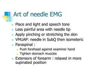
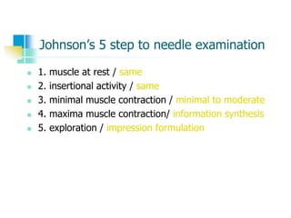
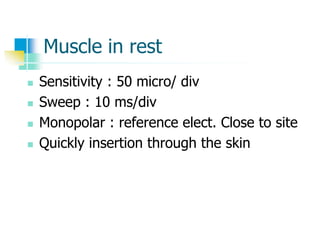
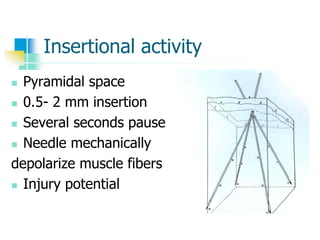
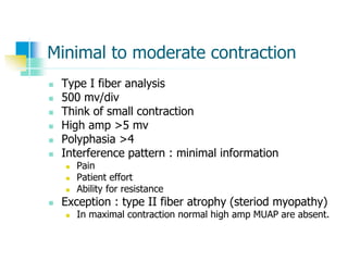
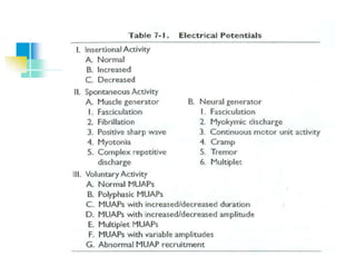
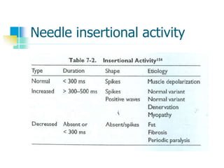
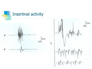
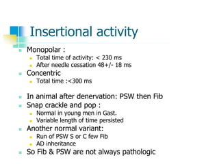
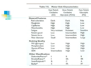
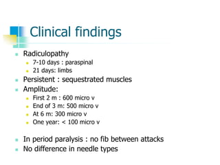
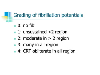
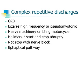
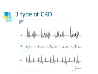
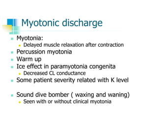
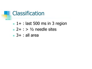
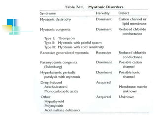
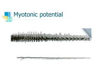
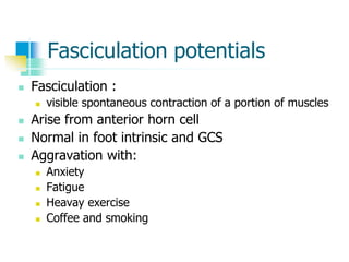
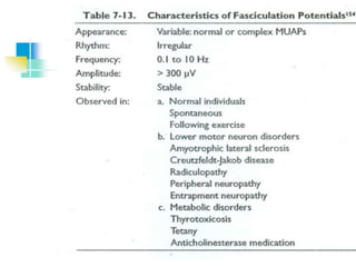
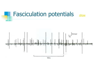
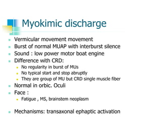
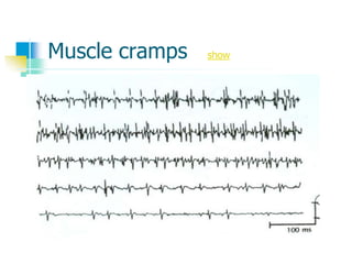
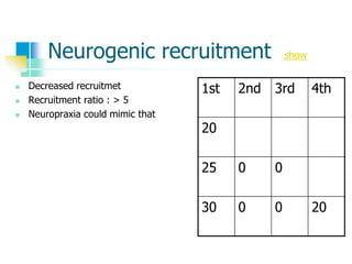
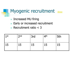
Needle Electromyography
DUMITRU
Chapter 7
Preparation
 Patient anxiety
 Do not show needle
 Gauze
 Muscle soreness 1-2 days
The examiner
 Disposable glove
 Sodium hypochloride 15-20 min
 Distraction technique for entrance
Art of needle EMG
 Place and light and speech tone
 Less painful area with needle tip
 Apply pinching or stretching the skin
 VMUAP: needle in SubQ then isometeric
 Paraspinal :
 Push forehead against examiner hand
 Tighten stomach muscles
 Extensors of forearm : relaxed in more
supinated position
Johnson’s 5 step to needle examination
 1. muscle at rest / same
 2. insertional activity / same
 3. minimal muscle contraction / minimal to moderate
 4. maxima muscle contraction/ information synthesis
 5. exploration / impression formulation
Muscle in rest
 Sensitivity : 50 micro/ div
 Sweep : 10 ms/div
 Monopolar : reference elect. Close to site
 Quickly insertion through the skin
Insertional activity
 Pyramidal space
 0.5- 2 mm insertion
 Several seconds pause
 Needle mechanically
depolarize muscle fibers
 Injury potential
Minimal to moderate contraction
 Type I fiber analysis
 500 mv/div
 Think of small contraction
 High amp >5 mv
 Polyphasia >4
 Interference pattern : minimal information
 Pain
 Patient effort
 Ability for resistance
 Exception : type II fiber atrophy (steriod myopathy)
 In maximal contraction normal high amp MUAP are absent.
Information synthesis
 EDX examination differ from reading
EKG in isolation from patient.
 Finally impression formulation
Electrophysiology
action potential generation
 Membrane potential depolarized from -
80 mV to -50 to -60 mV ( threshold)
 Voltage gated Na channel activated 0.5 ms
 Then depolarization to +40 mV
 Delayed increased K permeability with
inactivation of Na channel 2.0 ms
Isopotential lines
Needle insertional activity
Insertinal activity
Insertional activity
 Monopolar :
 Total time of activity: < 230 ms
 After needle cessation 48+/- 18 ms
 Concentric
 Total time :<300 ms
 In animal after denervation: PSW then Fib
 Snap crackle and pop :
 Normal in young men in Gast.
 Variable length of time persisted
 Another normal variant:
 Run of PSW S or C few Fib
 AD inheritance
 So Fib & PSW are not always pathologic
Spontaneous activity
 After placing a needle into endplate
 Miniature endplate potential (MEPPs)
 Endplate spike
Difference between MEEPs and endplates
Innervation ratio
Single muscle fiber
 It’s a triphasic wave (+ - +)
 Biphasic (positive-negative) seen in:
 Positive terminal wave is small and lost
 Musculotendinous junction
 Endplate zone
MUAPs
Anatomy
 Motor unit definition
 Types:
 Alpha: skeletomotor
 Beta : skeletofusimotor
 Gamma: fusiomotor
 Alpha situated in ventrolateral horn
 Limbs motor units located laterally in comparison with neck and
trunk muscle.
 They have axon collateral to synapse with Renshaw interneourons
and other alpha motor neurons
 One MU may be distributed over 100 muscle fiber
 MUs dos not extended between 2 muscle
 One single fiber innerveted by only one MN
 Sometimes 2 NMJ seen but its from one MN
 Electrophysiolgic counting
 Incremental motor unit counting
 Max CMAP divided by mean incremental amplitude
 PAS staining
 Circular region about 20-30% muscle volume
 Higher density in enter of motor unit
 Upper limb MU territory: circular to oval 5-7 mm
 Lower limb MU territory: m7-12 mm
Periodic acid shift staining
Physiology
 MU classifications
 Fast twitch
 More force innervated by fact conductive neurons
 Slow twitch
 Small force by slow conductive neurons
 Speed of contraction
 Specific contractile proteins
 Rate of calcium uptake
 Fast twitches have
 More extensive sarcoplasmic reticulum
 Richer in Ca-Mg ATPase
Motor units classification
 Fast fatigue (FF)
 Readily fatigue
 High tetanus tension
 Fast contraction time
 Slow (s)
 Low tetanus tension
 Long contraction time
 Resistant to fatigue
 Third group
 Moderate tetanic tension
 Fast twitch time
 Relatively fatigue resistant (FR)
 Forth small intermediate population (F int)
 Between FF and FR
Histochemical technique
 Fatigue resistant: higher activity of
 Mitochondrial enzymes
 Succinate dehydrogenase
 Fatigue sensitive: higher activity in
 Phosphorylase activity (myosin ATPase
Recruitment principles
 Henneman size principle
 First MU activated is weakest
 Then large MUs activated
 So contraction in smooth and
appropriate to need
Parameters
 In one territory
 10 MU type I and more than 30 (I&II)
 Interdigited muscle fibers
 Active and inactive fibers
 Muscle tissue act as LPF (HFF)
 Reduce peck amplitude
MUAP parameters
figure
 Amplitude
 Rise time
 Duration
 Phase
 Spike duration
 Initial and terminal portion
Phase
 Polyphay : 5 or more
 Monopolar:30% nl
 Concentric: 15% nl
 Main spike arise from
 1-12 muscle fiber
 <500 mic m distance
 Distant potential :After filtering
 low frequency initial and terminal
 Small negative spike
Instrumentation factor
 Reduction in MUAP amplitude
 Reduced HFF
 Concentric needle
 Large electrode lead off surface
 Rotation on concentric needle
 Removal of Teflon in monopolar
Effect of temperature on MUAP
 Controversial
 Authors opinion:
 Reduction of temp
 Increased initially
 More dec. in temp.: AP failure and MUAP decreased
 In >65 Y : MUAP increased
MUAP duration
 Duration depend on:
 Width of Endplate Zone
 Lesser: terminal axon conduction velocity
 Inc LFF:
 Deletion of initial and terminal
 Dec. duration
 Concentric needle :
 Reduced duration
 Cooling
 Increased duration
Phase
 Synchronocity
 Aging and temperature reducing:
 Increased phase
 Elevating LFF : inc phasicity
 Monopolar have more polyphasic potential
Recruitment
 Principles:
 First MUAP : small and slow type I
 Minimal to moderate force needed
 Late type II MUAPs in not simple to analyze
 Subtle pathology my be miss
 Increasing fire rate of first MUAP with more force
 Slow and constant contraction is sufficient for analysis
Others
 Equipment :
 LFF: 10-30
 HFF: 10,000 or more
 Sweep speed 20 ms/div
 Techniques :
 Identify first MUAP and measure firing rate
Recruitment
 Recruitment frequency:
 Frequency of MUAP A when MUAP B begin fireing
 Recognition:
 Observing for second MU to appear
 Change in sound
 Recruitment interval
 Distance between 2 peaks of MUAP
 Normal muscle
 Recruitment frequency: 10 Hz
 Recruitment interval: 100 ms
 Facial muscles: short interval and high frequency
Normal recruitment
1st 2nd 3rd 4th
5 Hz
10 Hz 5 Hz
15 Hz 10 Hz 5 Hz
20 Hz 15 Hz 10 Hz 5 Hz
Recruitment ratio
 Recruitment ratio:
 Frequency of fastest MUAP divided by number of
Different MUAP in screen
 Should be close to 5
 If near 10: too few MU presented (neurogenic)
 If below 4 : too many MU existed (myogenic)
 Predicting firing rate:
Insertional activity
 Decreased I.A :
 Fibrosis
 Sub Q
 Attack of periodic paralysis
 Improper electrode connection
 Increased IA:
 Muscle denervation
 Other membrane instability
Fibrillation potential
 4-6 or longer days
 Threshold level and regularly repetition
 Previous endplate
 Decreased with:
 Decreased temperature
 Ischemia
 D-tubocurarine
 50 % regular
 High pitched: crackling cellophane or rain
Positive sharp waves
 Positive wave terminated to base line
 Monophasic
 Same ethiology
 Purely demyelinatig disease have PSWs
but no fibrilation
 Combination of Fib/PSW
Clinical findings
 Radiculopathy
 7-10 days : paraspinal
 21 days: limbs
 Persistent : sequestrated muscles
 Amplitude:
 First 2 m : 600 micro v
 End of 3 m: 500 micro v
 At 6 m: 300 micro v
 One year: < 100 micro v
 In period paralysis : no fib between attacks
 No difference in needle types
Grading of fibrillation potentials
 0: no fib
 1: unsustained <2 region
 2: moderate in > 2 region
 3: many in all region
 4: CRT obliterate in all region
Complex repetitive discharges
 CRD
 Bizarre high frequency or pseudomyotonic
 Heavy machinery or idling motorcycle
 Hallmark : start and stop abruptly
 Not stop with nerve block
 Ephaptical pathway
3 type of CRD
Myotonic discharge
 Myotonia:
 Delayed muscle relaxation after contraction
 Percussion myotonia
 Warm up
 Ice effect in paramyotonia congenita
 Decreased CL conductance
 Some patient severity related with K level
 Sound dive bomber ( waxing and waning)
 Seen with or without clinical myotonia
Classification
 1+ : last 500 ms in 3 region
 2+ : > ½ needle sites
 3+ : all area
Myotonic potential
Fasciculation potentials
 Fasciculation :
 visible spontaneous contraction of a portion of muscles
 Arise from anterior horn cell
 Normal in foot intrinsic and GCS
 Aggravation with:
 Anxiety
 Fatigue
 Heavay exercise
 Coffee and smoking
Fasciculation potentials show
Myokimic discharge
 Vermicular movement movement
 Burst of normal MUAP with interburst silence
 Sound : low power motor boat engine
 Difference with CRD:
 No regularity in burst of MUs
 No typical start and stop abruptly
 They are group of MU but CRD single muscle fiber
 Normal in orbic. Oculi
 Face :
 Fatigue , MS, brainstem neoplasm
 Mechanisms: transaxonal ephaptic activation
Myokymic potentials
Continuous muscle fiber activity
 CNS and PNS are involved
 Stiff man syndrome:
 CNS: so inhibited with
 Peripheral and NMJ and spinal block
 General anesthesia and sleep
 Diazepam but not phenytion and CMZ
 Isaacs syndrome (neuromyotonia):
 PNS: so inhibited with
 Nm block
 But no peripheral or spinal block or
 General anesthesia or sleep not diminished
 Auto Ab against nerve voltage gated K channels
Cramps
 Sustained or painful contraction of multiple MU
 Exercise or malpositioning
 Needle: multiple MU synchronously firing 40-50 or 300 Hz
 Induced by :
 Hyponatremia
 Hypocalcemia
 Vitamin deficiency
 Ischemia
 Arise from peripheral portion of MU
 Cramp with electrical silence
 Mc Ardle’s disease
Muscle cramps show
Multiplet discharge
 Rapid firing of single MU
 Doublets, triplets or multiplet
 Tetany:
 Muscle twitching or Carpopedal spasm
 Association with:
 Alkalosis
 Hypocalcemia and magnesiumemia
 Hyperkalemia
 Local ischemia
 Chvostek’s sign facial nerve
 Peroneal sign : fibular head
 Trousseau’s sign : limb ischemia
Doublet and triplet
Neural loss
 2 process after denervation:
 Re-growth 2-3 mm/day
 Collateral sprouting
 Point of node of Ranvier
 Bunds of bunger : remaining Schwan cell sheaths
 Each MN : support 4-5 times
 So 20 % remaining could cover others
 Fiber types grouping
 2 weeks
 MU territory remain unchanged
Muscle loss
 Muscle fiber loss
 Muscle fibers approximated but no
electrical summation
 Fiber type grouping also occur
MUAP findings in muscle disease
 MUAP duration decreased
 Increase number of phase
 Fiber splitting and slow conduction:
 Satellite potential
 10% in normal
 12 % in neuropathy
 45 % in myopathy
 Responsible for abnormal long duration 60 ms
 Large MUAP: needle near hypertrophic fibers
 Segmental necrosis : Fib/PSW
Neurogenic recruitment show
 Decreased recruitmet
 Recruitment ratio : > 5
 Neuropraxia could mimic that
1st 2nd 3rd 4th
20
25 0 0
30 0 0 20
Myogenic recruitment show
 Increased MU firing
 Early or increased recruitment
 Recruitment ratio < 3
1st 2nd 3rd 4th 5th
15 15 15 15 15
Other entity
 CVA
 First MU 7-8 firing rate
 Second MU 10-15
 50-60 Hz
 EKG 1Hz
 Pacemaker
Pain
 Cervical paraspinal
 Lumbosacral paraspinal
 Intrinsic hand muscle
Contraindication
 No absolute
 Relative :
 Needle in coagulopathy
 Increased bleeding in:
 Plt < 50.000
 PT & PTT > 1.5 – 2 times nl
 Hemophilia: optimized clotting factors
 Lymphedema:
 Infection : iodine solution use
 Alcohol : questionable
 HIV chance : 0.5 % after needling
 Pneumothorax
 Cervical if needle far from mid line inserted
Other entity
 Muscle biopsy
 CK level 1.5 nl return 48 H to base line

Needle electromyography

  • 1.
  • 2.
    Preparation  Patient anxiety Do not show needle  Gauze  Muscle soreness 1-2 days
  • 3.
    The examiner  Disposableglove  Sodium hypochloride 15-20 min  Distraction technique for entrance
  • 4.
    Art of needleEMG  Place and light and speech tone  Less painful area with needle tip  Apply pinching or stretching the skin  VMUAP: needle in SubQ then isometeric  Paraspinal :  Push forehead against examiner hand  Tighten stomach muscles  Extensors of forearm : relaxed in more supinated position
  • 5.
    Johnson’s 5 stepto needle examination  1. muscle at rest / same  2. insertional activity / same  3. minimal muscle contraction / minimal to moderate  4. maxima muscle contraction/ information synthesis  5. exploration / impression formulation
  • 6.
    Muscle in rest Sensitivity : 50 micro/ div  Sweep : 10 ms/div  Monopolar : reference elect. Close to site  Quickly insertion through the skin
  • 7.
    Insertional activity  Pyramidalspace  0.5- 2 mm insertion  Several seconds pause  Needle mechanically depolarize muscle fibers  Injury potential
  • 8.
    Minimal to moderatecontraction  Type I fiber analysis  500 mv/div  Think of small contraction  High amp >5 mv  Polyphasia >4  Interference pattern : minimal information  Pain  Patient effort  Ability for resistance  Exception : type II fiber atrophy (steriod myopathy)  In maximal contraction normal high amp MUAP are absent.
  • 9.
    Information synthesis  EDXexamination differ from reading EKG in isolation from patient.  Finally impression formulation
  • 10.
    Electrophysiology action potential generation Membrane potential depolarized from - 80 mV to -50 to -60 mV ( threshold)  Voltage gated Na channel activated 0.5 ms  Then depolarization to +40 mV  Delayed increased K permeability with inactivation of Na channel 2.0 ms
  • 12.
  • 14.
  • 15.
  • 16.
    Insertional activity  Monopolar:  Total time of activity: < 230 ms  After needle cessation 48+/- 18 ms  Concentric  Total time :<300 ms  In animal after denervation: PSW then Fib  Snap crackle and pop :  Normal in young men in Gast.  Variable length of time persisted  Another normal variant:  Run of PSW S or C few Fib  AD inheritance  So Fib & PSW are not always pathologic
  • 17.
    Spontaneous activity  Afterplacing a needle into endplate  Miniature endplate potential (MEPPs)  Endplate spike
  • 18.
  • 19.
  • 20.
    Single muscle fiber It’s a triphasic wave (+ - +)  Biphasic (positive-negative) seen in:  Positive terminal wave is small and lost  Musculotendinous junction  Endplate zone
  • 21.
    MUAPs Anatomy  Motor unitdefinition  Types:  Alpha: skeletomotor  Beta : skeletofusimotor  Gamma: fusiomotor  Alpha situated in ventrolateral horn  Limbs motor units located laterally in comparison with neck and trunk muscle.  They have axon collateral to synapse with Renshaw interneourons and other alpha motor neurons  One MU may be distributed over 100 muscle fiber  MUs dos not extended between 2 muscle  One single fiber innerveted by only one MN  Sometimes 2 NMJ seen but its from one MN
  • 22.
     Electrophysiolgic counting Incremental motor unit counting  Max CMAP divided by mean incremental amplitude  PAS staining  Circular region about 20-30% muscle volume  Higher density in enter of motor unit  Upper limb MU territory: circular to oval 5-7 mm  Lower limb MU territory: m7-12 mm
  • 23.
  • 24.
    Physiology  MU classifications Fast twitch  More force innervated by fact conductive neurons  Slow twitch  Small force by slow conductive neurons  Speed of contraction  Specific contractile proteins  Rate of calcium uptake  Fast twitches have  More extensive sarcoplasmic reticulum  Richer in Ca-Mg ATPase
  • 25.
    Motor units classification Fast fatigue (FF)  Readily fatigue  High tetanus tension  Fast contraction time  Slow (s)  Low tetanus tension  Long contraction time  Resistant to fatigue  Third group  Moderate tetanic tension  Fast twitch time  Relatively fatigue resistant (FR)  Forth small intermediate population (F int)  Between FF and FR
  • 26.
    Histochemical technique  Fatigueresistant: higher activity of  Mitochondrial enzymes  Succinate dehydrogenase  Fatigue sensitive: higher activity in  Phosphorylase activity (myosin ATPase
  • 28.
    Recruitment principles  Hennemansize principle  First MU activated is weakest  Then large MUs activated  So contraction in smooth and appropriate to need
  • 29.
    Parameters  In oneterritory  10 MU type I and more than 30 (I&II)  Interdigited muscle fibers  Active and inactive fibers  Muscle tissue act as LPF (HFF)  Reduce peck amplitude
  • 30.
    MUAP parameters figure  Amplitude Rise time  Duration  Phase  Spike duration  Initial and terminal portion
  • 31.
    Phase  Polyphay :5 or more  Monopolar:30% nl  Concentric: 15% nl  Main spike arise from  1-12 muscle fiber  <500 mic m distance  Distant potential :After filtering  low frequency initial and terminal  Small negative spike
  • 32.
    Instrumentation factor  Reductionin MUAP amplitude  Reduced HFF  Concentric needle  Large electrode lead off surface  Rotation on concentric needle  Removal of Teflon in monopolar
  • 33.
    Effect of temperatureon MUAP  Controversial  Authors opinion:  Reduction of temp  Increased initially  More dec. in temp.: AP failure and MUAP decreased  In >65 Y : MUAP increased
  • 34.
    MUAP duration  Durationdepend on:  Width of Endplate Zone  Lesser: terminal axon conduction velocity  Inc LFF:  Deletion of initial and terminal  Dec. duration  Concentric needle :  Reduced duration  Cooling  Increased duration
  • 35.
    Phase  Synchronocity  Agingand temperature reducing:  Increased phase  Elevating LFF : inc phasicity  Monopolar have more polyphasic potential
  • 36.
    Recruitment  Principles:  FirstMUAP : small and slow type I  Minimal to moderate force needed  Late type II MUAPs in not simple to analyze  Subtle pathology my be miss  Increasing fire rate of first MUAP with more force  Slow and constant contraction is sufficient for analysis
  • 37.
    Others  Equipment : LFF: 10-30  HFF: 10,000 or more  Sweep speed 20 ms/div  Techniques :  Identify first MUAP and measure firing rate
  • 38.
    Recruitment  Recruitment frequency: Frequency of MUAP A when MUAP B begin fireing  Recognition:  Observing for second MU to appear  Change in sound  Recruitment interval  Distance between 2 peaks of MUAP  Normal muscle  Recruitment frequency: 10 Hz  Recruitment interval: 100 ms  Facial muscles: short interval and high frequency
  • 39.
    Normal recruitment 1st 2nd3rd 4th 5 Hz 10 Hz 5 Hz 15 Hz 10 Hz 5 Hz 20 Hz 15 Hz 10 Hz 5 Hz
  • 40.
    Recruitment ratio  Recruitmentratio:  Frequency of fastest MUAP divided by number of Different MUAP in screen  Should be close to 5  If near 10: too few MU presented (neurogenic)  If below 4 : too many MU existed (myogenic)  Predicting firing rate:
  • 41.
    Insertional activity  DecreasedI.A :  Fibrosis  Sub Q  Attack of periodic paralysis  Improper electrode connection  Increased IA:  Muscle denervation  Other membrane instability
  • 42.
    Fibrillation potential  4-6or longer days  Threshold level and regularly repetition  Previous endplate  Decreased with:  Decreased temperature  Ischemia  D-tubocurarine  50 % regular  High pitched: crackling cellophane or rain
  • 43.
    Positive sharp waves Positive wave terminated to base line  Monophasic  Same ethiology  Purely demyelinatig disease have PSWs but no fibrilation  Combination of Fib/PSW
  • 44.
    Clinical findings  Radiculopathy 7-10 days : paraspinal  21 days: limbs  Persistent : sequestrated muscles  Amplitude:  First 2 m : 600 micro v  End of 3 m: 500 micro v  At 6 m: 300 micro v  One year: < 100 micro v  In period paralysis : no fib between attacks  No difference in needle types
  • 45.
    Grading of fibrillationpotentials  0: no fib  1: unsustained <2 region  2: moderate in > 2 region  3: many in all region  4: CRT obliterate in all region
  • 46.
    Complex repetitive discharges CRD  Bizarre high frequency or pseudomyotonic  Heavy machinery or idling motorcycle  Hallmark : start and stop abruptly  Not stop with nerve block  Ephaptical pathway
  • 47.
  • 48.
    Myotonic discharge  Myotonia: Delayed muscle relaxation after contraction  Percussion myotonia  Warm up  Ice effect in paramyotonia congenita  Decreased CL conductance  Some patient severity related with K level  Sound dive bomber ( waxing and waning)  Seen with or without clinical myotonia
  • 49.
    Classification  1+ :last 500 ms in 3 region  2+ : > ½ needle sites  3+ : all area
  • 51.
  • 52.
    Fasciculation potentials  Fasciculation:  visible spontaneous contraction of a portion of muscles  Arise from anterior horn cell  Normal in foot intrinsic and GCS  Aggravation with:  Anxiety  Fatigue  Heavay exercise  Coffee and smoking
  • 54.
  • 55.
    Myokimic discharge  Vermicularmovement movement  Burst of normal MUAP with interburst silence  Sound : low power motor boat engine  Difference with CRD:  No regularity in burst of MUs  No typical start and stop abruptly  They are group of MU but CRD single muscle fiber  Normal in orbic. Oculi  Face :  Fatigue , MS, brainstem neoplasm  Mechanisms: transaxonal ephaptic activation
  • 57.
  • 58.
    Continuous muscle fiberactivity  CNS and PNS are involved  Stiff man syndrome:  CNS: so inhibited with  Peripheral and NMJ and spinal block  General anesthesia and sleep  Diazepam but not phenytion and CMZ  Isaacs syndrome (neuromyotonia):  PNS: so inhibited with  Nm block  But no peripheral or spinal block or  General anesthesia or sleep not diminished  Auto Ab against nerve voltage gated K channels
  • 59.
    Cramps  Sustained orpainful contraction of multiple MU  Exercise or malpositioning  Needle: multiple MU synchronously firing 40-50 or 300 Hz  Induced by :  Hyponatremia  Hypocalcemia  Vitamin deficiency  Ischemia  Arise from peripheral portion of MU  Cramp with electrical silence  Mc Ardle’s disease
  • 60.
  • 61.
    Multiplet discharge  Rapidfiring of single MU  Doublets, triplets or multiplet  Tetany:  Muscle twitching or Carpopedal spasm  Association with:  Alkalosis  Hypocalcemia and magnesiumemia  Hyperkalemia  Local ischemia  Chvostek’s sign facial nerve  Peroneal sign : fibular head  Trousseau’s sign : limb ischemia
  • 62.
  • 63.
    Neural loss  2process after denervation:  Re-growth 2-3 mm/day  Collateral sprouting  Point of node of Ranvier  Bunds of bunger : remaining Schwan cell sheaths  Each MN : support 4-5 times  So 20 % remaining could cover others  Fiber types grouping  2 weeks  MU territory remain unchanged
  • 64.
    Muscle loss  Musclefiber loss  Muscle fibers approximated but no electrical summation  Fiber type grouping also occur
  • 65.
    MUAP findings inmuscle disease  MUAP duration decreased  Increase number of phase  Fiber splitting and slow conduction:  Satellite potential  10% in normal  12 % in neuropathy  45 % in myopathy  Responsible for abnormal long duration 60 ms  Large MUAP: needle near hypertrophic fibers  Segmental necrosis : Fib/PSW
  • 66.
    Neurogenic recruitment show Decreased recruitmet  Recruitment ratio : > 5  Neuropraxia could mimic that 1st 2nd 3rd 4th 20 25 0 0 30 0 0 20
  • 67.
    Myogenic recruitment show Increased MU firing  Early or increased recruitment  Recruitment ratio < 3 1st 2nd 3rd 4th 5th 15 15 15 15 15
  • 68.
    Other entity  CVA First MU 7-8 firing rate  Second MU 10-15  50-60 Hz  EKG 1Hz  Pacemaker
  • 69.
    Pain  Cervical paraspinal Lumbosacral paraspinal  Intrinsic hand muscle
  • 70.
    Contraindication  No absolute Relative :  Needle in coagulopathy  Increased bleeding in:  Plt < 50.000  PT & PTT > 1.5 – 2 times nl  Hemophilia: optimized clotting factors  Lymphedema:  Infection : iodine solution use  Alcohol : questionable  HIV chance : 0.5 % after needling  Pneumothorax  Cervical if needle far from mid line inserted
  • 71.
    Other entity  Musclebiopsy  CK level 1.5 nl return 48 H to base line