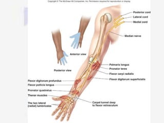
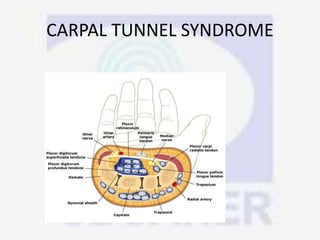
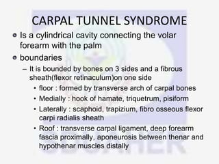
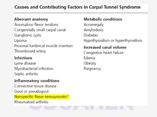
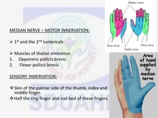
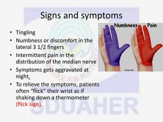
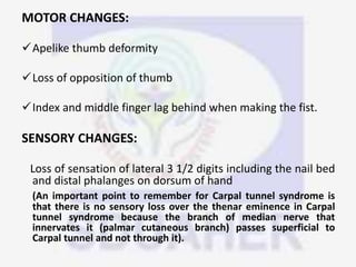
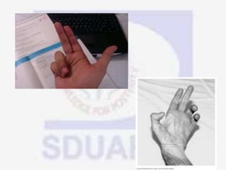
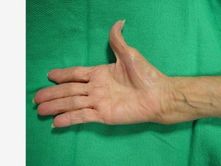
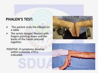
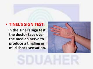
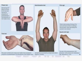
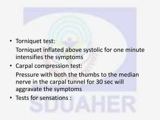
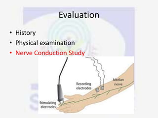
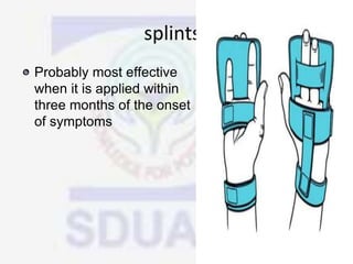
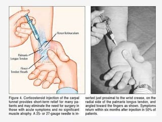
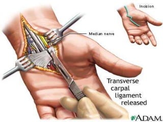
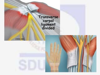
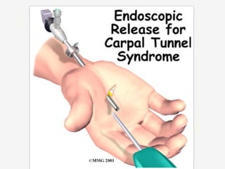
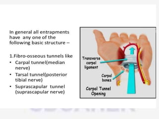
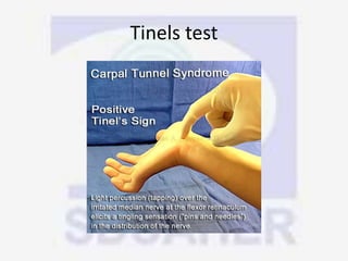
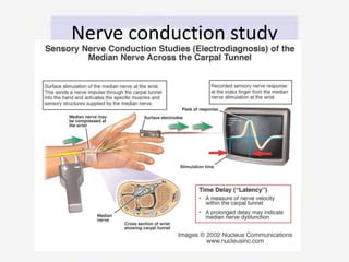
Nerve compression syndrome, also known as entrapment neuropathy, occurs when a peripheral nerve is compressed, causing mechanical damage. Carpal tunnel syndrome is a common example, where the median nerve is compressed as it passes through the carpal tunnel in the wrist. Symptoms include tingling, numbness, and pain in the fingers innervated by the median nerve that is worsened at night. Physical exams and tests like Phalen's maneuver, Tinel's sign, and nerve conduction studies can help diagnose CTS. Treatment involves splinting, medications, injections, or carpal tunnel release surgery if conservative measures fail.
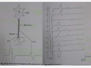
3. DEFINITION
⢠Nerve compression syndrome/ compression
neuropathy/ Entrapment Neuropathy is
defined as: Pressure or Pressure induced
injury to a segment of a peripheral nerve
secondary to anatomical or pathological
structures
4. INTRODUCTION
⢠The nerve is injured by
1. chronic direct compression,
2. angulations
3. stretching forces
causing mechanical damage to
the nerve.
8. Micro anatomy
⢠Each nerve fiber, or axon, is a direct extension of a
dorsal root ganglion cell (sensory), an anterior horn cell
(motor), or a postganglionic sympathetic nerve cell,
and it is either myelinated or unmyelinated.
⢠Sensory and motor nerves contain both unmyelinated
and myelinated fibers in a ratio of 4 to 1
⢠In the unmyelinated or sparsely myelinated fibers
several axons are wrapped by a single Schwann cell. In
the more heavily myelinated fibers the Schwann cell by
rotation forms a multilaminated structure that encloses
a myelin sheath about a single axon.
9.
10. ⢠The segment of myelinated nerve fiber enclosed by a single
Schwann cell is referred to as an in-ternode and varies in
length between 0.1 and 1.8 mm with the more heavily
myelinated fibers having the longer internodes
⢠The point at which one Schwann cell ends and the next begins
is relatively sparse in myelin and is called the nodal gap, or
node of Ranvier .
⢠The axon with its Schwann cell and myelin sheath is in turn
surrounded by a veil of delicate fibrous tissue called the
endoneurium. Seen longitudinally, the endoneurium forms a
tube encircling individually the Schwann cell sheaths that
cluster together to form a fascicle (or funicle as termed by
Sunderland). Each fascicle or separate group of sheathed
axons is in turn surrounded by a denser layer of perineurium.
11. ⢠The entire group of fascicles with their surrounding
perineurium is encased as a mixed spinal or peripheral
nerve in a denser epineurium
⢠The blood supply to the peripheral nerve enters through
the mesoneurium, which is loose connective tissue
extending from the epineurium to the surrounding tissues.
⢠There is both an extrinsic (segmental) as well as an intrinsic
(longitudinal) blood supply to each nerve.
⢠The intrinsic blood supply that runs longitudinally within
the epineurium, perineurium, and endoneurium is fairly
extensive and allows surgical mobilization without
complete devascularization over variable lengths of nerves.
14. PATHOPHYISOLOGY
⢠Focal slowing of Nerve conduction is the principal
electrophysiological feature of entrapment neuropathy
⢠Mild degrees of pressure(suprasystolic) applied to the nerve
for short periods produce reversible dysfunction d/t
ischemia(entrapped nerve more sensitive to ischemia than
normal nerve)
⢠Acute ischemia may be responsible for paresthesias and
dysethesias
⢠Prolonged ischemia may l/t neural tissue infarction
⢠Epineurium protects against compression
⢠Epineurium and perineurium protect against stretch
16. Double Crush and multiple crush syndromes
⢠A proximal level of nerve compression could cause more
distal sites to be susceptible to compression.
⢠The summation of compression along the nerve would result
in alterations of axoplasmic flow
⢠The possibility of a distal site of compression making the
more proximal nerve susceptible to secondary compression: A
reverse double crush.
⢠Systemic diseases such as obesity, diabetes, thyroid disease,
alcoholism, rheumatoid arthritis and neuropatthies lower the
threshold for the occurrence of a nerve compression and
alter axoplasmic transport rendering that nerve more
susceptible to develop compression neuropathy and act as a
âcrushâ.
18. Entrapment Neuropathy in Diabetes
⢠DM is a significant predisposing factor for entrapment
neuropathies .
⢠Metabolic and phenotypic abnormalities of endoneurial and
perineurial fibroblasts lies behind the vulnerability of DM
patients to entrapment neuropathy.
⢠In contrast to angiopathies, retinopathy, and nephropathy,
three representative complications of DM, mast cells do not
play significant roles in the onset or progression of the
entrapment neuropathy associated with DM.
19.
20. Classification of nerve injuries
Seddon Classification
Neuropraxia:
1.Minor contusion or compression with preservation of axis â cylinder of myelin
sheath.
2.Impulse transmission physiologically interrupted.
3.Complete recovery in a few days to weeks.
Axonotemesis :
1.More significant injury
2.Breakdown of axon and distal Wallerian degeneration but with preservation of
schwann cell & endoneurial tubes
3.Spontaneous regeneration with good functional recovery can be expected.
Neurotmesis
1.More severe injury
2.Complete anatomical severance, avulsion or crushing of nerve
3.Axon, Schwann cell & endoneurial tubes are completely disrupted
4.Spontaneous recovery cannot be expected unless surgically intervened
21. Sunderland Classification
⢠Each degree of injury suggesting a greater anatomical
disruption with its correspondingly altered prognosis.
⢠Anatomically various degrees (1st â 5th) represent injury to
Myelin
Axon
Endoneurial tube & itâs content
Perineurium
Entire nerve trunk
⢠Sixth degree (Mackinson) or mixed injuries occur in which
a nerve trunk is partially severed and remaining part of
trunk sustains 1st to 4th degree injury.
⢠Mixed recovery pattern depending on degree of injury to
each portion of nerve.
22.
23.
24. CLINICAL SCENARIO
Either or all
⢠Pain
⢠Numbness
⢠Tingling
⢠Burning
⢠Weakness
⢠Muscle wasting(severe cases)
in respective anatomical areas
26. General conditions associated that
lead to neuropathy
⢠Systemic
⢠Guillain-Barre syndrome
⢠Double crush syndrome
- A proximal level of nerve compression could cause
more distal sites to be susceptible to compression.
27. Physical examination
⢠Motor changes
⢠-deformity
⢠-loss of movements
⢠-lagging
⢠Sensory changes
⢠- areas of loss of sensation
⢠Autonomous
⢠-vasomotor
⢠-pilomotor
⢠-tropic
29. SENSORY EXAMINATIONS
1. MODALITY TEST : pain, temperature, touch,
pressure, vibration
2. FUNCTIONAL TEST : ability of the patient to
perform fine apprehensive task with the
discriminative levels.
3. OBJECTIVE TEST : tinels test, sweat test, skin
resistance test, wrinkle test.
32. ⢠Denervation fibrillations seen after 3 weeks of
axonotemesis and neuronotemesis which are
not seen in normal and neuropraxia.
⢠In neuropraxia minimal residual activity seen
which suggest continuity of the nerve fiber.
⢠Earliest sign of reinnervation is low ampitude
polyphasics, slow duration waves.
⢠High complexed poly phasic waves suggest
early nerve generation.
34. ⢠Normally the speed is 50-70 meters per
second
⢠Time interval between application of stimulus
to median nerve and response as seen in
abductor policis brevis is prolonged from 3
miliseconds to 7-10 miloseconds.
⢠Sensory conduction velocity can be measured
by stimulating distally and measuring
potential at wrist.
35. F WAVES
⢠To study the proximal segment late responces
the F WAVEES and H REFLEXES are used.
⢠F wave is a long latency muscle action
potential produced at the supramaximal
stimulus from antidromic stimulation of
mototr neurons involving the conduction to
and from the spinal cord.
⢠It occurs at interface between the PNS and
CNS.
36.
37. ⢠Properties of F waves:
⢠Latency, chronodispersion, amplitude,
persistence, F/M ratio, F estimation[ f
estimate = (2D/CV) X10 +1 +DL ]
⢠CLINICAL IMPORTANCE:
⢠Absent in sedated patient
⢠They study the entire course of nerve so they
are more reliable in neuropathies.
38. ⢠Slowing of the proxiaml Fwave conduction in
comparison the distal seen in GBS
⢠Absent F waves and normal M wave shows nerve
block.
⢠Fwave chronodispersion is prolongated in
polyneuropathies.
⢠F waves are absent in cases of spinal shock.
⢠All features of Fwaves are increased in patients
with long standing spasticity or upper motor
neuron syndromes.
44. SUPRASCAPULAR NERVE
ENTRAPMENT
⢠Throwers, other overhead athletes and weight-
lifters
⢠Arises from superior trunk of brachial plexus
⢠Innervates supraspinatus and infraspinatus
⢠Compression most commonly suprascapular or
spinoglenoid notch
45.
46.
47.
48. ETIOLOGY :
⢠compression due to ganglion cyst,
chondrosarcoma, ewings sarcoma, metastasis.
⢠Scapular frcatures
⢠Traction â forcible depression of shoulder,
repeated ext rotation of shoulders, sustained
adduction.
49. Predisposing factors:
⢠Deep scapular notch
⢠Hypertrophy of transverse scapular ligament,
spinoglenoid ligament.
Clinics:
⢠Dull ache in post. Shoulder which aggrevates on
shoulder adduction and external rotation.
⢠Pain over scapular notch, atropy of infraspinatus,
weakness of externl rotation and adbuction.
50.
51.
52. ⢠MRI may exclude rotator cuff tears, demonstrate atrophy and/or
reveal a ganglion or space-occupying lesion- if present, strongly
consider surgical excision
⢠NCS/EMG may assist with the diagnosis
⢠Typically begin with non-operative mgmt.
TREATMENT :
⢠Rest from repetitive hyperabduction
⢠NSAIDs and corticosteroid injections considered
⢠Nonresponders may benefit from a spinoglenoid notchplasty,
transverse scapular ligament release, nerve decompression or
surgical exploration
53. Suprascapular notch
With the patient prone, make an incision
parallel to and
about 3 cm superior to the scapular spine
Suprascapular
artery is above and suprascapular
nerve is beneath ligament
Elevate the trapezius subperiosteally, and
expose the
supraspinatus muscle.
â Identify the nerve by elevating the
supraspinatus
muscle and dissecting superior and inferior to
the
muscle.
â Identify the suprascapular notch, and
release the transverse
ligament.
57. CARPAL TUNNEL SYNDROME
Is a cylindrical cavity connecting the volar
forearm with the palm
boundaries
â It is bounded by bones on 3 sides and a fibrous
sheath(flexor retinaculum)on one side
⢠floor : formed by transverse arch of carpal bones
⢠Medially : hook of hamate, triquetrum, pisiform
⢠Laterally : scaphoid, trapizium, fibro osseous flexor
carpi radialis sheath
⢠Roof : transverse carpal ligament, deep forearm
fascia proximally, aponeurosis between thenar and
hypothenar muscles distally
58.
59. MEDIAN NERVE â MOTOR INNERVATION:
ď 1st and the 2nd lumbricals
ď Muscles of thenar eminence:
1. Opponens pollicis brevis
2. Flexor pollicis brevis
SENSORY INNERVATION:
ďśSkin of the palmar side of the thumb, index and
middle finger.
ďśHalf the ring finger and nail bed of these fingers.
60. Signs and symptoms
⢠Tingling
⢠Numbness or discomfort in the
lateral 3 1/2 fingers
⢠Intermittent pain in the
distribution of the median nerve
⢠Symptoms gets aggravated at
night.
⢠To relieve the symptoms, patients
often âflickâ their wrist as if
shaking down a thermometer
(flick sign).
61. MOTOR CHANGES:
ďźApelike thumb deformity
ďźLoss of opposition of thumb
ďźIndex and middle finger lag behind when making the fist.
SENSORY CHANGES:
Loss of sensation of lateral 3 1/2 digits including the nail bed
and distal phalanges on dorsum of hand
(An important point to remember for Carpal tunnel syndrome is
that there is no sensory loss over the thenar eminence in Carpal
tunnel syndrome because the branch of median nerve that
innervates it (palmar cutaneous branch) passes superficial to
Carpal tunnel and not through it).
62.
63.
64. VASOMOTOR CHANGES:
⢠Skin area with sensory loss is warmer
⢠Dry skin
TROPHIC CHANGES:
⢠Long standing cases leads to dry and scaly skin
⢠Nail crack easily
⢠Atrophy of the pulp of the fingers.
65. Physical Assessment Tests:
⢠Less sensitivity to pain where the median nerve runs to
the fingers
⢠Thumb weakness
⢠Inability to tell the difference between one and two
sharp points on the fingertips
⢠Flick Signal. The patient is asked, "What do you do
when your symptoms are worse?"
If the patient responds with a motion that resembles
shaking a thermometer, the doctor can strongly
suspect carpal tunnel.
66. PHALENâS TEST:
ď The patient rests the elbows on
a table
ď The wrists dangle( flexion) with
fingers pointing down and the
backs of the hands pressed
together.
POSITIVE: If symptoms develop
within a minute, CTS is
indicated.
67. ⢠TINELâS SIGN TEST:
In the Tinel's sign test,
the doctor taps over
the median nerve to
produce a tingling or
mild shock sensation.
68. o DURKAN TEST:
The doctor presses over the carpal tunnel for 30 seconds to
produce tingling or shock in the median nerve.
o HAND ELEVATION TEST:
The patient raises his or her hand overhead for 2 minutes
to produce symptoms of CTS.
69.
70. ⢠Torniquet test:
Torniquet inflated above systolic for one minute
intensifies the symptoms
⢠Carpal compression test:
Pressure with both the thumbs to the median
nerve in the carpal tunnel for 30 sec will
aggravate the symptoms
⢠Tests for sensations :
72. ⢠CONSERVATIVE TREATMENTS
â GENERAL MEASURES
â WRIST SPLINTS
â ORAL MEDICATIONS
â LOCAL INJECTION
â ULTRASOUND THERAPY
â Predicting the Outcome of Conservative
Treatment
⢠SURGERY
73. ⢠Avoid repetitive wrist and hand motions that
may exacerbate symptoms or make symptom
relief difficult to achieve.
⢠Not use vibratory tools
⢠Ergonomic measures to relieve symptoms
depending on the motion that needs to be
minimized
77. ⢠Nonsteroidal anti-inflammatory drugs
(NSAIDs)
⢠pyridoxine (vitamin B6)
⢠Orally administered corticosteroids
â Prednisolone
â 20 mg per day for two weeks
â followed by 10 mg per day for two weeks
78.
79.
80. ⢠Splinting is generally recommended after local
corticosteroid injection.
⢠If the first injection is successful, a repeat
injection can be considered after a few
months
⢠Surgery should be considered if a patient
needs more than two injections
81. Surgical management
⢠Should be considered in patients with
symptoms that do not respond to
conservative measures and in patients with
severe nerve entrapment as evidenced by
nerve conduction studies,thenar atrophy, or
motor weakness.
⢠It is important to note that surgery may be
effective even if a patient has normal nerve
conduction studies
83. ⢠Transverse incision proximal to the anterior
wrist crease between flexor carpi ulnaris and
flexor carpi radialis tendons. Distal
longitudinal incision made between proximal
palmar crease and 1 cm distal to hamate hook
in line with radial border of ring finger.
84.
85.
86.
87. ANTERIOR INTEROSSEOUS SYNDROME
&
PRONATOR SYNDROME
Site of compression essentially same for both
Pronator syndrome(PS) and Ant. Int. nerve
⢠Laceratus pronatus
⢠Between superficial and deep head of
pronator teres.
⢠Beneath FDS arch.
90. differential diagnosis of sites of
compression
PROVOCATIVE TESTS
⢠Flexion of elbow against resistance between 120-135
degrees
â struthers ligament
⢠Flexion of elbow with forearm pronation
-- lacertus fibrosus
⢠Pronation against resistance combined with wrist
flexion
- 2 heads of pronator teres
⢠Resisted flexion of FDS of middle finger
- musculotendinous arch of FDS
91. Anterior interosseous syndrome/
kiloh nevin syndrome
⢠Arises from posterior surface of the median nerve 5-8
cm distal to the
⢠Major branch supplying FDP, FPL, PQ.
⢠Etiology :
1. Thickened fascia between deep and superficial head
of PT.
2. Tendinous origin of deep head of PT and FDS.
3. An accessory tendon of FPL ( GANTZERâS MUSCLE )
4. Thrombosed ulnar collateral vessels.
5. Volkmanns ischemia.
92. ⢠Clinics:
⢠Acute pain in proxiaml forearm.
⢠No sensory deficits seen usually.
⢠Loss of pinching position due to absence of
pinching of DIP of index finger and
interphalangeal joint of thumb.( SPINNERS SIGN )
⢠Deterioration of handwriting.
⢠Weakness FDP, FPL, PQ.
96. ⢠Ulnar nerve gets entrapped at 2 common sites:
ďAt the elbow (cubital tunnel syndrome)
ďGuyonâs canal (ulnar tunnel syndrome)
97. CUBITAL TUNNEL SYNDROME
⢠Second commonest nerve entrapment of the upper limb
⢠ANATOMY: CUBITAL TUNNEL
ďStarts at the groove between the olecranon & the medial
epicondyle.
ďTunnel is formed by a fibrous arch connecting the 2 heads of
the flexor carpi ulnaris & lies just distal to the medial
epicondyle.
98.
99. CAUSES OF ENTRAPMENT
⢠ARCADE OF STRUTHERâS: Formed by superficial muscle
fibres of the medial head of triceps attaching to the medial
epicondyle ridge by a thickened condensation of fascia.
⢠Tight fascial band over the cubital tunnel.
⢠Medial head of triceps
⢠Aponeurosis of flexor carpi ulnaris
⢠Recurrent subluxation of ulnar nerve, results in neuritis.
⢠Osteophytic spurs
⢠Cubitus valgus following supra condylar fracture.
100. CLINICAL FEATURES
⢠Numbness involving the little finger & the ulnar half of the
ring finger.
⢠Hand weakness & clumsiness
⢠Tenderness over the ulnar nerve at the elbow.
⢠Tinelâs sign is positive: exacerbation of paraesthesiaâs with
light percussion over the ulnar nerve.
⢠Advanced cases : clawing of the ring & little fingers
101. TREATMENT
⢠NON OPERATIVE: Early stages
ďActivity modification
ďImmobilization of the elbow in 30 degrees of extension, followed by
periods of mobilization with elbow padding.
⢠SURGICAL:
ďDecompression of the nerve by dividing of the basic offending
structure.
ďAnterior transposition of the ulnar nerve
ďMedial epicondylectomy
102.
103. GUYONâS CANAL
⢠Ulnar nerve is compressed as it passes through
GUYONâS canal in the wrist.
⢠Less common than entrapment of the ulnar nerve at the
elbow.
104. ANATOMY:GUYONâS CANAL
â ROOF: composed of palmar carpal ligament blending into the
FCU tendon attaching to the pisiform & the pisiohamate
ligaments.
â Medial wall : pisiform & pisiohamate ligament.
â Lateral wall: hook of hamate & some fibres of the transverse
carpal ligament.
â Ulnar nerve enters guyonâs canal accompanied by ulnar A &
Ulnar V.
â Guyonâs canal lies in the space between flexor retinaculum &
volar carpal ligaments
105.
106. ⢠The anatomy of distal ulnar tunnel is divided into 3 zones.
⢠Zone 1:proximal to the bifurcation of the ulnar nerve &
consists of both sensory & motor fibres of the nerve.
⢠Zone 2: represents the motor branch of the ulnar N distal
to the bifurcation.
⢠Zone 3: represents the sensory branches of the ulnar
nerve beyond its bifurcation
107.
108. Clinical presentations:
⢠ZONE 1 LESIONS : Mixed sensory & motor loss.
⢠ZONE 2 LESIONS : Isolated motor deficit.
⢠ZONE 3 LESIONS : Isolated ulnar N sensory loss.
⢠Common Causes in zone 1 & 2: ganglions, fractures of the hook of
hamate.
⢠Zone 3: ulnar artery thrombosis
OTHER CAUSES:
⢠Malunited fracture of fourth/fifth metacarpal.
⢠Anomalous muscles
⢠Occupational trauma : Bicycling, videogames, computer mouse,
using palmof hand as hammer. Pizza cutter.
109. INVESTIGATIONS
⢠X RAY : Oblique/carpal tunnel views
ďDelineate bony anatomy to diagnose hook of hamate fractures.
⢠MRI: Ganglia, space occupying lesions
TREATMENT
⢠Operative release of the canal by reflecting the FCU, pisiform &
pisiohamate ligament ulnarly.
⢠Distal deep fascia of the forearm below the wrist crease should
be released.
⢠Resection of any space occupying lesion
⢠Treatment of hook of hamate fractures.
111. POSTERIOR INTEROSSEOUS NERVE
SYNDROME
ANATOMY
ďProximal to the elbow joint, the radial nerve branches into
the superficial radial nerve & the PIN.
ďThe PIN travels around the radial neck and through the
interval between the 2 heads of the supinator muscle.
ďAlso called as supinator tunnel syndrome.
ďThis opening which has an overlying compressive fibrous
arch is known as arcade of frosche.
TYPES :
Type 1 ; all muscle supplied by the nerve are papalysed : EDC,
EIP, EDQ, AbPL, EPB
TYPE 2: only few are paralysed
112.
113. Clinical features:
â Initially, presents with a dull ache in the proximal forearm.
â Later, there is difficulty in extending the fingers & the thumb.
â Partial wrist drop as ECRL is spared .
Etiology:
ď§ Fracture of elbow, Ganglion cyst, fibroma, lipoma, VIC
ď§ Proliferative synovitis (rheumatoid arthritis)
⢠Electro diagnostic testing may localize the site of
compression.
⢠Initially : observation & non operative treatment.
⢠Operative methods: exploration & appropriate division of
compressing structures.
114. RADIAL TUNNEL SYNDROME
⢠The PIN passes between the 2 heads of the supinator
muscle in the radial tunnel.
⢠Boundaries of radial tunnel
ďMedial: biceps tendon
ďLateral : brachioradialis & extensor
carpi radialis longus & brevis tendons
ďRoof: brachioradialis
ďfloor :deep head of the supinator
115. ⢠Pain is often acute & can mimic tennis elbow.
⢠Increased pain on resistive active forearm supination.
⢠Middle finger test : elbow extended and wrist
⢠Electrophysiological studies shows no abnormality.
⢠Treatment: non-operative: Activity modification, splinting,
NSAIDâS & rest.
⢠Surgical decompression is often combined with lateral
epicondyle release.
116.
117.
118. WARTENBERGâS SYNDROME
⢠Compression of the superficial branch of the radial nerve
can occur most commonly as it exits from beneath the
brachioradialis in the forearm.
⢠Nerve can get trapped b/w the ECRL & the
brachioradialis,( approx 7cm proximal to the radial styloid
process) especially with pronation in the forearm.
⢠Also called as CHEIRALGIA PARESTHETICA
⢠PRISONERS PALSY
⢠HAND CUFF DISEASE
131. LATERAL FEMORAL CUTANEOUS
NERVE /MERALGIA PARASTHETICA
It arises from L2 and L3 nerve root levels, emerges
from the lateral border of psoas muscle, crosses
iliacusand then passes through a tunnel formed by a
small slit in the latral end of the inguinal ligament.
Gives purely sensory supply to the antero lateral and
lateral aspect of the thigh.
143. PERONEAL NEUROPATHY
AT FIBULAR HEAD
⢠Usually involves both
deep and superficial
peroneal nerves
⢠Therefore weakness in
ankle df and eversion
⢠Sensory loss over
dorsum of the foot and
lat calf
⢠May be pain and Tinelâs
over fib neck
⢠Ankle inversion spared
as innervated by Tib
nerve.
144. Causes
⢠Habitual leg crossing
⢠Repetitive stretch from squatting
⢠Cresentric band at the origin of peroneus longus
muscle, beneath which the nerve travels ( fibular
tunnel syndrome )
⢠Ganglions, schwannomas, neurofibromas,
leprosy.
⢠Associated to ankle inversion injury including # fib
â Traction to nerve
â Prolonged immobilisation (especially sedated ptâs)
145. ⢠Differential diagnosis
⢠Sciatic neuropathy
⢠L5 radiculopathy
⢠Investigations
⢠EMG and NCS
⢠MRIâs in slowly progressing to check for
masses
146. Treatment
⢠Local injection
⢠AFO
⢠Stretches to prevent
contractures
⢠Gait rehab
⢠Eliminate offending
activities ie leg crossing
⢠Surgery rarely needed
except where extensive
nerve damage or mass
present
148. Tarsal tunnel syndrome
⢠Compression of the tibial
nerve branches under the
flexor retinaculum
⢠Pain medial ankle, burning,
numbness, tingling under foot
⢠More common women than
men
⢠Worst with weight bearing
⢠Possible wasting of intrinsics in
foot
⢠Tinelâs positive in tarsal tunnel
⢠Reduced light touch on soles
of foot
149. Physical examination :
⢠Tinel's test is performed by taping the area below the medial
malleolus. The test is positive when it results in tingling along the
nerve distribution.
⢠Alongside this test, the physiotherapist can use the âstraight leg raiseâ
test to provoke symptoms similar to a nerve problem.
⢠Another test that can be use is a dorsiflexion-eversion test if the test is
positive it may be due to the entrapment of the nerve in the tarsal
tunnel. In this test the distal posterior tibial nerve is stretched and
compressed. Diagnosis of tarsal tunnel syndrome is largely clinical.
This test can only give a suspicion of tarsal tunnel syndrome.
⢠The dorsiflexion-eversion test and the inversion test may both
increase symptoms. When performing dorsiflexion and eversion of the
foot, tension is applied on the nerve. The volume of the tarsal tunnel
is decreased when inversion of the foot is been performed. This shows
that either of these tests can reproduce pain or increase the
symptoms.
150. Differential diagnosis
⢠Plantar Fascitis
â DF with eversion then SLR
â Tinelâs not +ve in pf
â EMG/NCS
â High resolution US
⢠Fat pad atrophy
â More pain over fat pad
â Visible loss of fat pad
151. ⢠Rest
⢠NSAIDâs
⢠Steroid Injections
⢠Heel pads
⢠Orthoses
⢠Stretching exercises for PF and calf
⢠Surgical intervention : release of the flexor
retinaculum by incision behind and below medial
malleoli.
153. MORTONâS NEUROMA
(INTERDIGITAL NEUROPATHY)
⢠Compression of the Plantar
digital nerves in the space
between the metatarsal
heads
⢠Usually 3rd space followed by
2nd and rarely 1st or 4th space
⢠Can give pain which is
debilitating as mobility
severely limited
⢠Pain often relieved by
removing tight footwear
⢠May be accompanied by
numbness of toes adjacent to
pain
154. Differential diagnosis
⢠TTS
â Can be very difficult to differentiate
⢠Plantar fascitis
⢠TMT OA
â Both of these will have no neurological S&S
â Also compression of the MT heads should not be
exquisitely painful