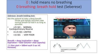
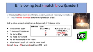
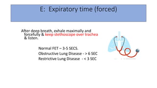
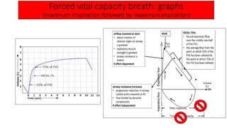
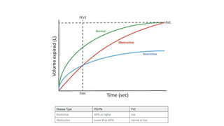
Pulmonary function test
Dr Kavita Kadyan
Assistant professor,
DNB Anesthesiology & Critical care
Pulmonary function test
Bedside PFT Traditional PFT
BEDSIDE PFT mnemonic
0 : hold: breath hold test (Seberese)
1: single breath test (Seberese)
A: ABG
B: Blowing test (match blow)(snider)
C: Cough test (GREENE)
D: Debono test (whistle)
E: Expiratory time (forced)
F: Flow meter(peak flow meter) (wrights)
0 : hold means no breathing
0 breathing: breath hold test (Seberese)
Sabrasez breath holding test:
Ask the patient to take a deep breath
inhale and exhale and then take
normal breath & hold it as long as
possible.
• >25 SEC.- NORMAL
Cardiopulmonary Reserve
• 15-25 SEC- LIMITED
• <15 SEC - VERY POOR
(breath = deep inspiration + deep
expiration + tidal volume= vital capacity)
( 1 litre start + 500ml each 5 sec VC
increase)
The length of time can crudely help to
quantify the Vital capacity
of the patient.
25- 30 SEC - 3500 ml VC
20 – 25 SEC - 3000 ml VC
15 - 20 SEC - 2500 ml VC
10 - 15 SEC - 2000 ml VC
5 - 10 SEC - 1500 ml VC
0 - 5 sec = 1000ml
1: single breath test (Seberese)
After deep breath, hold it and start
counting till the next breath.
 Normal- 30-40 COUNT
 It is a measure of the FRC.
>15 : normal
<15 : dec reserve
11-15 : mild impairment
5-10 : mod impaired
<5 : severe impairment
B: Blowing test (match blow)(snider)
 Measures Maximum Breathing Capacity/Maximum voluntary ventilation.
 Should take 6 attempts before interpretation of test
Ask to blow a match stick from a distance of 6” (15 cms) with
 Mouth wide open
 Chin rested/supported
 No pursed lips
 No head movement
 No air movement in the room
 Mouth and match stick at the same level
(Match Blow = Maximum Breathing : MB : MB)
MATCH TEST:
DISTANCE MBC (N-150-175 L/min)
9” >150 L/MIN.
6” >60 L/MIN.
3” >40 L/MIN.
C: Cough test
GREENE & BEROWITZ COUGH TEST:
DEEP BREATH F/B COUGH
ABILITY TO COUGH tells ..
 STRENGTH
 EFFECTIVENESS
INADEQUATE COUGH :
FVC <20 mL/Kg
FEV1 < 15 ml/Kg
PEFR < 200 L/min.
VC ~ 3 TIMES TV FOR EFFECTIVE COUGH.
A wet productive cough / self propagated paroxysms of coughing – patient susceptible for
pulmonary Complication.
D: Debono test (whistle)
measures PEFR.
 Patient blows down a wide bore tube at the end of which is
a whistle, on the side is a hole with adjustable knob.
 As the patient blows down the tube, the whistle sounds only
when the rate of airflow exceeds a certain value.
 The side leak hole is gradually increased till the sound of whistling
disappears.
The last position when the whistle can be appreciated is read off the
scale and taken as the PEFR.
E: Expiratory time (forced)
After deep breath, exhale maximally and
forcefully & keep stethoscope over trachea
& listen.
Normal FET – 3-5 SECS.
Obstructive Lung Disease - > 6 SEC
Restrictive Lung Disease - < 3 SEC
F: Flow meter(peak flow meter) (wrights)
Measures PEFR
N – MALES- 450-700 L/MIN.
FEMALES- 350-500 L/MIN.
<200 L/min.–inadequate cough efficiency
Wright ‘s Respirometer
measures VT (tidal volume ) and minute volume of patient
Simple and rapid
Instrument- compact, light and portable.
Disadvantage: It under- reads at low flow rates and
over- reads at high flow rates.
Can be connected to endo tracheal tube or face
mask
Prior explanation to patient is needed.
What we get from PFTs?
GOALS
 To predict presence of pulmonary dysfunction
To know the functional nature of disease.
To assess the severity of disease
To assess the progression of disease
To assess the response to treatment
Medicolegal- to assess lung impairment as a result
of occupational hazard.
To identify patients at perioperative risk of
pulmonary complications
Evaluates
• Pulmonary function tests is a general term used to indicate a series of
maneuvers or investigations that are performed to help measure lung
functions. It evaluates one or more aspects of the respiratory system.
• Pulmonary parenchymal function/ Gas exchange (size & integrity of
the pulmonary capillary bed)
• Respiratory mechanics
• Cardiopulmonary interaction
• ACP GUIDELINES FOR PREOPERATIVE SPIROMETRY (modified )
Lung resection
H/o smoking, dyspnoea
Cardiac surgery
Upper abdominal surgery
Lower abdominal surgery
Uncharacterized pulmonary disease (defined as history of pulmonary
Disease or symptoms and no PFT in last 60 days)
Indications of PFTs
• Evaluation of signs & symptoms like breathlessness, chronic cough,
exertional dyspnoea to detect the disease.
• Evaluate the extent and assess severity of disease.
• Screening at risk patients and follow their response to therapy
• Measure the effect of drugs on pulmonary function
• To assess preoperative risk
• Monitor pulmonary drug toxicity
Contraindications
• History of recent thoraco-abdominal surgery
• Oral or facial pain which may be exacerbated by mouthpiece
• Stress incontinence
• Recent eye surgery (cataract)
• Thoracic, abdominal and cerebral aneurysm
• Active haemoptysis
• Nausea, vomiting
• Pneumothorax
• Dementia or confused patient,
• History of myocardial infarction within one month /Unstable angina
Volumes of lung
How to measure non-measurable lung
volumes?
Spirometry : don’t tell RV, FRC, TLC
• N2 wash out
• Helium dilution test
• Total body plethysmography
Spirometry
Helium dilution technique
Nitrogen washout technique
https://doctorlib.info/physiology/medical/143.html
Plethysmography
Functional residual capacity
• Functional Residual Capacity (FRC):
• Sum of RV and ERV or the volume of air in the lungs at end-expiratory tidal position.
• (30-35 ml/kg) (2300-3300ml).
• Measured with multiple-breath closed-circuit helium dilution, multiple-breath open-circuit
nitrogen washout, or body plethysmography.
• It can not be measured by spirometry.
Functions of FRC:
• Oxygen store
• Buffer for maintaining a steady arterial po2
• Partial inflation helps prevent atelectasis
• Minimise the work of breathing
• Minimise pulmonary vascular resistance
• Minimised v/q mismatch
- only if closing capacity is less than FRC.
Spirometry tells us what?
Volume time graph Flow time graph
vc
Flow volume loop graph: measures Forced
VITAL CAPACITY(FVC)
• PEFR( peak expiratory flow rate): large airway feature+
patient effort dependent + <200ml is abnormal+
{after maximum inspiration→ Patient maximum expires}
• Maximum flow rate during an FVC maneuvers occurs
in initial 0.1 s and the expiratory flow at this time is
termed as PEFR
• FEF-25%: the flow rate at the 25% point of the total
volume (FVC) exhaled, large-medium sized airway
• FEF50% : the flow rate at the 50% point of the total
volume (FVC) exhaled, medium to small airway.
• FEF 75% : the flow rate at the 75% point of the total
volume (FVC) exhaled, small airway (<2mm)
• Forced Expiratory Flow at 25-75% of FVC (FEF25-
75%): It is the mid-half of the FVC, which is
independent of patient’s effort and an indicator for
obstruction in the small airways
• Cant measure RV, FRC, TLC
Maximum Voluntary Ventilation (MVV) or
maximum breathing capacity (MBC):
• It measures the speed and efficiency of filling &
emptying of the lungs during increased respiratory
effort.
• The maximum volume of air that can be breathed
in and out of the lungs in 1 minute by maximum
voluntary effort is MVV.
• It reflects peak ventilation in physiological
demands.
• The normal value is 150 ‐175 l/min.
• Subject is asked to breathe as quickly and as deeply as possible for 12 s.
• The measured volume is extrapolated to 1min ( i.e. measured volume X 5 = MVV in 1
min).
• As a general guide, the value should correlate closely to the FEV1 x 35.
• Periods longer than 15 s should not be allowed because prolonged hyperventilation leads
to fainting due to excessive lowering of arterial PCO2 and H+.
• MVV is markedly decreased in patients with Emphysema, Airway obstruction and with
poor respiratory muscle strength.
Lung Capacity
• Considered abnormal if <80% of predicted value.
• Physiological factors influencing VC:
 Height
 Sex
 Age
 Posture
 Strength of respiratory muscle
Factors affecting VC & FRC
VC = IRV+TV+ERV
FRC= RV+ ERV
 HEIGHT :directly proportional to ht.
• Tall : more lung volume
 SEX –
• more in males : large chest size, more muscle power, more BSA.
 POSTURE-
• decreases in supine position,
• increase in erect position
 ABDOMINAL CONDITIONS–
• Obesity: decrease in lung capacities
 MUSCLE PARALYSIS/WEAKNESS : decrease
 DISEASE OF RESPIRATORY system:
• Restrictive lung disease (e.g. fibrosis, Pregnancy) decrease,
• Emphysema : increase RV, FRC
 ANAESTHESIA : position + muscle paralysis + surgical abdominal
intervention + pain
 AGE – decreases with increasing age
 FRC does NOT change with age.
FRC does NOT change with age.
• Various lung volumes and capacities will
change when comparing the standing, sitting,
and supine (lying down) positions.
• Inspiratory and expiratory volumes will be
greater when sitting or standing than lying
down.
• This is explained by gravity pulling the
abdominal contents away from the diaphragm
when upright, therefore increasing the volume
of the thoracic cavity.
• In a supine position the patient’s TV, IRV and
ERV will decrease as the abdominal organs
rest against the diaphragm limiting its
movements.
• Thus, the vital capacity will decrease
Posture
Posture
Neonates
Age
https://derangedphysiology.com
Sex
Weight
Height
individuals with tall stature tend to have greater TLC than those with short
stature
DIFFERENT POSTURES AFFECTING VC
• POSITION
 TRENDELENBERG
LITHOTOMY
PRONE
RT. LATERAL
LT. LATERAL
• DECREASE IN VC
 14.5%
18%
10%
12%
10%
In post operative period if VC falls below 3 times VC–
artificial respiration is needed to maintain airway clear
of secretions.
Muscle of respiration: weakness leads to decreases in the lung capacities
Exercise
Spirometry paper graph
Forced vital capacity breath: graphs
(maximum inspiration followed by maximum expiration)
Common problems in spirometry
A flow-volume loop can have a sawtooth
profile. This can occur during exhalation or
inhalation. The sawtooth pattern is usually
generated either by airflow disturbances in
the upper airway or from tremors of the
respiratory muscles. It is associated with
sleep apnea, obesity, upper airway injury
and some neurological disorders but is not a
reliable diagnostic sign of these disorders
nor does its presence or absence indicate
the severity of the disorder.
A rounded peak flow can be an indication of an inadequate subject effort
An abrupt drop in
expiratory flow
usually indicates an
early termination of
exhalation.
A notch in a flow-
volume loop
indicates a cough or
other pause in
exhalation. Since
the FEV1/FVC ratio is
usually between 0.70
and 0.85, if this
notch appears during
the first three-
quarters of the
exhalation then the
pause likely affects
the FEV1.
A side-ways hop
at the beginning
of exhalation is an
indication of a
starting hesitation
and usually
indicates the
presence of
excessive back-
extrapolation.
DISEASES OF LUNG
OBSTRUCTIVE DISORDERS RESTRICTIVE DISORDERS
LUNG- SHRINKED
LUNG-
OVERINFLATED
EXHALE-LIMITED
INHALE-
NORMAL
EXHALE - LIMITED
INHALE-
RESTRICTED
OBSTRUCTIVE DISORDERS RESTRICTIVE DISORDERS
Inspiration Normal ↓
Expiration ↓ ↓
TLC INCREASED DECREASED
DISEASES OF LUNG
OBSTRUCTIVE DISORDERS RESTRICTIVE DISORDERS
Inspiration Normal ↓
Expiration ↓ ↓
TLC INCREASED DECREASED
PEFR ↓
N/↑
FEV1 ↓↓ N/↓
FVC ↓ ↓
FEV1/FVC LOW NORMAL
Lung Volumes and Obstructive and Restrictive
Disease?
Obstructive disorders
RESTRICTIVE
“R” word elongation,
i.e. tall and narrow
Peak flow PEFR is long,
VC is narrow
OBSTRUCTIVE
“O” word widening,
i.e. small and long
Peak flow PEFR is small,
VC is long
Overall Obstructive (O) vs Restrictive (R) ?
Write word OR
Same sequence … is the flow loop,
O is towards left,
R is towards right
Flow-volume loops typically become more
concave both with increasing age and with
an increase in the severity of airway
obstruction. This concavity has been
variously termed “scooping” or “coving”.
Normal vs. Obstructive vs. Restrictive
Upper airway obstruction patterns
• Detect obstructive lesion in the major
airways.
Characterize the lesion:
• LOCATION : Intra-thoracic or Extra-thoracic
• BEHAVIOUR OF LESION WITH
RESPIRATION: Fixed or Variable
During Exhalation During Inhalation
Upper airway obstruction patterns
Intrathoracic flow limitations will appear only
during exhalation
Extrathoracic flow limitations only appear
during inhalation
During Exhalation During Inhalation
Upper airway obstruction patterns
Intrathoracic flow limitations will appear only
during exhalation
Extrathoracic flow limitations only appear
during inhalation
Airway obstruction
Variable extra-thoracic obstruction
• A forced inspiration causes
negative transmural pressure
inside the airway which tends
to collapse it.
• During expiration, positive
pressure in airway decreases
obstruction.
• Hence, inspiratory flow is
reduced to a greater extent
than expiratory flow.
• Bilateral and unilateral vocal
cord paralysis, Vocal cord
constriction, Chronic
neuromuscular disorders,
Airway burns and Obstructive
sleep apnoea.
Variable intra-thoracic obstruction
• During forced expiration,
high pleural pressure develop
causing an increased
intrathoracic pressure,
thereby decreasing airway
diameter.
• The flow volume loop shows
a greater reduction in the
expiratory phase.
• During inspiration, lower
pleural pressure around
airway tends to decrease
obstruction.
• A flattening of expiratory limb
is observed.
• eg. Tracheomalacia,
Polychondritis, Tumors of
Fixed obstruction
• There is a constant airflow
limitation on inspiration
and expiration
• Benign stricture of
trachea, Goiter,
Endotracheal neoplasms,
and bronchial stenosis.
Tracheomalacia can cause the
largest airways to collapse
during exhalation. Although
this can be said to cause an
expiratory plateau the pattern
has some differences in that
the amount of collapse tends
to increase during exhalation
making it somewhat variable
Some other findings of PFT
• Bd response
• DLCO
Bronchodilator (Bd) response on PFT
Salbutamol or levo-salbutamol 4 puffs of
100Ug, 15-20 min before
DL-CO {DIFFUSING CAPACITY CARBON
MONOXIDE}
DL-CO and diseases
DL-CO and diseases
DL-CO and diseases
PFT values
SPIROMETRY INTERPRETATION
What do the numbers mean?
FVC
• Interpretation of % predicted:
• 80-120% Normal
• 70-79% Mild reduction
• 50%-69% Moderate reduction
• <50% Severe reduction
FEV1
• Interpretation of % predicted:
• 80-120% Normal
• 70-75% Mild
• 60-69% Moderate
• 50-59% Moderately Severe
• <49-35 % Severe
• <35% Very Severe
RESULTS are expressed as % predicted of a predicted normal value of a person with the same age, sex
and height.
SPIROMETRY INTERPRETATION
What do the numbers mean?
FEF 25-75%
• Interpretation of % predicted:
• >79% Normal
• 60-79% Mild obstruction
• 40-59% Moderate obstruction
• <40% Severe obstruction
FEV1/FVC
• Interpretation of absolute value:
• ≥ 80 Normal
• ≤ 79 Abnormal
RESULTS are expressed as % predicted of a predicted normal value of a person with the same age, sex
and height.
GOLD Classification for COPD severity
Stepwise approach
Steps:
• calculate FEV1
• Calculate FVC
• Calculate FEV1/FVC
• Calculate TLC
• Calculate DLCO
• See Bd reversibility
Stepwise analysis
Examples
Pulmonary function test Dr Kavita.pptx

Pulmonary function test Dr Kavita.pptx

  • 1.
    Pulmonary function test DrKavita Kadyan Assistant professor, DNB Anesthesiology & Critical care
  • 2.
  • 3.
    BEDSIDE PFT mnemonic 0: hold: breath hold test (Seberese) 1: single breath test (Seberese) A: ABG B: Blowing test (match blow)(snider) C: Cough test (GREENE) D: Debono test (whistle) E: Expiratory time (forced) F: Flow meter(peak flow meter) (wrights)
  • 4.
    0 : holdmeans no breathing 0 breathing: breath hold test (Seberese) Sabrasez breath holding test: Ask the patient to take a deep breath inhale and exhale and then take normal breath & hold it as long as possible. • >25 SEC.- NORMAL Cardiopulmonary Reserve • 15-25 SEC- LIMITED • <15 SEC - VERY POOR (breath = deep inspiration + deep expiration + tidal volume= vital capacity) ( 1 litre start + 500ml each 5 sec VC increase) The length of time can crudely help to quantify the Vital capacity of the patient. 25- 30 SEC - 3500 ml VC 20 – 25 SEC - 3000 ml VC 15 - 20 SEC - 2500 ml VC 10 - 15 SEC - 2000 ml VC 5 - 10 SEC - 1500 ml VC 0 - 5 sec = 1000ml
  • 5.
    1: single breathtest (Seberese) After deep breath, hold it and start counting till the next breath.  Normal- 30-40 COUNT  It is a measure of the FRC. >15 : normal <15 : dec reserve 11-15 : mild impairment 5-10 : mod impaired <5 : severe impairment
  • 6.
    B: Blowing test(match blow)(snider)  Measures Maximum Breathing Capacity/Maximum voluntary ventilation.  Should take 6 attempts before interpretation of test Ask to blow a match stick from a distance of 6” (15 cms) with  Mouth wide open  Chin rested/supported  No pursed lips  No head movement  No air movement in the room  Mouth and match stick at the same level (Match Blow = Maximum Breathing : MB : MB) MATCH TEST: DISTANCE MBC (N-150-175 L/min) 9” >150 L/MIN. 6” >60 L/MIN. 3” >40 L/MIN.
  • 7.
    C: Cough test GREENE& BEROWITZ COUGH TEST: DEEP BREATH F/B COUGH ABILITY TO COUGH tells ..  STRENGTH  EFFECTIVENESS INADEQUATE COUGH : FVC <20 mL/Kg FEV1 < 15 ml/Kg PEFR < 200 L/min. VC ~ 3 TIMES TV FOR EFFECTIVE COUGH. A wet productive cough / self propagated paroxysms of coughing – patient susceptible for pulmonary Complication.
  • 8.
    D: Debono test(whistle) measures PEFR.  Patient blows down a wide bore tube at the end of which is a whistle, on the side is a hole with adjustable knob.  As the patient blows down the tube, the whistle sounds only when the rate of airflow exceeds a certain value.  The side leak hole is gradually increased till the sound of whistling disappears. The last position when the whistle can be appreciated is read off the scale and taken as the PEFR.
  • 9.
    E: Expiratory time(forced) After deep breath, exhale maximally and forcefully & keep stethoscope over trachea & listen. Normal FET – 3-5 SECS. Obstructive Lung Disease - > 6 SEC Restrictive Lung Disease - < 3 SEC
  • 10.
    F: Flow meter(peakflow meter) (wrights) Measures PEFR N – MALES- 450-700 L/MIN. FEMALES- 350-500 L/MIN. <200 L/min.–inadequate cough efficiency
  • 11.
    Wright ‘s Respirometer measuresVT (tidal volume ) and minute volume of patient Simple and rapid Instrument- compact, light and portable. Disadvantage: It under- reads at low flow rates and over- reads at high flow rates. Can be connected to endo tracheal tube or face mask Prior explanation to patient is needed.
  • 13.
    What we getfrom PFTs?
  • 14.
    GOALS  To predictpresence of pulmonary dysfunction To know the functional nature of disease. To assess the severity of disease To assess the progression of disease To assess the response to treatment Medicolegal- to assess lung impairment as a result of occupational hazard. To identify patients at perioperative risk of pulmonary complications
  • 15.
    Evaluates • Pulmonary functiontests is a general term used to indicate a series of maneuvers or investigations that are performed to help measure lung functions. It evaluates one or more aspects of the respiratory system. • Pulmonary parenchymal function/ Gas exchange (size & integrity of the pulmonary capillary bed) • Respiratory mechanics • Cardiopulmonary interaction
  • 16.
    • ACP GUIDELINESFOR PREOPERATIVE SPIROMETRY (modified ) Lung resection H/o smoking, dyspnoea Cardiac surgery Upper abdominal surgery Lower abdominal surgery Uncharacterized pulmonary disease (defined as history of pulmonary Disease or symptoms and no PFT in last 60 days)
  • 17.
    Indications of PFTs •Evaluation of signs & symptoms like breathlessness, chronic cough, exertional dyspnoea to detect the disease. • Evaluate the extent and assess severity of disease. • Screening at risk patients and follow their response to therapy • Measure the effect of drugs on pulmonary function • To assess preoperative risk • Monitor pulmonary drug toxicity
  • 18.
    Contraindications • History ofrecent thoraco-abdominal surgery • Oral or facial pain which may be exacerbated by mouthpiece • Stress incontinence • Recent eye surgery (cataract) • Thoracic, abdominal and cerebral aneurysm • Active haemoptysis • Nausea, vomiting • Pneumothorax • Dementia or confused patient, • History of myocardial infarction within one month /Unstable angina
  • 19.
  • 20.
    How to measurenon-measurable lung volumes? Spirometry : don’t tell RV, FRC, TLC • N2 wash out • Helium dilution test • Total body plethysmography
  • 21.
  • 23.
  • 24.
  • 25.
  • 26.
    Functional residual capacity •Functional Residual Capacity (FRC): • Sum of RV and ERV or the volume of air in the lungs at end-expiratory tidal position. • (30-35 ml/kg) (2300-3300ml). • Measured with multiple-breath closed-circuit helium dilution, multiple-breath open-circuit nitrogen washout, or body plethysmography. • It can not be measured by spirometry.
  • 27.
    Functions of FRC: •Oxygen store • Buffer for maintaining a steady arterial po2 • Partial inflation helps prevent atelectasis • Minimise the work of breathing • Minimise pulmonary vascular resistance • Minimised v/q mismatch - only if closing capacity is less than FRC.
  • 28.
    Spirometry tells uswhat? Volume time graph Flow time graph vc
  • 30.
    Flow volume loopgraph: measures Forced VITAL CAPACITY(FVC) • PEFR( peak expiratory flow rate): large airway feature+ patient effort dependent + <200ml is abnormal+ {after maximum inspiration→ Patient maximum expires} • Maximum flow rate during an FVC maneuvers occurs in initial 0.1 s and the expiratory flow at this time is termed as PEFR • FEF-25%: the flow rate at the 25% point of the total volume (FVC) exhaled, large-medium sized airway • FEF50% : the flow rate at the 50% point of the total volume (FVC) exhaled, medium to small airway. • FEF 75% : the flow rate at the 75% point of the total volume (FVC) exhaled, small airway (<2mm) • Forced Expiratory Flow at 25-75% of FVC (FEF25- 75%): It is the mid-half of the FVC, which is independent of patient’s effort and an indicator for obstruction in the small airways • Cant measure RV, FRC, TLC
  • 31.
    Maximum Voluntary Ventilation(MVV) or maximum breathing capacity (MBC): • It measures the speed and efficiency of filling & emptying of the lungs during increased respiratory effort. • The maximum volume of air that can be breathed in and out of the lungs in 1 minute by maximum voluntary effort is MVV. • It reflects peak ventilation in physiological demands. • The normal value is 150 ‐175 l/min. • Subject is asked to breathe as quickly and as deeply as possible for 12 s. • The measured volume is extrapolated to 1min ( i.e. measured volume X 5 = MVV in 1 min). • As a general guide, the value should correlate closely to the FEV1 x 35. • Periods longer than 15 s should not be allowed because prolonged hyperventilation leads to fainting due to excessive lowering of arterial PCO2 and H+. • MVV is markedly decreased in patients with Emphysema, Airway obstruction and with poor respiratory muscle strength.
  • 32.
    Lung Capacity • Consideredabnormal if <80% of predicted value. • Physiological factors influencing VC:  Height  Sex  Age  Posture  Strength of respiratory muscle
  • 33.
    Factors affecting VC& FRC VC = IRV+TV+ERV FRC= RV+ ERV  HEIGHT :directly proportional to ht. • Tall : more lung volume  SEX – • more in males : large chest size, more muscle power, more BSA.  POSTURE- • decreases in supine position, • increase in erect position  ABDOMINAL CONDITIONS– • Obesity: decrease in lung capacities  MUSCLE PARALYSIS/WEAKNESS : decrease  DISEASE OF RESPIRATORY system: • Restrictive lung disease (e.g. fibrosis, Pregnancy) decrease, • Emphysema : increase RV, FRC  ANAESTHESIA : position + muscle paralysis + surgical abdominal intervention + pain  AGE – decreases with increasing age  FRC does NOT change with age.
  • 34.
    FRC does NOTchange with age.
  • 35.
    • Various lungvolumes and capacities will change when comparing the standing, sitting, and supine (lying down) positions. • Inspiratory and expiratory volumes will be greater when sitting or standing than lying down. • This is explained by gravity pulling the abdominal contents away from the diaphragm when upright, therefore increasing the volume of the thoracic cavity. • In a supine position the patient’s TV, IRV and ERV will decrease as the abdominal organs rest against the diaphragm limiting its movements. • Thus, the vital capacity will decrease Posture
  • 36.
  • 37.
  • 38.
  • 39.
  • 40.
  • 41.
    Height individuals with tallstature tend to have greater TLC than those with short stature
  • 42.
    DIFFERENT POSTURES AFFECTINGVC • POSITION  TRENDELENBERG LITHOTOMY PRONE RT. LATERAL LT. LATERAL • DECREASE IN VC  14.5% 18% 10% 12% 10% In post operative period if VC falls below 3 times VC– artificial respiration is needed to maintain airway clear of secretions.
  • 43.
    Muscle of respiration:weakness leads to decreases in the lung capacities
  • 44.
  • 45.
  • 46.
    Forced vital capacitybreath: graphs (maximum inspiration followed by maximum expiration)
  • 47.
  • 48.
  • 49.
    A flow-volume loopcan have a sawtooth profile. This can occur during exhalation or inhalation. The sawtooth pattern is usually generated either by airflow disturbances in the upper airway or from tremors of the respiratory muscles. It is associated with sleep apnea, obesity, upper airway injury and some neurological disorders but is not a reliable diagnostic sign of these disorders nor does its presence or absence indicate the severity of the disorder.
  • 50.
    A rounded peakflow can be an indication of an inadequate subject effort
  • 51.
    An abrupt dropin expiratory flow usually indicates an early termination of exhalation.
  • 52.
    A notch ina flow- volume loop indicates a cough or other pause in exhalation. Since the FEV1/FVC ratio is usually between 0.70 and 0.85, if this notch appears during the first three- quarters of the exhalation then the pause likely affects the FEV1.
  • 53.
    A side-ways hop atthe beginning of exhalation is an indication of a starting hesitation and usually indicates the presence of excessive back- extrapolation.
  • 54.
    DISEASES OF LUNG OBSTRUCTIVEDISORDERS RESTRICTIVE DISORDERS LUNG- SHRINKED LUNG- OVERINFLATED EXHALE-LIMITED INHALE- NORMAL EXHALE - LIMITED INHALE- RESTRICTED
  • 56.
    OBSTRUCTIVE DISORDERS RESTRICTIVEDISORDERS Inspiration Normal ↓ Expiration ↓ ↓ TLC INCREASED DECREASED
  • 58.
    DISEASES OF LUNG OBSTRUCTIVEDISORDERS RESTRICTIVE DISORDERS Inspiration Normal ↓ Expiration ↓ ↓ TLC INCREASED DECREASED PEFR ↓ N/↑ FEV1 ↓↓ N/↓ FVC ↓ ↓ FEV1/FVC LOW NORMAL
  • 61.
    Lung Volumes andObstructive and Restrictive Disease?
  • 63.
  • 65.
    RESTRICTIVE “R” word elongation, i.e.tall and narrow Peak flow PEFR is long, VC is narrow OBSTRUCTIVE “O” word widening, i.e. small and long Peak flow PEFR is small, VC is long Overall Obstructive (O) vs Restrictive (R) ? Write word OR Same sequence … is the flow loop, O is towards left, R is towards right
  • 66.
    Flow-volume loops typicallybecome more concave both with increasing age and with an increase in the severity of airway obstruction. This concavity has been variously termed “scooping” or “coving”.
  • 67.
    Normal vs. Obstructivevs. Restrictive
  • 72.
    Upper airway obstructionpatterns • Detect obstructive lesion in the major airways. Characterize the lesion: • LOCATION : Intra-thoracic or Extra-thoracic • BEHAVIOUR OF LESION WITH RESPIRATION: Fixed or Variable
  • 73.
    During Exhalation DuringInhalation Upper airway obstruction patterns Intrathoracic flow limitations will appear only during exhalation Extrathoracic flow limitations only appear during inhalation
  • 74.
    During Exhalation DuringInhalation Upper airway obstruction patterns Intrathoracic flow limitations will appear only during exhalation Extrathoracic flow limitations only appear during inhalation
  • 75.
  • 76.
    Variable extra-thoracic obstruction •A forced inspiration causes negative transmural pressure inside the airway which tends to collapse it. • During expiration, positive pressure in airway decreases obstruction. • Hence, inspiratory flow is reduced to a greater extent than expiratory flow. • Bilateral and unilateral vocal cord paralysis, Vocal cord constriction, Chronic neuromuscular disorders, Airway burns and Obstructive sleep apnoea.
  • 77.
    Variable intra-thoracic obstruction •During forced expiration, high pleural pressure develop causing an increased intrathoracic pressure, thereby decreasing airway diameter. • The flow volume loop shows a greater reduction in the expiratory phase. • During inspiration, lower pleural pressure around airway tends to decrease obstruction. • A flattening of expiratory limb is observed. • eg. Tracheomalacia, Polychondritis, Tumors of
  • 78.
    Fixed obstruction • Thereis a constant airflow limitation on inspiration and expiration • Benign stricture of trachea, Goiter, Endotracheal neoplasms, and bronchial stenosis.
  • 79.
    Tracheomalacia can causethe largest airways to collapse during exhalation. Although this can be said to cause an expiratory plateau the pattern has some differences in that the amount of collapse tends to increase during exhalation making it somewhat variable
  • 81.
    Some other findingsof PFT • Bd response • DLCO
  • 82.
    Bronchodilator (Bd) responseon PFT Salbutamol or levo-salbutamol 4 puffs of 100Ug, 15-20 min before
  • 84.
    DL-CO {DIFFUSING CAPACITYCARBON MONOXIDE}
  • 85.
  • 86.
  • 87.
  • 92.
  • 93.
    SPIROMETRY INTERPRETATION What dothe numbers mean? FVC • Interpretation of % predicted: • 80-120% Normal • 70-79% Mild reduction • 50%-69% Moderate reduction • <50% Severe reduction FEV1 • Interpretation of % predicted: • 80-120% Normal • 70-75% Mild • 60-69% Moderate • 50-59% Moderately Severe • <49-35 % Severe • <35% Very Severe RESULTS are expressed as % predicted of a predicted normal value of a person with the same age, sex and height.
  • 94.
    SPIROMETRY INTERPRETATION What dothe numbers mean? FEF 25-75% • Interpretation of % predicted: • >79% Normal • 60-79% Mild obstruction • 40-59% Moderate obstruction • <40% Severe obstruction FEV1/FVC • Interpretation of absolute value: • ≥ 80 Normal • ≤ 79 Abnormal RESULTS are expressed as % predicted of a predicted normal value of a person with the same age, sex and height.
  • 95.
  • 96.
  • 97.
    Steps: • calculate FEV1 •Calculate FVC • Calculate FEV1/FVC • Calculate TLC • Calculate DLCO • See Bd reversibility Stepwise analysis
  • 100.