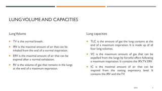
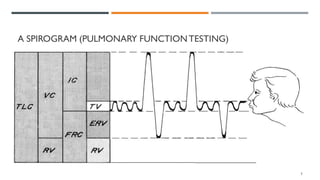
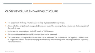
PULMONARY FUNCTIONTEST
Shilpasree Saha,
BPT (TIPS, Agartala), MPT-Cardio-Thoracic Disorders (MMIPR, MM(DU, Mullana)
Assistant Professor, NIHS
INTRODUCTION
• Pulmonary function tests
(PFTs) help in the evaluation of
the mechanical function of the
lungs.
• PFTs are useful in assessing the
functional status of the
respiratory system both in
physiological and pathological
condition.
• It is carried out by using a
spirometer.
2
BASICS OF PFT
• They are based on researched norms taking into
account gender, height, and age to see if he falls within
the "normal" range, or has a restrictive or obstructive
component based on the tests.
• When the patient performs the test actual results
(observed) will be compared with the predicted value.
• If the patient is not within the normal range, a
bronchodilator is given, and the test will be repeated to
see if there is significant improvement with medication.
• Basically, the pulmonary function tests are categorized
as volume, flow, or diffusion studies.
20XX 3
LUNGVOLUME AND CAPACITIES
LungVolume
 TV is the normal breath.
 IRV is the maximal amount of air that can be
inhaled from the end of a normal inspiration.
 ERV is the maximal amount of air that can be
expired after a normal exhalation.
 RV is the volume of gas that remains in the lungs
at the end of a maximum expiration.
Lung capacities
 TLC is the amount of gas the lung contains at the
end of a maximum inspiration. It is made up of all
four lung volumes.
 VC is the maximum amount of gas that can be
expelled from the lungs by forceful effort following
a maximum inspiration. It contains the IRV,TV, ERV.
 IC is the maximal amount of air that can be
inspired from the resting expiratory level. It
contains the IRV and theTV.
20XX 4
A SPIROGRAM (PULMONARY FUNCTIONTESTING)
5
DEAD SPACE
• There is a series of conducting airways in the lungs
from the trachea down to the terminal bronchi,
which do not participate in respiration but only
move the gases to the alveoli. This is the volume
known as anatomic dead space.
• Generally, the anatomic dead space is appropriately
equal to the adult body weight. For example, in a
150-lb. person, there is an approximately 150 mL
anatomic dead space.
• The physiological dead space is defined as including
anatomical dead space and alveolar dead space
components.
6
 Normal tidal volume (TV), the breath normally taken, needs to be large enough to reach the alveoli well past the
anatomic dead space. In a normal adult, the TV is generally 450 to 600 mL.
 The anatomic dead space would thus represent about one thirdTV volume.
 The rest of the breath would reach the alveoli and be considered "alveolar ventilation."
 With many neurologically impaired patients who have a limitedTV, it is important to note that little alveolar
ventilation may be taking place when the patient is breathing in a rapid and shallow pattern.
 For example, if a patient's TV was 200 mL, 150 mL would be anatomic dead space and only 50 mL of each breath
would be alveolar ventilation.
20XX 7
INDICATION
 Investigation of patients with
symptoms/signs/investigations that suggest
pulmonary disease e.g. Cough, Wheeze,
Breathlessness, Crackles,Abnormal chest x-ray.
 Monitoring patients with known pulmonary
disease for progression and response to treatment
e.g. Interstitial fibrosis, COPD, Asthma, Pulmonary
vascular disease.
 Investigation of patients with disease that may have
a respiratory complications e.g. Connective tissue
disorders, Neuromuscular diseases.
 Preoperative evaluation prior to e.g. Lung resection,Abdominal surgery, Cardiothoracic surgery.
 Evaluation patients a risk of lung diseases e.g. Exposure to pulmonary toxins such a radiation, medication, or
environmental or occupational exposure
 Surveillance following lung transplantation to assess for Infection.
20XX 9
CONTRA-INDICATION
 Myocardial infarction within the last month
 Unstable angina
 Recent thoraco-abdominal surgery
 Recent ophthalmic surgery
 Thoracic or abdominal aneurysm
 Current pneumothorax
SAMPLE FOOTER TEXT 20XX 10
PROCEDURE
 Spirometry is the most frequently used measure
of lung function and is a measure of volume against
time.
 It is a simple and quick procedure to perform:
patients are asked to take a maximal inspiration
and then to forcefully expel air through mouth
into the mouthpiece for as long and as quickly as
possible against closed nostril.
20XX 11
Measurements that are made include:
 Forced expiratory volume in one second (FEV1)
 Forced vital capacity (FVC)
 The ratio of the two volumes (FEV1/FVC)
20XX 12
AIR FLOW MEASUREMENTS
 When patients perform aVC maneuver, it can either be slow or fast.
 During exhalation, the amount of air exhaled over time can be measured. In a slowVC a patient with emphysema
can take a great deal of time to empty his lungs.
 In a forcedVC a normal individual can exhale 75% of theVC in the first second of exhalation (FEV I).
SAMPLE FOOTER TEXT 20XX 13
FLOWVOLUME CURVE
 The flow volume curve is helpful in diagnosing lung
disease.
 The curve demonstrates that flow rises to a high
value and then declines over most of expiration
 In restrictive lung disease, the maximum flow rate
is reduced.
 In obstructive lung disease, the flow rate is low in
relation to lung volume, and a scooped-out
appearance is often seen.
20XX 14
FLOWVOLUME LOOP
 Another diagnostic test that uses forced
expiration is the flow volume loop.
 It is a graphical analysis of the flow generated
during a forced expiratory volume maneuver
followed by a forced inspiratory volume maneuver.
 This graph offers a pictorial representation of data
(e.g., peak inspiratory and expiratory flow rates
FVC, and FEV1).
 The shape of the graph may also be helpful in
diagnosing disease, again seeing a more scooped-
out appearance with obstructive disease.
SAMPLE FOOTER TEXT 20XX 15
SAMPLE FOOTER TEXT 20XX 16
CLOSINGVOLUME AND AIRWAY CLOSURE
 The assessment of closing volume is used to help diagnose small airway disease.
 A test called the single breath nitrogen (N2) washout is used for assessing closing volume and closing capacity of
the small airways.
 In this test, the patient takes a singleVC breath of 100% oxygen.
 During complete exhalation, the N2 concentration can be measured.
 The characteristic tracing of N2 concentration can be measured.The characteristic tracing of N2 concentration
vs. lung volume reflects sequential emptying of differentially ventilated lung units, resulting in different expiratory
N2 concentrations.
SAMPLE FOOTER TEXT 20XX 17
FOUR PHASES CAN BE IDENTIFIED:
 Phase I contains pure dead space and virtually none of
the potential N2 from the RV.
 Phase Il is associated with an increasing N2
concentration of a mixture of gas from the dead space
and alveoli.
 The plateau in N2 concentration observed in Phase III
reflects pure alveolar gas emanating from the bases and
middle lung zones.
 Phase IV occurs toward the end of expiration and is
characterized by an abrupt increase in N2 concentration.
 This high N2 concentration reflects closure of airways at
the base of the lungs and expiration of gas from the
upper lung zones, because in the single breath of 100%
oxygen, less oxygen was initially directed to this area.
 Closing volume is the lung volume at which the inflection
of Phase IV, the marked increase in N2 concentration
after the plateau, is observed.
 Closing capacity refers to closing volume and RV.
SAMPLE FOOTER TEXT 20XX 18
 The closing volume is 10% of the vital capacity in young, healthy individuals. It increases with age and is 40% of the
vital capacity at age 65.
 Closing volume is used as an aid in the diagnosis of small airway disease and as a means of evaluating treatment or
drug response.
SAMPLE FOOTER TEXT 20XX 19
MAXIMALVOLUNTARYVENTILATION
 Maximal voluntary ventilation measures the maximal breathing capacity of the patient. It reflects strengths and
endurance of the respiratory muscles.
 The patient is asked to pant for 15 seconds into the spirometer tubing.
 This is often examined preoperatively with the other results to determine a patient's prognosis for success after
surgery, such as his or her ability to cough, to take deep breaths, and to enhance airway clearance.
SAMPLE FOOTER TEXT 20XX 20
REFERENCES
 Ranu H,Wilde M, Madden B. Pulmonary function tests. Ulster Med J. 2011 May;80(2):84-90. PMID: 22347750;
PMCID: PMC3229853.
 Principles And Practice Of Cardiopulmonary Physical Therapy, Donna Frownfelter, Third Edition.
20XX 21
THANKYOU
SAMPLE FOOTER TEXT

PULMONARY FUNCTION TEST.pdf

  • 1.
    PULMONARY FUNCTIONTEST Shilpasree Saha, BPT(TIPS, Agartala), MPT-Cardio-Thoracic Disorders (MMIPR, MM(DU, Mullana) Assistant Professor, NIHS
  • 2.
    INTRODUCTION • Pulmonary functiontests (PFTs) help in the evaluation of the mechanical function of the lungs. • PFTs are useful in assessing the functional status of the respiratory system both in physiological and pathological condition. • It is carried out by using a spirometer. 2
  • 3.
    BASICS OF PFT •They are based on researched norms taking into account gender, height, and age to see if he falls within the "normal" range, or has a restrictive or obstructive component based on the tests. • When the patient performs the test actual results (observed) will be compared with the predicted value. • If the patient is not within the normal range, a bronchodilator is given, and the test will be repeated to see if there is significant improvement with medication. • Basically, the pulmonary function tests are categorized as volume, flow, or diffusion studies. 20XX 3
  • 4.
    LUNGVOLUME AND CAPACITIES LungVolume TV is the normal breath.  IRV is the maximal amount of air that can be inhaled from the end of a normal inspiration.  ERV is the maximal amount of air that can be expired after a normal exhalation.  RV is the volume of gas that remains in the lungs at the end of a maximum expiration. Lung capacities  TLC is the amount of gas the lung contains at the end of a maximum inspiration. It is made up of all four lung volumes.  VC is the maximum amount of gas that can be expelled from the lungs by forceful effort following a maximum inspiration. It contains the IRV,TV, ERV.  IC is the maximal amount of air that can be inspired from the resting expiratory level. It contains the IRV and theTV. 20XX 4
  • 5.
    A SPIROGRAM (PULMONARYFUNCTIONTESTING) 5
  • 6.
    DEAD SPACE • Thereis a series of conducting airways in the lungs from the trachea down to the terminal bronchi, which do not participate in respiration but only move the gases to the alveoli. This is the volume known as anatomic dead space. • Generally, the anatomic dead space is appropriately equal to the adult body weight. For example, in a 150-lb. person, there is an approximately 150 mL anatomic dead space. • The physiological dead space is defined as including anatomical dead space and alveolar dead space components. 6
  • 7.
     Normal tidalvolume (TV), the breath normally taken, needs to be large enough to reach the alveoli well past the anatomic dead space. In a normal adult, the TV is generally 450 to 600 mL.  The anatomic dead space would thus represent about one thirdTV volume.  The rest of the breath would reach the alveoli and be considered "alveolar ventilation."  With many neurologically impaired patients who have a limitedTV, it is important to note that little alveolar ventilation may be taking place when the patient is breathing in a rapid and shallow pattern.  For example, if a patient's TV was 200 mL, 150 mL would be anatomic dead space and only 50 mL of each breath would be alveolar ventilation. 20XX 7
  • 8.
    INDICATION  Investigation ofpatients with symptoms/signs/investigations that suggest pulmonary disease e.g. Cough, Wheeze, Breathlessness, Crackles,Abnormal chest x-ray.  Monitoring patients with known pulmonary disease for progression and response to treatment e.g. Interstitial fibrosis, COPD, Asthma, Pulmonary vascular disease.  Investigation of patients with disease that may have a respiratory complications e.g. Connective tissue disorders, Neuromuscular diseases.
  • 9.
     Preoperative evaluationprior to e.g. Lung resection,Abdominal surgery, Cardiothoracic surgery.  Evaluation patients a risk of lung diseases e.g. Exposure to pulmonary toxins such a radiation, medication, or environmental or occupational exposure  Surveillance following lung transplantation to assess for Infection. 20XX 9
  • 10.
    CONTRA-INDICATION  Myocardial infarctionwithin the last month  Unstable angina  Recent thoraco-abdominal surgery  Recent ophthalmic surgery  Thoracic or abdominal aneurysm  Current pneumothorax SAMPLE FOOTER TEXT 20XX 10
  • 11.
    PROCEDURE  Spirometry isthe most frequently used measure of lung function and is a measure of volume against time.  It is a simple and quick procedure to perform: patients are asked to take a maximal inspiration and then to forcefully expel air through mouth into the mouthpiece for as long and as quickly as possible against closed nostril. 20XX 11
  • 12.
    Measurements that aremade include:  Forced expiratory volume in one second (FEV1)  Forced vital capacity (FVC)  The ratio of the two volumes (FEV1/FVC) 20XX 12
  • 13.
    AIR FLOW MEASUREMENTS When patients perform aVC maneuver, it can either be slow or fast.  During exhalation, the amount of air exhaled over time can be measured. In a slowVC a patient with emphysema can take a great deal of time to empty his lungs.  In a forcedVC a normal individual can exhale 75% of theVC in the first second of exhalation (FEV I). SAMPLE FOOTER TEXT 20XX 13
  • 14.
    FLOWVOLUME CURVE  Theflow volume curve is helpful in diagnosing lung disease.  The curve demonstrates that flow rises to a high value and then declines over most of expiration  In restrictive lung disease, the maximum flow rate is reduced.  In obstructive lung disease, the flow rate is low in relation to lung volume, and a scooped-out appearance is often seen. 20XX 14
  • 15.
    FLOWVOLUME LOOP  Anotherdiagnostic test that uses forced expiration is the flow volume loop.  It is a graphical analysis of the flow generated during a forced expiratory volume maneuver followed by a forced inspiratory volume maneuver.  This graph offers a pictorial representation of data (e.g., peak inspiratory and expiratory flow rates FVC, and FEV1).  The shape of the graph may also be helpful in diagnosing disease, again seeing a more scooped- out appearance with obstructive disease. SAMPLE FOOTER TEXT 20XX 15
  • 16.
  • 17.
    CLOSINGVOLUME AND AIRWAYCLOSURE  The assessment of closing volume is used to help diagnose small airway disease.  A test called the single breath nitrogen (N2) washout is used for assessing closing volume and closing capacity of the small airways.  In this test, the patient takes a singleVC breath of 100% oxygen.  During complete exhalation, the N2 concentration can be measured.  The characteristic tracing of N2 concentration can be measured.The characteristic tracing of N2 concentration vs. lung volume reflects sequential emptying of differentially ventilated lung units, resulting in different expiratory N2 concentrations. SAMPLE FOOTER TEXT 20XX 17
  • 18.
    FOUR PHASES CANBE IDENTIFIED:  Phase I contains pure dead space and virtually none of the potential N2 from the RV.  Phase Il is associated with an increasing N2 concentration of a mixture of gas from the dead space and alveoli.  The plateau in N2 concentration observed in Phase III reflects pure alveolar gas emanating from the bases and middle lung zones.  Phase IV occurs toward the end of expiration and is characterized by an abrupt increase in N2 concentration.  This high N2 concentration reflects closure of airways at the base of the lungs and expiration of gas from the upper lung zones, because in the single breath of 100% oxygen, less oxygen was initially directed to this area.  Closing volume is the lung volume at which the inflection of Phase IV, the marked increase in N2 concentration after the plateau, is observed.  Closing capacity refers to closing volume and RV. SAMPLE FOOTER TEXT 20XX 18
  • 19.
     The closingvolume is 10% of the vital capacity in young, healthy individuals. It increases with age and is 40% of the vital capacity at age 65.  Closing volume is used as an aid in the diagnosis of small airway disease and as a means of evaluating treatment or drug response. SAMPLE FOOTER TEXT 20XX 19
  • 20.
    MAXIMALVOLUNTARYVENTILATION  Maximal voluntaryventilation measures the maximal breathing capacity of the patient. It reflects strengths and endurance of the respiratory muscles.  The patient is asked to pant for 15 seconds into the spirometer tubing.  This is often examined preoperatively with the other results to determine a patient's prognosis for success after surgery, such as his or her ability to cough, to take deep breaths, and to enhance airway clearance. SAMPLE FOOTER TEXT 20XX 20
  • 21.
    REFERENCES  Ranu H,WildeM, Madden B. Pulmonary function tests. Ulster Med J. 2011 May;80(2):84-90. PMID: 22347750; PMCID: PMC3229853.  Principles And Practice Of Cardiopulmonary Physical Therapy, Donna Frownfelter, Third Edition. 20XX 21
  • 22.