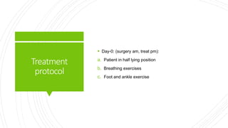
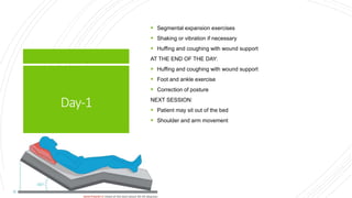
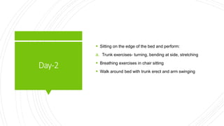
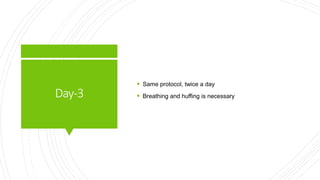
Physiotherapy in Pulmonary
surgery
Dr. Shilpasree Saha (PT)
Assistant Professor, NIHS
Thoracotomy
 Thoracotomy describes an incision made in the chest wall
to access the contents of the thoracic cavity (lung.
Bronchi, heart, esophagus).
 Thoracotomies typically can be divided into two:
1. Lateral (Anterolateral thoracotomies, Posterolateral
thoracotomies).
2. Anterior (transverse, vertical)
These can be further subdivided into supra-mammary and
infra-mammary
IndicationsFor
Surgery
I. Malignancy
II. Inflammatory conditions
III. Trauma: Stab wounds, gunshot wounds.
IV. Degenerative: Large lung bullae in selected patients
where there is compression of normal lung.
V. Congenital: Arterio-venous fistula, sequestrated lobe,
lobar emphysema.
VI. Diseases/infections
 Vertebral border of scapula and line of ribs (5,6,7,8) to the
anterior angle of costal margin.
 Trapezius, latissimus dorsi, rhomboids, serratus anterior,
intercostals and erector spinae are cutted.
 A rib may be removed so that when the other ribs are
retracted there is sufficient space for access to the thorax.
 Mainly used for lung operations.
Postero-lateral thoracotomy
Antero-lateral
thoracotomy  An antero-lateral thoracotomy is the standard approach
for a closed mitral valvotomy and is used by some
surgeons for pleurectomy.
 Serratus anterior, latissimus dorsi, pectoralis, intercostal
muscle are cutted.
 Patients are placed in the supine position with a small roll
under the ipsilateral shoulder. The patient’s arms are
tucked.
 The skin incision begins in close to the midline in front ,
follows the line of rib below the breast to the posterior
axillary line.
 Used for mitral valvotomy or pleurectomy.
Median
sternotomy
 Median sternotomy is the most widely used incision in
cardiac surgery.
 The skin incision should extend from just below the
sternal notch to a few centimeters below the xiphoid
process.
 Careful dissection behind sternal notch and xiphoid
process should be required to prevent accidental adjacent
vessels injuries.
 The sternotomy should be made on the midline of the
sternum after detecting the lateral margin of the sternum
by dipping the thumb and the index finger into the
intercostal space.
Usedforopenheart
surgery.
Indication of MS
Thoraco-laparotomy
incision
 Along the line of 7th or 8th rib and there may be an
abdominal incision as well.
 Used for access to the oesophagus.
TypesOfLung
Surgeries
 Pneumonectomy
 Lobectomy
 Wedge resection
Complications
1. Respiratory complication:
a. Infection
b. Collapse of remaining lung tissue
c. Pneumothorax
d. Broncho-pleural fistula
2. Circulatory complication:
a. DVT
b. Cardiac arrhythmia
c. Cardiac tamponade
d. Haemorrhage
Pneumonectomy
 The entire lung is removed.
 In a radical pneumonectomy, mediastinal lymph nodes and part of the
chest wall may also be removed.
 The resulting cavity is filled by protein rich fluid and fibrin.
 The cavity size is reduced by lateral shift of the trachea and heart, upward
shift of the diaphragm, and reduction of the intercostal spaces on the
operated side.
 Occasionally, and later, a scoliosis may develop.
Indications
 Carcinoma
 Bronchiectasis
 Tuberculosis
 Incision: postero-lateral thoracotomy
Physiotherapy
treatment
a. Gain patient confidence
b. Clear lung field
c. Teach breathing control
d. Inspiratory holding
e. Teach postural awareness
f. Arm, trunk and leg exercise
g. Mobility from the bed
Postoperative
Physiotherapy
 Goals:
a. Clear secretion from remaining lung
b. Retain full expansion from lung tissue
c. Prevent circulatory complication
d. Prevent wound complication
e. Regain arm and spine movement
f. Maintain good posture
g. Restore exercise tolerance
Treatment
protocol
 Day-0: (surgery am, treat pm):
a. Patient in half lying position
b. Breathing exercises
c. Foot and ankle exercise
Day-1
 Segmental expansion exercises
 Shaking or vibration if necessary
 Huffing and coughing with wound support
AT THE END OF THE DAY:
 Huffing and coughing with wound support
 Foot and ankle exercise
 Correction of posture
NEXT SESSION:
 Patient may sit out of the bed
 Shoulder and arm movement
Day-2
 Sitting on the edge of the bed and perform:
a. Trunk exercises- turning, bending at side, stretching
 Breathing exercises in chair sitting
 Walk around bed with trunk erect and arm swinging
Day-3
 Same protocol, twice a day
 Breathing and huffing is necessary
Day-4to
discharge
 Introduce to group therapy.
 Self care activities
 Walks more distance
 At 7th post op day, stair climbing with breathing control
 Breathing exercises
 Limb and trunk exercises
 Usually within 7-10 days, stitches come out.
 The patient is discharged after 2 weeks with strict home
exercise protocol.
Lobectomy
 Any of the five lobes may be removed.
 If a tumour in an upper lobe protrudes into the main
bronchus a cuff of main bronchus can be removed with
the lobe and the remaining lung and bronchus is joined to
the trachea. This is termed a sleeve lobectomy
Indications
 Bronchiectasis
 Tuberculosis
 Lung abcess
 Pneumonia
Preoperative
Physiotherapy
 BEGINS 48 HOURS TO A WEEK BEFORE SURGERY.
 Gain patient confidence
 Clear lung field
 Teach breathing exercises to expand lung tissue
 Inspiratory holding
 Teach postural awareness
 Arm, trunk and leg exercise
 Mobility from the bed
Postoperative
Physiotherapy
1. Half lying position with support.
2. Breathing exercises to expand all parts of remaining
lung.
3. Vibration over unoperated side.
4. Huffing with incision support.
5. Segmental expansion exercises.
6. Postural drainage by elevating the foot of the bed, if
necessary.
Day-0 (Treatment after given
analgesia)
Day-1
 Half lying position
 Foot and ankle exercises.
 Quadriceps contraction.
 Hip and knee bending and stretching.
 Assisted arm movement of operated side.
 Breathing exercises with inspiratory holding.
 Adding vibration to the operated side plus percussion as
required.
 Side-lying on the unoperated side, with no obstruction in
drains and upper arm supported by pillow and one more
pillow under the knee.
Day-2
 As first day
 Self supported huffing
 Arm exercises with full range.
 Trunk exercises.
 Abdominal contraction
 Discourage sitting crossed leg, as it occludes the popliteal
artery and vein and may result in DVT.
 At the end of 2nd day drains should removed.
 Fluid drain may be left in if there is less than 200 ml drained
in 24 hours.
 The patient can go for a short walk if necessary, with the
drainage bottle in a trolley.
Day-3and4
 One or two session per day.
 May introduce into group therapy.
 Trunk and arm exercises continued.
 Walking distance should increased.
 Self care activities.
 Stair climbing can be started.
Subsequent
treatment
 Bilateral breathing exercises
 Stitches may taken out between 7-10 days.
 Discharge between 10-12 days.
Segmental
resection
 A bronchopulmonary segment is removed with its
segmental artery and bronchus.
 This used to be indicated for tuberculosis but is now
rarely performed.
Wedgeresection
 Removal of small part of lung tissue.
Thoracoplasty
 This operation is performed to produce permanent
collapse of lung.
 Used to treat pulmonary TB, or chronic empyema.
 Resection of a varying number of ribs, leaving the
periosteum in position.
 4-10 ribs may be removed.
Complications
 Deformity
 Paradoxical breathing
Postoperative
Physiotherapy
 Breathing exercises
 Cough and huff using firm pressure over apical area of
thorax.
Day-0 (Treatment after given
analgesia)
Day-1
 Posture correction
 Active assisted arm movement.
Day-2
 Continue with breathing exercise and coughing.
 Posture correction.
Day-3and4
DAY -3
 Patient will be up and about.
 Manual resistance training for shoulder girdlearm on
effected side.
DAY -4
 Trunk exercises in sitting is added.
Day5-discharge
DAY-5 TO 7
 Trunk exercises in standing.
 Posture correction in walking.
DAY- 8 TO DISCHARGE
 Trunk mobility
 Thoracic cage mobility
 Posture correction should maintain for atleast 3 months
after discharge.
Operationof
pleura
 Pleurectomy
 Abrasion pleurodesis
 Decortication of lung
Pleurectomy
DEFINATION
 Removal of parietal pleura from an area of the chest wall
leaving a raw surface to which visceral layer sticks and is
performed for pneumothorax.
PT- MANAGEMENT
 Important to emphasize expansion breathing exercises
for effected side.
Abrasion
pleurodesis
DEFINITION
 Insertion of a powder into the pleural cavity, acts as an
irritant to the pleural surfaces, causing them to adhere to
each other.
 Performed in spontaneous pneumothorax and malignant
pleural effusion.
PT- MANAGEMENT
 Important to emphasize expansion breathing exercises
for effected side.
Decortication
ABRASION PLEURODESIS
 Stripping of layers of pleura that have become thickened due to
chronic inflammation from pleurisy which restricts movement of chest
wall and expansion of lung.
 In case of Unresolved empyema, whole pleura is removed to clear
away the chronic pus filled area plus surrounding fibrous tissue.
PT- MANAGEMENT
 Important to emphasize expansion breathing exercises
for effected side.
Drainsand
tubes
 Chest drains also known as under water sealed drains
(UWSD) are inserted to allow draining of the pleural spaces
of air, blood or fluid, allowing expansion of the lungs and
restoration of negative pressure in the thoracic cavity.
 Negative pressure is disrupted when air, or fluid and air,
enters the pleural space and separates the visceral pleura
from the parietal pleura, preventing the lung from collapsing
and compressing at the end of exhalation.
 The underwater seal also prevents backflow of air or fluid
into the pleural cavity.
 Appropriate chest drain management is required to maintain
respiratory function and haemodynamic stability.
 Two types:
a) Open
b) Closed
Opendrainage
 Include corrugated rubbers or plastic sheets.
 Drain fluid collects in gauze pads or stoma bags.
 They increase the risk of infection.
 E.G.- Penrose drain
Closeddrainage
 Consists of tubes draining into a bag or bottles.
 They include chest or abdominal drains.
 The risk of infection is less.
 Enables the lung to re-expand following pneumothorax and
lobectomy.
 Fluid is removed after any thoracic surgery to prevent pleural effusion
(expect pneumonectomy)
Equipments
 A tube
 A bottle with sterile water
 A suction pump
 The tube passes from inside the pleural cavity down
through a tight-fitting cap at the neck of the bottle to
below the level of water- under water seal.
 Air may be allowed to escape freely from a second tube
positioned high above the water in the neck of the bottle
or a suction machine may be attached to this tube.
 It is a single bottle, open to air. The patient's chest tube is
submerged under a level of water (usually about 2cm)
which acts as a one-way valve. When the patient's pleural
pressure exceeds the level of water (i.e. it is greater than
2cm H2O), the air in the tube will bubble out and escape
into the atmosphere. When the patient takes a breath in,
the negative intrapleural pressure will suck drain water up
the tube, but no additional air can enter.
Single Bottle Drainage
Disadvantages
 It is unsuitable for draining pleural fluid. Air will vent out of
the single bottle effortlessly, but any fluid drained will
collect in the bottle, increasing the fluid level. As the fluid
level rises, the pressure required to force air and fluid out
of the chest cavity increases; i.e. the more fluid drains out
of the patient, the deeper the tip of the tube, and the more
pressure will be required to force further fluid/gas out of
the pleural cavity.
 Fluid may reflux into the patient's chest cavity. As long as
this bottle remains well below the level of the patient's
pleural space, no fluid will get sucked up into the chest. If
the bottle is held above the level of the chest, everything
inside it may regurgitate back into the pleural cavity, with
non-hilarious consequences.
Two-chamber
underwaterseal
pleuraldrain
 This system separates the fluid collection chamber from
the water seal chamber. That way, there is still an
underwater seal to prevent the re-entrainment of air, and
pleural fluid can collect in the first chamber without
affecting the depth of the underwater seal.
Thethree-
chamber
underwater seal
drain
 This system is much like the two-bottle system, but with
an added chamber to help regulate the suction pressure,
i.e. it is specifically designed to be used with suction.
Pointstobenoted:
 Measured by calibrated scale in the bottle.
 200 ml of fluid being usual in first 24 hours.
 Air drainage can be seen as bubble in water.
 No bubble indicates either air in cavity has been removed
or the tube is kinked or blocked.
 Bottle draining should not placed higher than the chest
level because water could pour into pleural cavity.
1. Amount and type of drainage
Airleaks
 If bubble continue after the lung re-expansion, may be
due to air leak arising from a hole in the tube.
 If the patient is asked to take deep breath and cough,
bubbles appear if there is air leak.
Swingof water
 Water level remain same when suction is applied.
 No suction- water level falls in bottle on inspiration and
rises on expiration.
 Swing stops- lung fully expanded or tube is kinked or
blocked.
suction
 Tends to pull water level in bottles creating a negative
pressure which pulls water in the tube down thus creating
a suction effect.
Tubes  Fixedto thoracic wall by stitch.
Clamp
 Used on the tubes when bottle is to be changed or
moved.

Physiotherapy in pulmonary_surgery[1].pptx

  • 1.
    Physiotherapy in Pulmonary surgery Dr.Shilpasree Saha (PT) Assistant Professor, NIHS
  • 2.
    Thoracotomy  Thoracotomy describesan incision made in the chest wall to access the contents of the thoracic cavity (lung. Bronchi, heart, esophagus).  Thoracotomies typically can be divided into two: 1. Lateral (Anterolateral thoracotomies, Posterolateral thoracotomies). 2. Anterior (transverse, vertical) These can be further subdivided into supra-mammary and infra-mammary
  • 3.
    IndicationsFor Surgery I. Malignancy II. Inflammatoryconditions III. Trauma: Stab wounds, gunshot wounds. IV. Degenerative: Large lung bullae in selected patients where there is compression of normal lung. V. Congenital: Arterio-venous fistula, sequestrated lobe, lobar emphysema. VI. Diseases/infections
  • 4.
     Vertebral borderof scapula and line of ribs (5,6,7,8) to the anterior angle of costal margin.  Trapezius, latissimus dorsi, rhomboids, serratus anterior, intercostals and erector spinae are cutted.  A rib may be removed so that when the other ribs are retracted there is sufficient space for access to the thorax.  Mainly used for lung operations. Postero-lateral thoracotomy
  • 5.
    Antero-lateral thoracotomy  Anantero-lateral thoracotomy is the standard approach for a closed mitral valvotomy and is used by some surgeons for pleurectomy.  Serratus anterior, latissimus dorsi, pectoralis, intercostal muscle are cutted.
  • 6.
     Patients areplaced in the supine position with a small roll under the ipsilateral shoulder. The patient’s arms are tucked.  The skin incision begins in close to the midline in front , follows the line of rib below the breast to the posterior axillary line.  Used for mitral valvotomy or pleurectomy.
  • 7.
    Median sternotomy  Median sternotomyis the most widely used incision in cardiac surgery.  The skin incision should extend from just below the sternal notch to a few centimeters below the xiphoid process.  Careful dissection behind sternal notch and xiphoid process should be required to prevent accidental adjacent vessels injuries.  The sternotomy should be made on the midline of the sternum after detecting the lateral margin of the sternum by dipping the thumb and the index finger into the intercostal space.
  • 8.
  • 9.
    Thoraco-laparotomy incision  Along theline of 7th or 8th rib and there may be an abdominal incision as well.  Used for access to the oesophagus.
  • 10.
  • 11.
    Complications 1. Respiratory complication: a.Infection b. Collapse of remaining lung tissue c. Pneumothorax d. Broncho-pleural fistula 2. Circulatory complication: a. DVT b. Cardiac arrhythmia c. Cardiac tamponade d. Haemorrhage
  • 13.
    Pneumonectomy  The entirelung is removed.  In a radical pneumonectomy, mediastinal lymph nodes and part of the chest wall may also be removed.  The resulting cavity is filled by protein rich fluid and fibrin.  The cavity size is reduced by lateral shift of the trachea and heart, upward shift of the diaphragm, and reduction of the intercostal spaces on the operated side.  Occasionally, and later, a scoliosis may develop.
  • 14.
    Indications  Carcinoma  Bronchiectasis Tuberculosis  Incision: postero-lateral thoracotomy
  • 15.
    Physiotherapy treatment a. Gain patientconfidence b. Clear lung field c. Teach breathing control d. Inspiratory holding e. Teach postural awareness f. Arm, trunk and leg exercise g. Mobility from the bed
  • 16.
    Postoperative Physiotherapy  Goals: a. Clearsecretion from remaining lung b. Retain full expansion from lung tissue c. Prevent circulatory complication d. Prevent wound complication e. Regain arm and spine movement f. Maintain good posture g. Restore exercise tolerance
  • 17.
    Treatment protocol  Day-0: (surgeryam, treat pm): a. Patient in half lying position b. Breathing exercises c. Foot and ankle exercise
  • 18.
    Day-1  Segmental expansionexercises  Shaking or vibration if necessary  Huffing and coughing with wound support AT THE END OF THE DAY:  Huffing and coughing with wound support  Foot and ankle exercise  Correction of posture NEXT SESSION:  Patient may sit out of the bed  Shoulder and arm movement
  • 19.
    Day-2  Sitting onthe edge of the bed and perform: a. Trunk exercises- turning, bending at side, stretching  Breathing exercises in chair sitting  Walk around bed with trunk erect and arm swinging
  • 20.
    Day-3  Same protocol,twice a day  Breathing and huffing is necessary
  • 21.
    Day-4to discharge  Introduce togroup therapy.  Self care activities  Walks more distance  At 7th post op day, stair climbing with breathing control  Breathing exercises  Limb and trunk exercises  Usually within 7-10 days, stitches come out.  The patient is discharged after 2 weeks with strict home exercise protocol.
  • 22.
    Lobectomy  Any ofthe five lobes may be removed.  If a tumour in an upper lobe protrudes into the main bronchus a cuff of main bronchus can be removed with the lobe and the remaining lung and bronchus is joined to the trachea. This is termed a sleeve lobectomy
  • 23.
  • 24.
    Preoperative Physiotherapy  BEGINS 48HOURS TO A WEEK BEFORE SURGERY.  Gain patient confidence  Clear lung field  Teach breathing exercises to expand lung tissue  Inspiratory holding  Teach postural awareness  Arm, trunk and leg exercise  Mobility from the bed
  • 25.
    Postoperative Physiotherapy 1. Half lyingposition with support. 2. Breathing exercises to expand all parts of remaining lung. 3. Vibration over unoperated side. 4. Huffing with incision support. 5. Segmental expansion exercises. 6. Postural drainage by elevating the foot of the bed, if necessary. Day-0 (Treatment after given analgesia)
  • 26.
    Day-1  Half lyingposition  Foot and ankle exercises.  Quadriceps contraction.  Hip and knee bending and stretching.  Assisted arm movement of operated side.  Breathing exercises with inspiratory holding.  Adding vibration to the operated side plus percussion as required.  Side-lying on the unoperated side, with no obstruction in drains and upper arm supported by pillow and one more pillow under the knee.
  • 27.
    Day-2  As firstday  Self supported huffing  Arm exercises with full range.  Trunk exercises.  Abdominal contraction  Discourage sitting crossed leg, as it occludes the popliteal artery and vein and may result in DVT.  At the end of 2nd day drains should removed.  Fluid drain may be left in if there is less than 200 ml drained in 24 hours.  The patient can go for a short walk if necessary, with the drainage bottle in a trolley.
  • 28.
    Day-3and4  One ortwo session per day.  May introduce into group therapy.  Trunk and arm exercises continued.  Walking distance should increased.  Self care activities.  Stair climbing can be started.
  • 29.
    Subsequent treatment  Bilateral breathingexercises  Stitches may taken out between 7-10 days.  Discharge between 10-12 days.
  • 30.
    Segmental resection  A bronchopulmonarysegment is removed with its segmental artery and bronchus.  This used to be indicated for tuberculosis but is now rarely performed.
  • 31.
    Wedgeresection  Removal ofsmall part of lung tissue.
  • 32.
    Thoracoplasty  This operationis performed to produce permanent collapse of lung.  Used to treat pulmonary TB, or chronic empyema.  Resection of a varying number of ribs, leaving the periosteum in position.  4-10 ribs may be removed.
  • 33.
  • 34.
    Postoperative Physiotherapy  Breathing exercises Cough and huff using firm pressure over apical area of thorax. Day-0 (Treatment after given analgesia)
  • 35.
    Day-1  Posture correction Active assisted arm movement.
  • 36.
    Day-2  Continue withbreathing exercise and coughing.  Posture correction.
  • 37.
    Day-3and4 DAY -3  Patientwill be up and about.  Manual resistance training for shoulder girdlearm on effected side. DAY -4  Trunk exercises in sitting is added.
  • 38.
    Day5-discharge DAY-5 TO 7 Trunk exercises in standing.  Posture correction in walking. DAY- 8 TO DISCHARGE  Trunk mobility  Thoracic cage mobility  Posture correction should maintain for atleast 3 months after discharge.
  • 39.
    Operationof pleura  Pleurectomy  Abrasionpleurodesis  Decortication of lung
  • 40.
    Pleurectomy DEFINATION  Removal ofparietal pleura from an area of the chest wall leaving a raw surface to which visceral layer sticks and is performed for pneumothorax. PT- MANAGEMENT  Important to emphasize expansion breathing exercises for effected side.
  • 41.
    Abrasion pleurodesis DEFINITION  Insertion ofa powder into the pleural cavity, acts as an irritant to the pleural surfaces, causing them to adhere to each other.  Performed in spontaneous pneumothorax and malignant pleural effusion. PT- MANAGEMENT  Important to emphasize expansion breathing exercises for effected side.
  • 42.
    Decortication ABRASION PLEURODESIS  Strippingof layers of pleura that have become thickened due to chronic inflammation from pleurisy which restricts movement of chest wall and expansion of lung.  In case of Unresolved empyema, whole pleura is removed to clear away the chronic pus filled area plus surrounding fibrous tissue. PT- MANAGEMENT  Important to emphasize expansion breathing exercises for effected side.
  • 43.
    Drainsand tubes  Chest drainsalso known as under water sealed drains (UWSD) are inserted to allow draining of the pleural spaces of air, blood or fluid, allowing expansion of the lungs and restoration of negative pressure in the thoracic cavity.  Negative pressure is disrupted when air, or fluid and air, enters the pleural space and separates the visceral pleura from the parietal pleura, preventing the lung from collapsing and compressing at the end of exhalation.  The underwater seal also prevents backflow of air or fluid into the pleural cavity.  Appropriate chest drain management is required to maintain respiratory function and haemodynamic stability.  Two types: a) Open b) Closed
  • 44.
    Opendrainage  Include corrugatedrubbers or plastic sheets.  Drain fluid collects in gauze pads or stoma bags.  They increase the risk of infection.  E.G.- Penrose drain
  • 45.
    Closeddrainage  Consists oftubes draining into a bag or bottles.  They include chest or abdominal drains.  The risk of infection is less.  Enables the lung to re-expand following pneumothorax and lobectomy.  Fluid is removed after any thoracic surgery to prevent pleural effusion (expect pneumonectomy)
  • 46.
    Equipments  A tube A bottle with sterile water  A suction pump
  • 47.
     The tubepasses from inside the pleural cavity down through a tight-fitting cap at the neck of the bottle to below the level of water- under water seal.  Air may be allowed to escape freely from a second tube positioned high above the water in the neck of the bottle or a suction machine may be attached to this tube.  It is a single bottle, open to air. The patient's chest tube is submerged under a level of water (usually about 2cm) which acts as a one-way valve. When the patient's pleural pressure exceeds the level of water (i.e. it is greater than 2cm H2O), the air in the tube will bubble out and escape into the atmosphere. When the patient takes a breath in, the negative intrapleural pressure will suck drain water up the tube, but no additional air can enter. Single Bottle Drainage
  • 48.
    Disadvantages  It isunsuitable for draining pleural fluid. Air will vent out of the single bottle effortlessly, but any fluid drained will collect in the bottle, increasing the fluid level. As the fluid level rises, the pressure required to force air and fluid out of the chest cavity increases; i.e. the more fluid drains out of the patient, the deeper the tip of the tube, and the more pressure will be required to force further fluid/gas out of the pleural cavity.  Fluid may reflux into the patient's chest cavity. As long as this bottle remains well below the level of the patient's pleural space, no fluid will get sucked up into the chest. If the bottle is held above the level of the chest, everything inside it may regurgitate back into the pleural cavity, with non-hilarious consequences.
  • 49.
    Two-chamber underwaterseal pleuraldrain  This systemseparates the fluid collection chamber from the water seal chamber. That way, there is still an underwater seal to prevent the re-entrainment of air, and pleural fluid can collect in the first chamber without affecting the depth of the underwater seal.
  • 51.
    Thethree- chamber underwater seal drain  Thissystem is much like the two-bottle system, but with an added chamber to help regulate the suction pressure, i.e. it is specifically designed to be used with suction.
  • 53.
    Pointstobenoted:  Measured bycalibrated scale in the bottle.  200 ml of fluid being usual in first 24 hours.  Air drainage can be seen as bubble in water.  No bubble indicates either air in cavity has been removed or the tube is kinked or blocked.  Bottle draining should not placed higher than the chest level because water could pour into pleural cavity. 1. Amount and type of drainage
  • 54.
    Airleaks  If bubblecontinue after the lung re-expansion, may be due to air leak arising from a hole in the tube.  If the patient is asked to take deep breath and cough, bubbles appear if there is air leak.
  • 55.
    Swingof water  Waterlevel remain same when suction is applied.  No suction- water level falls in bottle on inspiration and rises on expiration.  Swing stops- lung fully expanded or tube is kinked or blocked.
  • 56.
    suction  Tends topull water level in bottles creating a negative pressure which pulls water in the tube down thus creating a suction effect.
  • 57.
    Tubes  Fixedtothoracic wall by stitch.
  • 58.
    Clamp  Used onthe tubes when bottle is to be changed or moved.