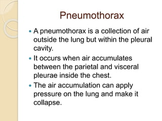
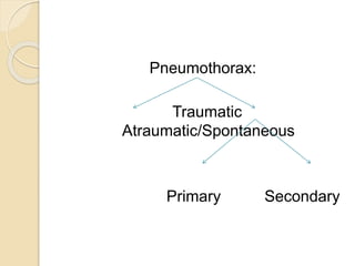
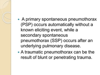
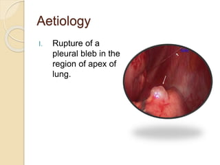
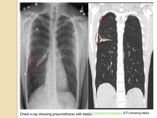
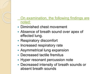
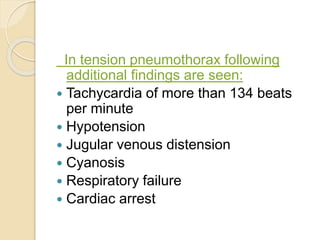
Pneumothorax : Physiotherapy
Assessment Include Goals And
Treatment
Ms. Shilpasree Saha
BPT, MPT (Cardio Thoracic Disorders)
MIAP
Pneumothorax
 A pneumothorax is a collection of air
outside the lung but within the pleural
cavity.
 It occurs when air accumulates
between the parietal and visceral
pleurae inside the chest.
 The air accumulation can apply
pressure on the lung and make it
collapse.
 Air can enter the pleural space by two
mechanisms, either by trauma causing
communication through the chest wall
or from the lung by rupture of the
visceral pleura.
Pneumothorax:
Traumatic
Atraumatic/Spontaneous
Primary Secondary
 A primary spontaneous pneumothorax
(PSP) occurs automatically without a
known eliciting event, while a
secondary spontaneous
pneumothorax (SSP) occurs after an
underlying pulmonary disease.
 A traumatic pneumothorax can be the
result of blunt or penetrating trauma.
 A simple pneumothorax does not shift
the mediastinal structures, as does a
tension pneumothorax.
 Open pneumothorax is an open
wound in the chest wall through which
air moves in and out.
Aetiology
I. Rupture of a
pleural bleb in the
region of apex of
lung.
Computed tomography (CT) showing blebs.
Chest x-ray showing pneumothorax with blebs.
II. Rupture of emphysematous bulla.
III. Rupture of lung abscess or
tuberculosis lesion into pleural
cavity.
a The chest radiography image shows a large lucency occupying the right upper thorax with mediastinal shift and
patchy opacities of the left apical area. b The coronal computed tomography image shows giant bullae occupying
the right thorax and multiple opacities over the left upper lung
Other causes
 Penetrating rib fracture
 Penetrating wounds
 Accidental opening of pleural cavity
during abdominal surgery
 Penetration of pleura during central
venous pressure catheter insertion.
Physical findings
 The most common symptoms are:
 Chest pain
 Shortness of breath.
 The chest pain is pleuritic, sharp,
severe, and radiates to the ipsilateral
shoulder.
On examination, the following findings are
noted:
 Diminished chest movement
 Absence of breath sound over apex of
effected lung.
 Respiratory discomfort
 Increased respiratory rate
 Asymmetrical lung expansion
 Decreased tactile fremitus
 Hyper resonant percussion note
 Decreased intensity of breath sounds or
absent breath sounds
In tension pneumothorax following
additional findings are seen:
 Tachycardia of more than 134 beats
per minute
 Hypotension
 Jugular venous distension
 Cyanosis
 Respiratory failure
 Cardiac arrest
Treatment
 A small pneumothorax requires few
days of bed rest until it resolves.
 A large pneumothorax (›25 percent of
pleural space is filled with air) is
treated with needle aspiration or by an
intercostal drain.
 Pleurodesis or pleurectomy may
indicated for recurrent pneumothorax.
PT-Treatment
 Expansion breathing exercise to
reexpand the lung.
 Shoulder ROM exercises to maintain
shoulder, shoulder girdle and thoracic
mobility

Pneumothorax and Physiotherapy management .pptx

  • 1.
    Pneumothorax : Physiotherapy AssessmentInclude Goals And Treatment Ms. Shilpasree Saha BPT, MPT (Cardio Thoracic Disorders) MIAP
  • 2.
    Pneumothorax  A pneumothoraxis a collection of air outside the lung but within the pleural cavity.  It occurs when air accumulates between the parietal and visceral pleurae inside the chest.  The air accumulation can apply pressure on the lung and make it collapse.
  • 3.
     Air canenter the pleural space by two mechanisms, either by trauma causing communication through the chest wall or from the lung by rupture of the visceral pleura.
  • 4.
  • 5.
     A primaryspontaneous pneumothorax (PSP) occurs automatically without a known eliciting event, while a secondary spontaneous pneumothorax (SSP) occurs after an underlying pulmonary disease.  A traumatic pneumothorax can be the result of blunt or penetrating trauma.
  • 6.
     A simplepneumothorax does not shift the mediastinal structures, as does a tension pneumothorax.  Open pneumothorax is an open wound in the chest wall through which air moves in and out.
  • 7.
    Aetiology I. Rupture ofa pleural bleb in the region of apex of lung.
  • 8.
    Computed tomography (CT)showing blebs. Chest x-ray showing pneumothorax with blebs.
  • 9.
    II. Rupture ofemphysematous bulla. III. Rupture of lung abscess or tuberculosis lesion into pleural cavity.
  • 10.
    a The chestradiography image shows a large lucency occupying the right upper thorax with mediastinal shift and patchy opacities of the left apical area. b The coronal computed tomography image shows giant bullae occupying the right thorax and multiple opacities over the left upper lung
  • 11.
    Other causes  Penetratingrib fracture  Penetrating wounds  Accidental opening of pleural cavity during abdominal surgery  Penetration of pleura during central venous pressure catheter insertion.
  • 12.
    Physical findings  Themost common symptoms are:  Chest pain  Shortness of breath.  The chest pain is pleuritic, sharp, severe, and radiates to the ipsilateral shoulder.
  • 13.
    On examination, thefollowing findings are noted:  Diminished chest movement  Absence of breath sound over apex of effected lung.  Respiratory discomfort  Increased respiratory rate  Asymmetrical lung expansion  Decreased tactile fremitus  Hyper resonant percussion note  Decreased intensity of breath sounds or absent breath sounds
  • 14.
    In tension pneumothoraxfollowing additional findings are seen:  Tachycardia of more than 134 beats per minute  Hypotension  Jugular venous distension  Cyanosis  Respiratory failure  Cardiac arrest
  • 15.
    Treatment  A smallpneumothorax requires few days of bed rest until it resolves.  A large pneumothorax (›25 percent of pleural space is filled with air) is treated with needle aspiration or by an intercostal drain.  Pleurodesis or pleurectomy may indicated for recurrent pneumothorax.
  • 16.
    PT-Treatment  Expansion breathingexercise to reexpand the lung.  Shoulder ROM exercises to maintain shoulder, shoulder girdle and thoracic mobility