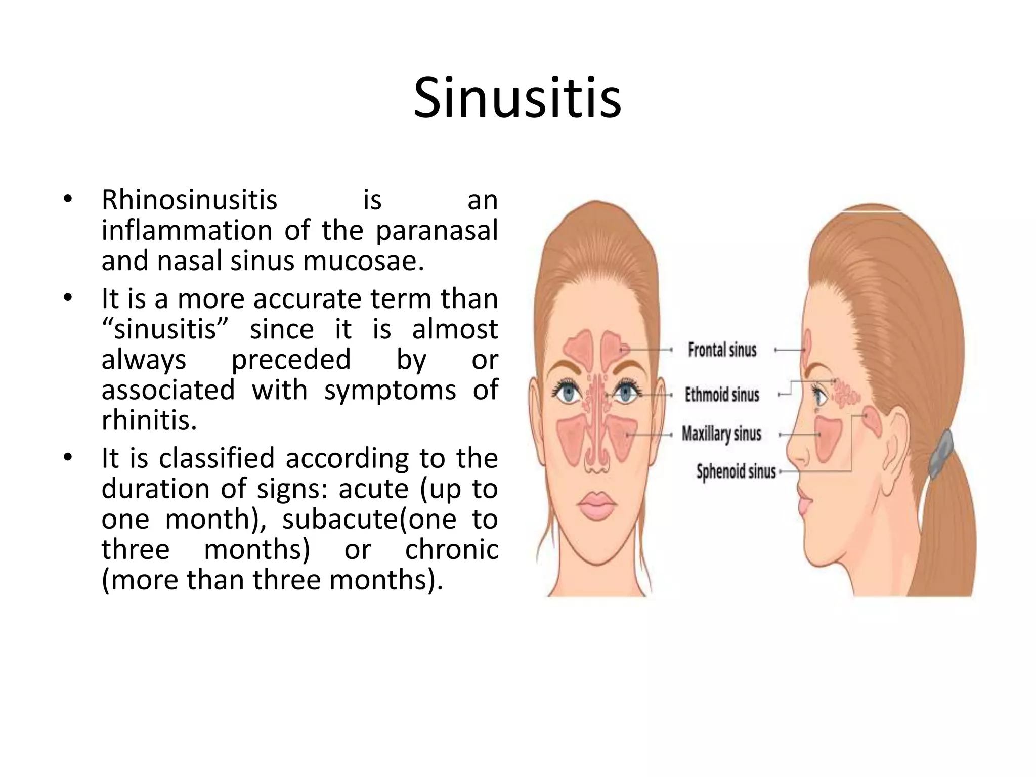
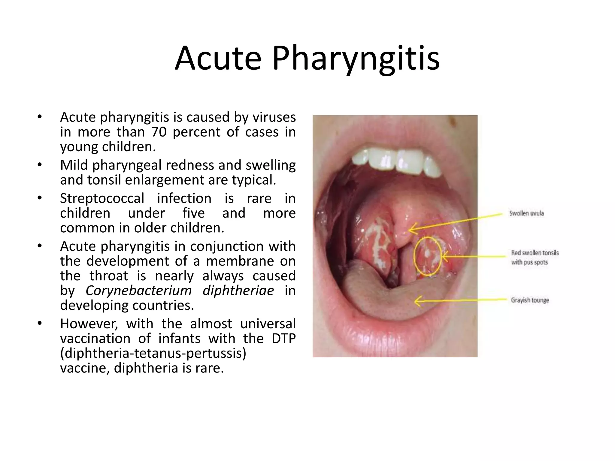
Respiratory Infections in Children
Ms.ShilpasreeSaha,
BPT,MPT(CardioThoracicDisorders)
• Acute respiratory infections (ARIs) are classified
as upper respiratory tract infections (URIs) or
lower respiratory tract infections (LRIs).
• The upper respiratory tract consists of the
airways from the nostrils to the vocal cords in the
larynx, including the paranasal sinuses and the
middle ear.
• The lower respiratory tract covers the
continuation of the airways from the trachea and
bronchi to the bronchioles and the alveoli.
Upper Respiratory Tract Infections
• URIs are the most common infectious
diseases.
• They include rhinitis (common cold), sinusitis,
ear infections, acute pharyngitis or
tonsillopharyngitis, epiglottitis, and
laryngitis—of which ear infections and
pharyngitis cause the more severe
complications (deafness and acute rheumatic
fever, respectively).
Sinusitis
• Rhinosinusitis is an
inflammation of the paranasal
and nasal sinus mucosae.
• It is a more accurate term than
“sinusitis” since it is almost
always preceded by or
associated with symptoms of
rhinitis.
• It is classified according to the
duration of signs: acute (up to
one month), subacute(one to
three months) or chronic
(more than three months).
• Acute bacterial rhinosinusitis is diagnosed in a
child based on several criteria: persistent upper
respiratory tract symptoms more than 10 days
(cough or nasal discharge or both); or recurrence
of symptoms after initial improvement: fever,
worsening cough, or worsening or new purulent
rhinorrhea ; or severe onset of symptoms like
fever or purulent nasal discharge lasting more
than three consecutive days associated with facial
tenderness or headache.
• Common pathogens involved in ABRS are Streptococcus
pneumonia, Haemophilus influenza, and
Moraxellacatarrhalis.
• The management of CRS in children consists primarily of
medical treatment to eradicate bacterial infection and
reduce underlying sinonasal inflammation.
• Surgical interventions, such as sinus puncture and lavage,
adenoidectomy, balloon sinuplasty, endoscopic sinus
surgery, open surgical approaches and turbinate reduction
are reserved for patients who fail medical management.
• Such procedures are designed to both eradicate potential
bacterial reservoirs and enhance sinonasal aeration and
drainage
Acute Pharyngitis
• Acute pharyngitis is caused by viruses
in more than 70 percent of cases in
young children.
• Mild pharyngeal redness and swelling
and tonsil enlargement are typical.
• Streptococcal infection is rare in
children under five and more
common in older children.
• Acute pharyngitis in conjunction with
the development of a membrane on
the throat is nearly always caused
by Corynebacterium diphtheriae in
developing countries.
• However, with the almost universal
vaccination of infants with the DTP
(diphtheria-tetanus-pertussis)
vaccine, diphtheria is rare.
Acute Ear Infection
• Chronic ear infection following repeated
episodes of acute ear infection is common in
developing countries, affecting 2 to 6 percent
of school-age children.
• The associated hearing loss may be disabling
and may affect learning.
• Repeated ear infections may lead to
mastoiditis, which in turn may spread
infection to the meninges
Lower Respiratory Tract Infections
• The common LRIs in children are pneumonia
and bronchiolitis.
• The respiratory rate is a valuable clinical sign
for diagnosing acute LRI in children who are
coughing and breathing rapidly.
• The presence of lower chest wall indrawing
identifies more severe disease.
Pneumonia
• Causative organism: Both bacteria and viruses
can cause pneumonia.
• Bacterial pneumonia is often caused
by Streptococcus pneumoniae (pneumococcus)
or Haemophilus influenzae, mostly type b (Hib),
and occasionally by Staphylococcus aureus or
other streptococci.
• Other pathogens, such as Mycoplasma
pneumoniae .
• Bacterial cultures of lung aspirate specimens
are often considered the gold standard, but
they are not practical for field application.
• Pneumonia is an invasion of the lower respiratory tract,
below the larynx by pathogens either by inhalation,
aspiration, respiratory epithelium invasion, or
hematogenous spread.
• There are barriers to infection that include anatomical
structures (nasal hairs, turbinates, epiglottis, cilia), and
humoral and cellular immunity.
• Once these barriers are breached, infection, either by
fomite/droplet spread (mostly viruses) or
nasopharyngeal colonization (mostly bacterial), results
in inflammation and injury or death of surrounding
epithelium and alveoli.
Stages
• There are four stages of lobar pneumonia.
1. The first stage occurs within 24 hours and is
characterized by alveolar edema and vascular
congestion. Both bacteria and neutrophils are
present.
2. Red hepatization is the second stage, and it has
the consistency of the liver. The stage is
characterized by neutrophils, red blood cells,
and desquamated epithelial cells. Fibrin deposits
in the alveoli are common.
• 3. The third stage of gray hepatization stage
occurs 2-3 days later, and the lung appears dark
brown. There is an accumulation of hemosiderin
and hemolysis of red cells.
• 4. The fourth stage is the resolution stage, where
the cellula infiltrates is resorbed, and the
pulmonary architecture is restored. If the healing
is not ideal, then it may lead to parapneumonic
effusions and pleural adhesions.
History & Physical findings
• In many cases, complaints associated with
pneumonia are nonspecific, including cough,
fever, tachypnea, and difficulty breathing.
• Young children may present with abdominal pain.
• Important history to obtain includes the duration
of symptoms, exposures, travel, sick contacts,
baseline health of the child, chronic diseases,
recurrent symptoms, choking, immunization
history, maternal health, or birth complications in
neonates
• Physical exam should include observation for
signs of respiratory distress, including
tachypnea, nasal flaring, lower chest in-
drawing, or hypoxia on room air.
• Infants may present with reported inability to
tolerate feeds, with grunting or apnea.
• Auscultation for rales or rhonchi may present
in all lung fields.
• Laboratory evaluation in children suspected of having
pneumonia should ideally start with non-invasive, rapid
bedside testing including nasopharyngeal swab assays
to identify causative organism. This can help minimize
unnecessary imaging and antibiotic treatment in
children with influenza or bronchiolitis.
• Children who present with severe disease and appear
toxic should have complete blood count (CBC),
electrolytes, renal/hepatic function testing, and blood
cultures performed. These tests are generally not
required in children who present with mild disease.
• Serology is being used to determine the
presence of mycoplasma, and chlamydia
species. PCR is becoming available in most
hospitals, but still, the results take 24-48
hours.
• Chest x-ray can be helpful in diagnosis and
confirmation of pneumonia,
Treatment / Management
• Supportive and symptomatic management is
key and includes supplemental oxygen for
hypoxia, antipyretics for fever, and fluids for
dehydration. This is especially important for
non-infectious pneumonitis and viral
pneumonia for which antibiotics are not
indicated.

Respiratory Infections in Children.pptx

  • 1.
    Respiratory Infections inChildren Ms.ShilpasreeSaha, BPT,MPT(CardioThoracicDisorders)
  • 2.
    • Acute respiratoryinfections (ARIs) are classified as upper respiratory tract infections (URIs) or lower respiratory tract infections (LRIs). • The upper respiratory tract consists of the airways from the nostrils to the vocal cords in the larynx, including the paranasal sinuses and the middle ear. • The lower respiratory tract covers the continuation of the airways from the trachea and bronchi to the bronchioles and the alveoli.
  • 3.
    Upper Respiratory TractInfections • URIs are the most common infectious diseases. • They include rhinitis (common cold), sinusitis, ear infections, acute pharyngitis or tonsillopharyngitis, epiglottitis, and laryngitis—of which ear infections and pharyngitis cause the more severe complications (deafness and acute rheumatic fever, respectively).
  • 4.
    Sinusitis • Rhinosinusitis isan inflammation of the paranasal and nasal sinus mucosae. • It is a more accurate term than “sinusitis” since it is almost always preceded by or associated with symptoms of rhinitis. • It is classified according to the duration of signs: acute (up to one month), subacute(one to three months) or chronic (more than three months).
  • 5.
    • Acute bacterialrhinosinusitis is diagnosed in a child based on several criteria: persistent upper respiratory tract symptoms more than 10 days (cough or nasal discharge or both); or recurrence of symptoms after initial improvement: fever, worsening cough, or worsening or new purulent rhinorrhea ; or severe onset of symptoms like fever or purulent nasal discharge lasting more than three consecutive days associated with facial tenderness or headache.
  • 6.
    • Common pathogensinvolved in ABRS are Streptococcus pneumonia, Haemophilus influenza, and Moraxellacatarrhalis. • The management of CRS in children consists primarily of medical treatment to eradicate bacterial infection and reduce underlying sinonasal inflammation. • Surgical interventions, such as sinus puncture and lavage, adenoidectomy, balloon sinuplasty, endoscopic sinus surgery, open surgical approaches and turbinate reduction are reserved for patients who fail medical management. • Such procedures are designed to both eradicate potential bacterial reservoirs and enhance sinonasal aeration and drainage
  • 7.
    Acute Pharyngitis • Acutepharyngitis is caused by viruses in more than 70 percent of cases in young children. • Mild pharyngeal redness and swelling and tonsil enlargement are typical. • Streptococcal infection is rare in children under five and more common in older children. • Acute pharyngitis in conjunction with the development of a membrane on the throat is nearly always caused by Corynebacterium diphtheriae in developing countries. • However, with the almost universal vaccination of infants with the DTP (diphtheria-tetanus-pertussis) vaccine, diphtheria is rare.
  • 8.
    Acute Ear Infection •Chronic ear infection following repeated episodes of acute ear infection is common in developing countries, affecting 2 to 6 percent of school-age children. • The associated hearing loss may be disabling and may affect learning. • Repeated ear infections may lead to mastoiditis, which in turn may spread infection to the meninges
  • 9.
    Lower Respiratory TractInfections • The common LRIs in children are pneumonia and bronchiolitis. • The respiratory rate is a valuable clinical sign for diagnosing acute LRI in children who are coughing and breathing rapidly. • The presence of lower chest wall indrawing identifies more severe disease.
  • 10.
    Pneumonia • Causative organism:Both bacteria and viruses can cause pneumonia. • Bacterial pneumonia is often caused by Streptococcus pneumoniae (pneumococcus) or Haemophilus influenzae, mostly type b (Hib), and occasionally by Staphylococcus aureus or other streptococci. • Other pathogens, such as Mycoplasma pneumoniae .
  • 11.
    • Bacterial culturesof lung aspirate specimens are often considered the gold standard, but they are not practical for field application.
  • 12.
    • Pneumonia isan invasion of the lower respiratory tract, below the larynx by pathogens either by inhalation, aspiration, respiratory epithelium invasion, or hematogenous spread. • There are barriers to infection that include anatomical structures (nasal hairs, turbinates, epiglottis, cilia), and humoral and cellular immunity. • Once these barriers are breached, infection, either by fomite/droplet spread (mostly viruses) or nasopharyngeal colonization (mostly bacterial), results in inflammation and injury or death of surrounding epithelium and alveoli.
  • 13.
    Stages • There arefour stages of lobar pneumonia. 1. The first stage occurs within 24 hours and is characterized by alveolar edema and vascular congestion. Both bacteria and neutrophils are present. 2. Red hepatization is the second stage, and it has the consistency of the liver. The stage is characterized by neutrophils, red blood cells, and desquamated epithelial cells. Fibrin deposits in the alveoli are common.
  • 14.
    • 3. Thethird stage of gray hepatization stage occurs 2-3 days later, and the lung appears dark brown. There is an accumulation of hemosiderin and hemolysis of red cells. • 4. The fourth stage is the resolution stage, where the cellula infiltrates is resorbed, and the pulmonary architecture is restored. If the healing is not ideal, then it may lead to parapneumonic effusions and pleural adhesions.
  • 15.
    History & Physicalfindings • In many cases, complaints associated with pneumonia are nonspecific, including cough, fever, tachypnea, and difficulty breathing. • Young children may present with abdominal pain. • Important history to obtain includes the duration of symptoms, exposures, travel, sick contacts, baseline health of the child, chronic diseases, recurrent symptoms, choking, immunization history, maternal health, or birth complications in neonates
  • 16.
    • Physical examshould include observation for signs of respiratory distress, including tachypnea, nasal flaring, lower chest in- drawing, or hypoxia on room air. • Infants may present with reported inability to tolerate feeds, with grunting or apnea. • Auscultation for rales or rhonchi may present in all lung fields.
  • 17.
    • Laboratory evaluationin children suspected of having pneumonia should ideally start with non-invasive, rapid bedside testing including nasopharyngeal swab assays to identify causative organism. This can help minimize unnecessary imaging and antibiotic treatment in children with influenza or bronchiolitis. • Children who present with severe disease and appear toxic should have complete blood count (CBC), electrolytes, renal/hepatic function testing, and blood cultures performed. These tests are generally not required in children who present with mild disease.
  • 18.
    • Serology isbeing used to determine the presence of mycoplasma, and chlamydia species. PCR is becoming available in most hospitals, but still, the results take 24-48 hours. • Chest x-ray can be helpful in diagnosis and confirmation of pneumonia,
  • 19.
    Treatment / Management •Supportive and symptomatic management is key and includes supplemental oxygen for hypoxia, antipyretics for fever, and fluids for dehydration. This is especially important for non-infectious pneumonitis and viral pneumonia for which antibiotics are not indicated.