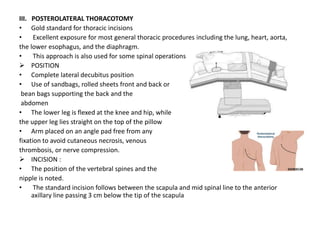
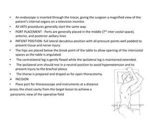
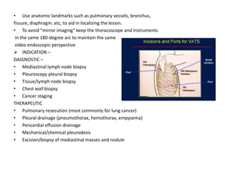
JAMIA MILLIA ISLAMIIA
CENTER FOR PHYSIOTHERAPIST AND REHABILITATION SCIENCE
THORACIC SURGERY
SUBMITTED BY – ANHA ALI BPT 4TH YEAR
SUBMITTED TO – DR. JAMAL MOIZ
DATE OF PRESENTATION – 19-01-21
SUBJECT – PHYSIPTHERAPY IN CARDIOPULMONARY CONDITION (BPT402)
 PURPOSE OF THORACIC SURGERY
• Diagnose by endoscopic or open biopsy
• Treat disease by repair of tissue
• Correct structural deformity
• Traumatic injury of lungs, heart repair
 TYPES OF THORACIC SURGERY INCISION
• Median sternotomy
• Postero lateral thoracotomy
• Anterolateral thoracotomy
• Bilateral thoraco-sternotomy
• Thoraco – laprotomy
• VATS
I. MEDIAN STERNOTOMY
• Most common thoracic incision. It is an incision down the midline of the entire
sternum.
• It offers excellent exposure of the heart pericardium, great vessels, thymus, anterior
mediastinal structures, lower trachea, and carina and is well suited for bilateral
pulmonary procedures such as resection of bilateral pulmonary metastasis
 INDICATION :
• For surgeries in the chest involving lungs, heart, esophagus
• Cardiac operations as open heart surgeries, heart transplant, corrective surgery for
congenital heart disease
• Anterior mediastinal lesions
• Bilateral lung procedures, lung transplant
• Removal of large tumors in thoracic cavity (mediastinal tumors)
 DISADVANTAGE OF MEDIAN STERNOTOMY INCISION :
• Scar formation
• Chronic chest pain.
• Injury of Brachial plexus
• Speed in opening and closing
 POSITION : Supine position and with both arms padded and draped on patient’s side.
 INSCION :
• Below the suprasternal notch to a point between the xiphoid process and umbilicus.
• An electric saw with a vertical blade is used.
• Bone wax is a useful tool to control bleeding from the sternum.
• Sternal retractor is used in lower third of the sternum and gradually opened
• Stainless steel wire is at present the standard suture in median sternotomy. At least two
wires in the manubrium and four or more wires in the body of the sternum are required
for a tight secure closure.
• Bleeding from an internal thoracic vessel occurs, it is controlled with a figure-eight stitch
II. PARTIAL STERNOTOMY
• A common variation of the median sternotomy, this incision is often combined with a
transverse collar neck incision to provide wide cervico mediastinal exposure.
• Can be extended supraclavicularly, usually on the right side for exposure of the innominate,
subclavian, and carotid vessels
 TWO TYPES
• J type – common, incision in the sternum is extended into the third or fourth inter costal
space as a controlled cut
• T junction – rare
 INDICATION-
• Isolated aortic valve replacement
• Aortic valve replacement with concomitant aortic surgery
• J type - particularly applicable to the elderly and those with impaired respiratory function
 CONTRAINDICATION
• Chest wall deformities
• Concomitant coronary surgery or other valve surgery
 POSITION – supine with both arms padded
 INCISIONS
• A straight skin incision of approximately 2.5 to 3 inches (7 to 8 cm) is made from the level of
the head of the second rib in the midline over the sternum and extended down to the level
of the head of the fourth rib.
• A regular (pendulum) saw or an oscillating saw (particularly for reoperations) can be used
• The right internal thoracic artery (ITA) is usually 1 cm away from the sternal edge and can
be protected by placing a forceps around the sternal edge to push the right ITA laterally away
from the saw
• A Finocchietto retractor is inserted and the mediastinal tissues exposed
• With help of cannulas cardiopulmonary bypass performed , after successful weaning
cannulas are clamped and removed
• Bleeding control with the help of 4 wires stitch, additional wire for the horizontal limb of the
J-incision ensures sternal stability
III. POSTEROLATERAL THORACOTOMY
• Gold standard for thoracic incisions
• Excellent exposure for most general thoracic procedures including the lung, heart, aorta,
the lower esophagus, and the diaphragm.
• This approach is also used for some spinal operations
 POSITION
• Complete lateral decubitus position
• Use of sandbags, rolled sheets front and back or
bean bags supporting the back and the
abdomen
• The lower leg is flexed at the knee and hip, while
the upper leg lies straight on the top of the pillow
• Arm placed on an angle pad free from any
fixation to avoid cutaneous necrosis, venous
thrombosis, or nerve compression.
 INCISION :
• The position of the vertebral spines and the
nipple is noted.
• The standard incision follows between the scapula and mid spinal line to the anterior
axillary line passing 3 cm below the tip of the scapula
 SURGERY
• Skin incision -> Latissimus dorsi and serratus anterior muscles incised -> Posteriorly, the
muscle layers of rhomboid and trapezius are incised ->The pleural space entered incising
the musculature between the ribs or via an osteotomy -> transect the muscles on the
superior border of the ribs to avoid injuring the neurovascular bundle ->ribs may be
transected or resected
• After the surgery, drainage tubes must be placed ->The rib approximator is closed ->
ordinary silk sutures are to be avoided as it increases post operative pain -> the cut ends of
trapezius and latissimus dorsi muscles are then approximated and sutured -> subcutaneous
tissue is closed using absorbable sutures -> The skin is closed using surgical clips or a
subcuticular stitch such as monocryl.
 DISADVANTAGES
• Increased potential for blood loss and moderate time
requirement for opening and closing the incision
• Prolonged ipsilateral shoulder and arm dysfunctions
• Compromised pulmonary function and chronic
post thoracotomy pain syndromes
• Scoliosis have been described in children
IV. AXILLARY THORACOTOMY
• Lateral decubitus position -> homolateral arm is abducted at 900at the shoulder level, and
flexed at the elbow
• Choice in majority of pulmonary resections, PDA ligation
 ADVANTAGES: muscle sparing, ease and speed, good cosmetic result.
 DISADVANTAGES: limited exposure
V. BILATERAL THORACO STERNOTOMY
• Less often used now a days
• Incision along the infra-mammary creases and across the sternum
4 th and 5th intercostal space
• Poor healing of the wound.
VII. ANTEROLATERAL THORACOTOMY
• Useful in variety of operation on heart, pulmonary resection and surgery of eosophagus.
 POSITION - Supine and operation site elevated to 300
 INCISION - Incision from lateral border of sternum to mid-axillary line at 4 th or 5 th
interspace.
 INDICATION –
• Emergency procedure to allow direct access to heart after cardiac arrest
• Unilateral lung thoracotomy
• Open lung biopsy
VII. LEFT THORACOLAPROTOMY –
• left thoraco abdominal incision give excellent exposure of distal esophagus or proximal
stomach
 INDICATION- lower or middle third carcinoma esophagus resection
 POSITION – same as in VATS
 INSCION – Oblique incision starting 2 finger below scapular tip then brought parallel to ribs
and cross coastal arc (8th intercostal space) then slight inferior curvature of incision as it
approach to midline abdomen
 COMPLICATION - incisional hernia, fistula formation, burst abdomen
INTRAPLEURAL TUBES (ICD)
• Whenever thoracotomy has been done
• Exit of fluids and air and monitoring of blood loss
• Inserted through a separate incision
VIII. VIDEO ASSISTED THORACIC SURGERY (VATS)
• It is a minimally invasive surgical procedure, used to diagnose and treat illness or injury to
the lung and other organs in thorax
• Miniaturization of video equipment
• In VATS procedures, surgeons operate through 2 to 4 tiny openings between the ribs. Each
opening is less than one inch in diameter, whereas 6- to 10-inch incisions are not
uncommon in open thoracic surgery.
• An endoscope is inserted through the trocar, giving the surgeon a magnified view of the
patient’s internal organs on a television monitor.
• All VATS procedures generally start the same way.
• PORT PLACEMENT - Ports are generally placed in the middle (7th inter costal space),
anterior, and posterior axillary lines
 PATIENT POSITION- full lateral decubitus position with all pressure points well padded to
prevent tissue and nerve injury.
• The hips are placed below the break point of the table to allow opening of the intercostal
spaces as the table is angulated.
• The contralateral leg is gently flexed while the ipsilateral leg is maintained extended.
• The ipsilateral arm should rest in a neutral position to avoid hyperextension and to
prevent injury to the brachial plexus
• The thorax is prepared and draped as for open thoracotomy.
 INCISION
• Place port for thoracoscope and instruments at a distance
across the chest cavity from the target lesion to achieve a
panoramic view of the operative field
• Use anatomic landmarks such as pulmonary vessels, bronchus,
fissure, diaphragm. etc, to aid in localizing the lesion.
• To avoid "mirror imaging” keep the thoracoscope and instruments
in the same 180-degree arc to maintain the same
video endoscopic perspective
 INDICATION –
DAIGNOSTIC –
• Mediastinal lymph node biopsy
• Pleuroscopy pleural biopsy
• Tissue/lymph node biopsy
• Chest wall biopsy
• Cancer staging
THERAPEUTIC
• Pulmonary resecution (most commonly for lung cancer)
• Pleural drainage (pneumothorax, hemothorax, empysema)
• Pericardial effusion drainage
• Mechanical/chemical pleurodesis
• Excision/biopsy of mediastinal masses and nodule
 CONTRAINDICATIONS
• Inability to tolerate single-lung ventilation
• Pulmonary lesion invading the mediastinum or chest wall (relative)
• Large pulmonary lesions (5 cm) (relative)
• Inability to achieve ipsilateral pulmonary atelectasis.
 ADVANTAGE –
• Reduced acute pain
• Less blood loss
• Reduce hospital stay duration
• Low incidence of post thoracotomy pain
• Early return to ADLs
 CONS OF VATS –
• Limited to tumors of certain size, location
COMPLICATIONS OF THORACOTOMY :-
• Postural deformity
• Broncho-pleural fistula
• Post thoracotomy pain syndrome
• Blood clots – DVT, pulmonary emboli
• Infection
• Complications of general anesthesia
• Arrhythmias
LUNG RESECTION
 TYPES:-
• Pneumonectomy
• Lobectomy
• Segmental or wedge resection
I. PNEUMONECTOMY –
• Removal of entire lungs
• Radical Pneumonectomy along with that of entire lung mediastinal gland is also removed
 INCISION –
• Postero lateral incision
 INDICATION-
• Carcinoma, bronchiectasis, tuberculosis
 COMPLICATION –
• Damage to phrenic and laryngeal nerve
II. LOBECTOMY
• Lobe of the lung is removed
 INSCION –
• Thoracotomy, VATS, RATS ( robotic assisted thoracic surgery)
 INDICATION –
• Bronchiectasis, tuberculosis, carcinoma, lung abscess
 COMPLICATION –
• Lung infection, pneumothorax, postural deformaity, broncho-pleural fistula
III. SEGMENTAL RESECTION –
• A broncho pulmonary segment is removed with its segmental artery and bronchus
 INDICATION – tuberculosis
PREOPERATIVE PHYSIOTHERAPY MANAGEMENT
 GOALS :
• Give psychological support
• Build patient confident
• Teach postural awareness
• Teach active cycle of breathing technique
• Teach upper limb, lower limb and trunk mobility exercises
• Teach bed mobility
• Teach sub maximal aerobic exercises
 Measurements of vitals – measurement of BP, HR, SpO2, BORG Score, ATS scale before,
during and after pre-operative exercises
 PRE OPERATIVE PT EXERCISES
• 6 MINUTE WALK TEST – MCID (minimally clinically important difference) = 31m in surgical,
elderly
• Cardiopulmonary exercise testing to measure Vo2, minute ventilation, Pco2,
• ACBT techniques in sitting or postural drainage position for effective cough and to
improve ventilation of lungs
• Avoidance of excess muscle fatigue
• More frequent, shorter-duration exercise sessions if necessary
• Self assisted coughing technique
• Inspiratory muscle training (IMT) via deep breathing exercises, incentive spirometry for 10
min., for at least two weeks up to the day of surgery .
• Strength training of shoulder girdle abductors (middle trapezius and rhomboids) and
increase flexibility of pectoralis muscles for postural alignment
• Lower extremity training include stationary bicycle, walking and stair climbing (if capable)
for 10-15 min., for at least two week up to the date of surgery
• Shoulder mobilization exercises – instruct patient to touch shoulder with hand so that
elbow point forward and form big circles with elbow , repeat 3-4 times twice a day for at
least two weeks up to the day of surgery (fig.below)
 Trunk mobility exercises –
1. Seated trunk lateral flexion - instruct patient to sit in chair raise up your arm and bend
to the opposite side for a stretch, lay one hand on their waist and turn to left side hold
for 3-10 sec return to center then further turn to right side .
2. Seated trunk rotation with arm relax – instruct patient to sit on chair cross your arm over
your chest and keeping hip and neck in neutral position turn left side hold for 10-30 sec
then turn to right side, repeat 3 -5 times on each side a day, exhaling while turning trunk
and inhale when return to the center
3. Seated trunk rotation stretch - While in a seated position, rotate your body to the side
and hold for a stretch. Hold for 10-30 sec repeat 3-5 times a day
SUMMARY :-
• Purpose of thoracic surgery
• Types along with its indication, position, incision, disadvantange – Median
sternotomy, Partial sternotomy, Antero- lateral thoracotomy, Postero- lateral
thoracotomy, Thoraco- laprotomy, VATS
• Complication of thoracotomy
• Lung resection and its type
• Pre operative physiotherapy management
REFERENCES -
• Cash Textbook of General Medical And a Surgical Conditions For
Physiotherapists By Joan E. Cash
• Dalton, M. L., & Connally, S. R. (1993). Median sternotomy. Surgery, gynecology
& obstetrics, 176(6), 615–624.
• Kulkarni, S. R., Fletcher, E., McConnell, A. K., Poskitt, K. R., & Whyman, M. R. (2010). Pre-
operative inspiratory muscle training preserves postoperative inspiratory muscle strength
following major abdominal surgery - a randomised pilot study. Annals of the Royal College
of Surgeons of England, 92(8), 700–707.
https://doi.org/10.1308/003588410X12771863936648
• Guidelines for pulmonary rehabilitation programs (4th edition) AACPR

thoracic surgery

  • 1.
    JAMIA MILLIA ISLAMIIA CENTERFOR PHYSIOTHERAPIST AND REHABILITATION SCIENCE THORACIC SURGERY SUBMITTED BY – ANHA ALI BPT 4TH YEAR SUBMITTED TO – DR. JAMAL MOIZ DATE OF PRESENTATION – 19-01-21 SUBJECT – PHYSIPTHERAPY IN CARDIOPULMONARY CONDITION (BPT402)
  • 2.
     PURPOSE OFTHORACIC SURGERY • Diagnose by endoscopic or open biopsy • Treat disease by repair of tissue • Correct structural deformity • Traumatic injury of lungs, heart repair  TYPES OF THORACIC SURGERY INCISION • Median sternotomy • Postero lateral thoracotomy • Anterolateral thoracotomy • Bilateral thoraco-sternotomy • Thoraco – laprotomy • VATS
  • 3.
    I. MEDIAN STERNOTOMY •Most common thoracic incision. It is an incision down the midline of the entire sternum. • It offers excellent exposure of the heart pericardium, great vessels, thymus, anterior mediastinal structures, lower trachea, and carina and is well suited for bilateral pulmonary procedures such as resection of bilateral pulmonary metastasis  INDICATION : • For surgeries in the chest involving lungs, heart, esophagus • Cardiac operations as open heart surgeries, heart transplant, corrective surgery for congenital heart disease • Anterior mediastinal lesions • Bilateral lung procedures, lung transplant • Removal of large tumors in thoracic cavity (mediastinal tumors)  DISADVANTAGE OF MEDIAN STERNOTOMY INCISION : • Scar formation • Chronic chest pain. • Injury of Brachial plexus • Speed in opening and closing
  • 4.
     POSITION :Supine position and with both arms padded and draped on patient’s side.  INSCION : • Below the suprasternal notch to a point between the xiphoid process and umbilicus. • An electric saw with a vertical blade is used. • Bone wax is a useful tool to control bleeding from the sternum. • Sternal retractor is used in lower third of the sternum and gradually opened • Stainless steel wire is at present the standard suture in median sternotomy. At least two wires in the manubrium and four or more wires in the body of the sternum are required for a tight secure closure. • Bleeding from an internal thoracic vessel occurs, it is controlled with a figure-eight stitch
  • 5.
    II. PARTIAL STERNOTOMY •A common variation of the median sternotomy, this incision is often combined with a transverse collar neck incision to provide wide cervico mediastinal exposure. • Can be extended supraclavicularly, usually on the right side for exposure of the innominate, subclavian, and carotid vessels  TWO TYPES • J type – common, incision in the sternum is extended into the third or fourth inter costal space as a controlled cut • T junction – rare  INDICATION- • Isolated aortic valve replacement • Aortic valve replacement with concomitant aortic surgery • J type - particularly applicable to the elderly and those with impaired respiratory function  CONTRAINDICATION • Chest wall deformities • Concomitant coronary surgery or other valve surgery  POSITION – supine with both arms padded
  • 6.
     INCISIONS • Astraight skin incision of approximately 2.5 to 3 inches (7 to 8 cm) is made from the level of the head of the second rib in the midline over the sternum and extended down to the level of the head of the fourth rib. • A regular (pendulum) saw or an oscillating saw (particularly for reoperations) can be used • The right internal thoracic artery (ITA) is usually 1 cm away from the sternal edge and can be protected by placing a forceps around the sternal edge to push the right ITA laterally away from the saw • A Finocchietto retractor is inserted and the mediastinal tissues exposed • With help of cannulas cardiopulmonary bypass performed , after successful weaning cannulas are clamped and removed • Bleeding control with the help of 4 wires stitch, additional wire for the horizontal limb of the J-incision ensures sternal stability
  • 7.
    III. POSTEROLATERAL THORACOTOMY •Gold standard for thoracic incisions • Excellent exposure for most general thoracic procedures including the lung, heart, aorta, the lower esophagus, and the diaphragm. • This approach is also used for some spinal operations  POSITION • Complete lateral decubitus position • Use of sandbags, rolled sheets front and back or bean bags supporting the back and the abdomen • The lower leg is flexed at the knee and hip, while the upper leg lies straight on the top of the pillow • Arm placed on an angle pad free from any fixation to avoid cutaneous necrosis, venous thrombosis, or nerve compression.  INCISION : • The position of the vertebral spines and the nipple is noted. • The standard incision follows between the scapula and mid spinal line to the anterior axillary line passing 3 cm below the tip of the scapula
  • 8.
     SURGERY • Skinincision -> Latissimus dorsi and serratus anterior muscles incised -> Posteriorly, the muscle layers of rhomboid and trapezius are incised ->The pleural space entered incising the musculature between the ribs or via an osteotomy -> transect the muscles on the superior border of the ribs to avoid injuring the neurovascular bundle ->ribs may be transected or resected • After the surgery, drainage tubes must be placed ->The rib approximator is closed -> ordinary silk sutures are to be avoided as it increases post operative pain -> the cut ends of trapezius and latissimus dorsi muscles are then approximated and sutured -> subcutaneous tissue is closed using absorbable sutures -> The skin is closed using surgical clips or a subcuticular stitch such as monocryl.  DISADVANTAGES • Increased potential for blood loss and moderate time requirement for opening and closing the incision • Prolonged ipsilateral shoulder and arm dysfunctions • Compromised pulmonary function and chronic post thoracotomy pain syndromes • Scoliosis have been described in children
  • 9.
    IV. AXILLARY THORACOTOMY •Lateral decubitus position -> homolateral arm is abducted at 900at the shoulder level, and flexed at the elbow • Choice in majority of pulmonary resections, PDA ligation  ADVANTAGES: muscle sparing, ease and speed, good cosmetic result.  DISADVANTAGES: limited exposure V. BILATERAL THORACO STERNOTOMY • Less often used now a days • Incision along the infra-mammary creases and across the sternum 4 th and 5th intercostal space • Poor healing of the wound. VII. ANTEROLATERAL THORACOTOMY • Useful in variety of operation on heart, pulmonary resection and surgery of eosophagus.  POSITION - Supine and operation site elevated to 300  INCISION - Incision from lateral border of sternum to mid-axillary line at 4 th or 5 th interspace.  INDICATION – • Emergency procedure to allow direct access to heart after cardiac arrest
  • 10.
    • Unilateral lungthoracotomy • Open lung biopsy VII. LEFT THORACOLAPROTOMY – • left thoraco abdominal incision give excellent exposure of distal esophagus or proximal stomach  INDICATION- lower or middle third carcinoma esophagus resection  POSITION – same as in VATS  INSCION – Oblique incision starting 2 finger below scapular tip then brought parallel to ribs and cross coastal arc (8th intercostal space) then slight inferior curvature of incision as it approach to midline abdomen  COMPLICATION - incisional hernia, fistula formation, burst abdomen INTRAPLEURAL TUBES (ICD) • Whenever thoracotomy has been done • Exit of fluids and air and monitoring of blood loss • Inserted through a separate incision VIII. VIDEO ASSISTED THORACIC SURGERY (VATS) • It is a minimally invasive surgical procedure, used to diagnose and treat illness or injury to the lung and other organs in thorax • Miniaturization of video equipment • In VATS procedures, surgeons operate through 2 to 4 tiny openings between the ribs. Each opening is less than one inch in diameter, whereas 6- to 10-inch incisions are not uncommon in open thoracic surgery.
  • 11.
    • An endoscopeis inserted through the trocar, giving the surgeon a magnified view of the patient’s internal organs on a television monitor. • All VATS procedures generally start the same way. • PORT PLACEMENT - Ports are generally placed in the middle (7th inter costal space), anterior, and posterior axillary lines  PATIENT POSITION- full lateral decubitus position with all pressure points well padded to prevent tissue and nerve injury. • The hips are placed below the break point of the table to allow opening of the intercostal spaces as the table is angulated. • The contralateral leg is gently flexed while the ipsilateral leg is maintained extended. • The ipsilateral arm should rest in a neutral position to avoid hyperextension and to prevent injury to the brachial plexus • The thorax is prepared and draped as for open thoracotomy.  INCISION • Place port for thoracoscope and instruments at a distance across the chest cavity from the target lesion to achieve a panoramic view of the operative field
  • 12.
    • Use anatomiclandmarks such as pulmonary vessels, bronchus, fissure, diaphragm. etc, to aid in localizing the lesion. • To avoid "mirror imaging” keep the thoracoscope and instruments in the same 180-degree arc to maintain the same video endoscopic perspective  INDICATION – DAIGNOSTIC – • Mediastinal lymph node biopsy • Pleuroscopy pleural biopsy • Tissue/lymph node biopsy • Chest wall biopsy • Cancer staging THERAPEUTIC • Pulmonary resecution (most commonly for lung cancer) • Pleural drainage (pneumothorax, hemothorax, empysema) • Pericardial effusion drainage • Mechanical/chemical pleurodesis • Excision/biopsy of mediastinal masses and nodule
  • 13.
     CONTRAINDICATIONS • Inabilityto tolerate single-lung ventilation • Pulmonary lesion invading the mediastinum or chest wall (relative) • Large pulmonary lesions (5 cm) (relative) • Inability to achieve ipsilateral pulmonary atelectasis.  ADVANTAGE – • Reduced acute pain • Less blood loss • Reduce hospital stay duration • Low incidence of post thoracotomy pain • Early return to ADLs  CONS OF VATS – • Limited to tumors of certain size, location COMPLICATIONS OF THORACOTOMY :- • Postural deformity • Broncho-pleural fistula • Post thoracotomy pain syndrome • Blood clots – DVT, pulmonary emboli • Infection • Complications of general anesthesia • Arrhythmias
  • 14.
    LUNG RESECTION  TYPES:- •Pneumonectomy • Lobectomy • Segmental or wedge resection I. PNEUMONECTOMY – • Removal of entire lungs • Radical Pneumonectomy along with that of entire lung mediastinal gland is also removed  INCISION – • Postero lateral incision  INDICATION- • Carcinoma, bronchiectasis, tuberculosis  COMPLICATION – • Damage to phrenic and laryngeal nerve II. LOBECTOMY • Lobe of the lung is removed  INSCION – • Thoracotomy, VATS, RATS ( robotic assisted thoracic surgery)  INDICATION – • Bronchiectasis, tuberculosis, carcinoma, lung abscess  COMPLICATION – • Lung infection, pneumothorax, postural deformaity, broncho-pleural fistula III. SEGMENTAL RESECTION – • A broncho pulmonary segment is removed with its segmental artery and bronchus  INDICATION – tuberculosis
  • 15.
    PREOPERATIVE PHYSIOTHERAPY MANAGEMENT GOALS : • Give psychological support • Build patient confident • Teach postural awareness • Teach active cycle of breathing technique • Teach upper limb, lower limb and trunk mobility exercises • Teach bed mobility • Teach sub maximal aerobic exercises  Measurements of vitals – measurement of BP, HR, SpO2, BORG Score, ATS scale before, during and after pre-operative exercises  PRE OPERATIVE PT EXERCISES • 6 MINUTE WALK TEST – MCID (minimally clinically important difference) = 31m in surgical, elderly • Cardiopulmonary exercise testing to measure Vo2, minute ventilation, Pco2, • ACBT techniques in sitting or postural drainage position for effective cough and to improve ventilation of lungs • Avoidance of excess muscle fatigue • More frequent, shorter-duration exercise sessions if necessary
  • 16.
    • Self assistedcoughing technique • Inspiratory muscle training (IMT) via deep breathing exercises, incentive spirometry for 10 min., for at least two weeks up to the day of surgery . • Strength training of shoulder girdle abductors (middle trapezius and rhomboids) and increase flexibility of pectoralis muscles for postural alignment • Lower extremity training include stationary bicycle, walking and stair climbing (if capable) for 10-15 min., for at least two week up to the date of surgery • Shoulder mobilization exercises – instruct patient to touch shoulder with hand so that elbow point forward and form big circles with elbow , repeat 3-4 times twice a day for at least two weeks up to the day of surgery (fig.below)
  • 17.
     Trunk mobilityexercises – 1. Seated trunk lateral flexion - instruct patient to sit in chair raise up your arm and bend to the opposite side for a stretch, lay one hand on their waist and turn to left side hold for 3-10 sec return to center then further turn to right side . 2. Seated trunk rotation with arm relax – instruct patient to sit on chair cross your arm over your chest and keeping hip and neck in neutral position turn left side hold for 10-30 sec then turn to right side, repeat 3 -5 times on each side a day, exhaling while turning trunk and inhale when return to the center 3. Seated trunk rotation stretch - While in a seated position, rotate your body to the side and hold for a stretch. Hold for 10-30 sec repeat 3-5 times a day
  • 18.
    SUMMARY :- • Purposeof thoracic surgery • Types along with its indication, position, incision, disadvantange – Median sternotomy, Partial sternotomy, Antero- lateral thoracotomy, Postero- lateral thoracotomy, Thoraco- laprotomy, VATS • Complication of thoracotomy • Lung resection and its type • Pre operative physiotherapy management
  • 19.
    REFERENCES - • CashTextbook of General Medical And a Surgical Conditions For Physiotherapists By Joan E. Cash • Dalton, M. L., & Connally, S. R. (1993). Median sternotomy. Surgery, gynecology & obstetrics, 176(6), 615–624. • Kulkarni, S. R., Fletcher, E., McConnell, A. K., Poskitt, K. R., & Whyman, M. R. (2010). Pre- operative inspiratory muscle training preserves postoperative inspiratory muscle strength following major abdominal surgery - a randomised pilot study. Annals of the Royal College of Surgeons of England, 92(8), 700–707. https://doi.org/10.1308/003588410X12771863936648 • Guidelines for pulmonary rehabilitation programs (4th edition) AACPR