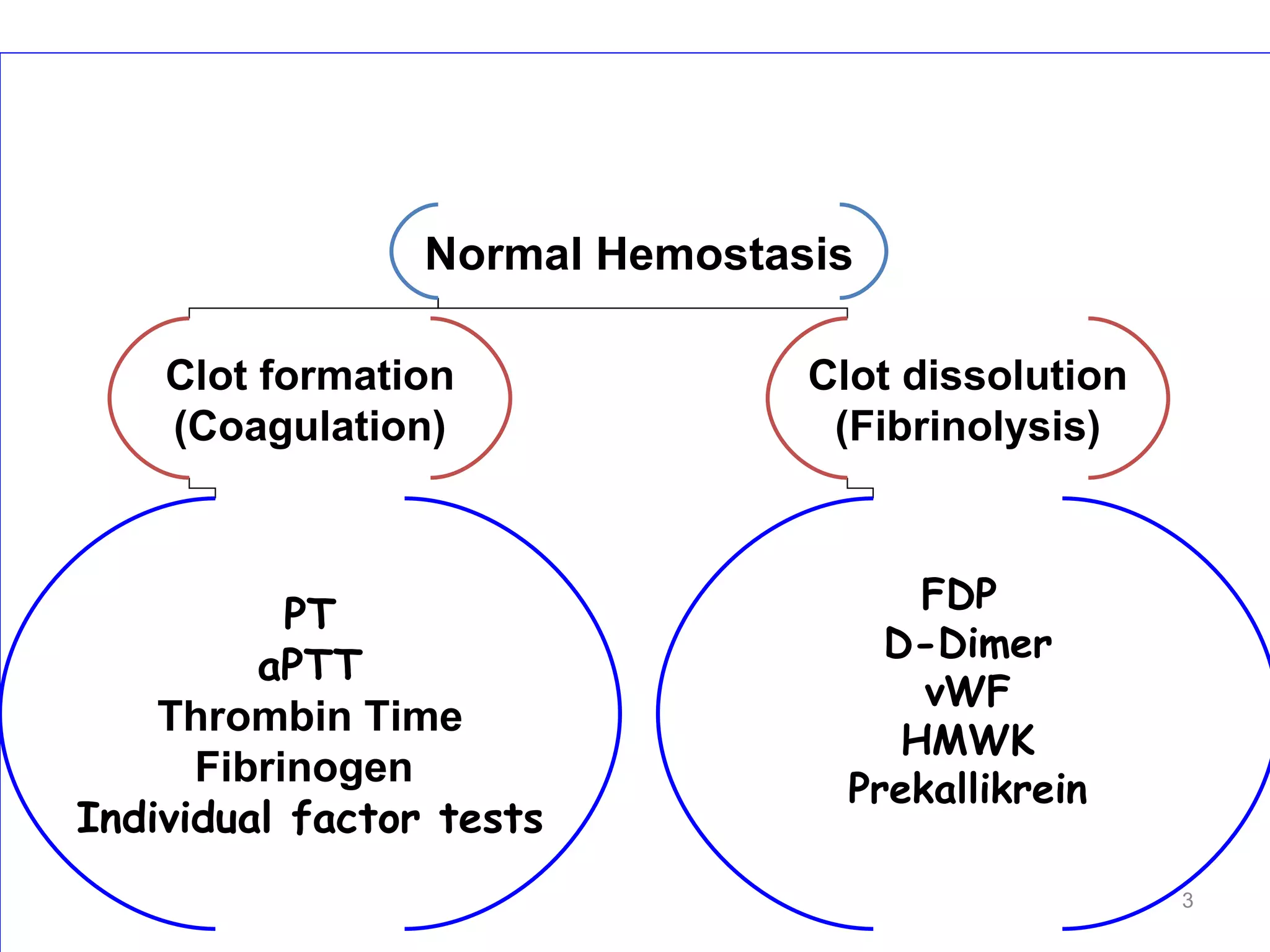
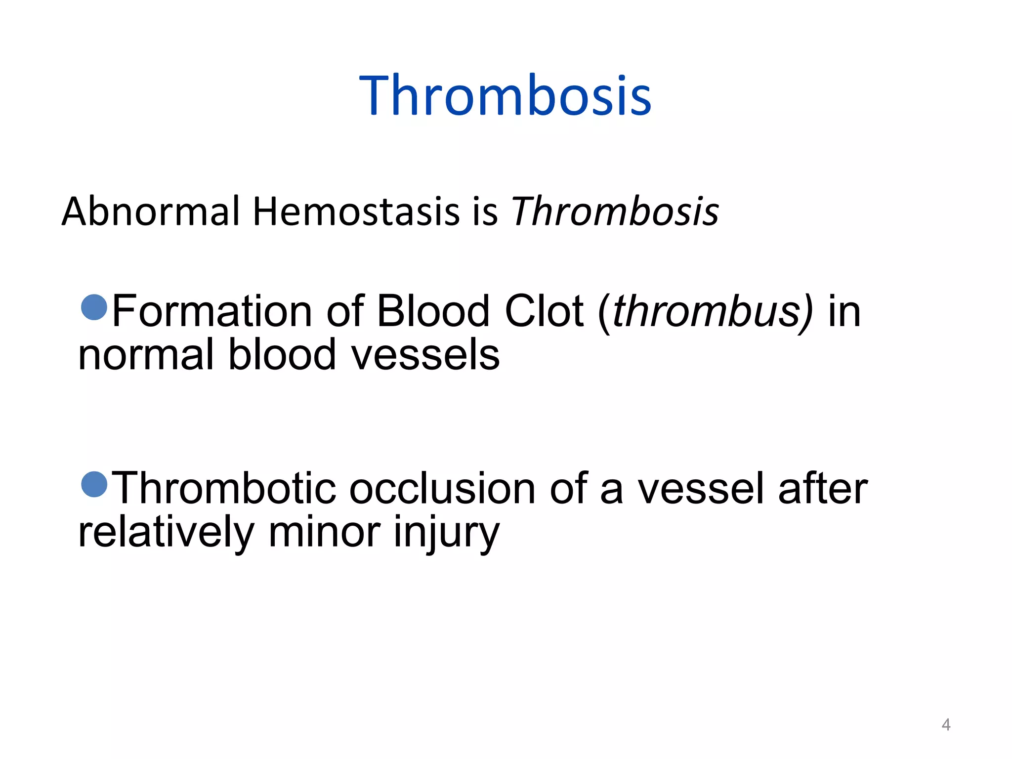
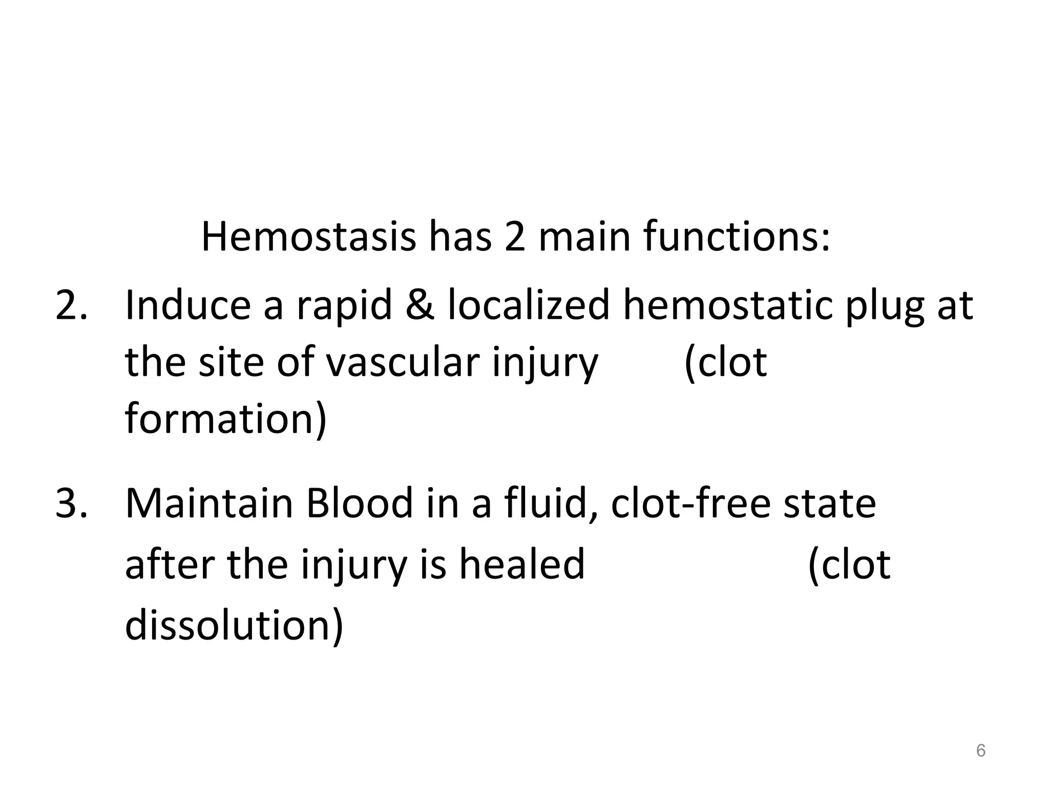
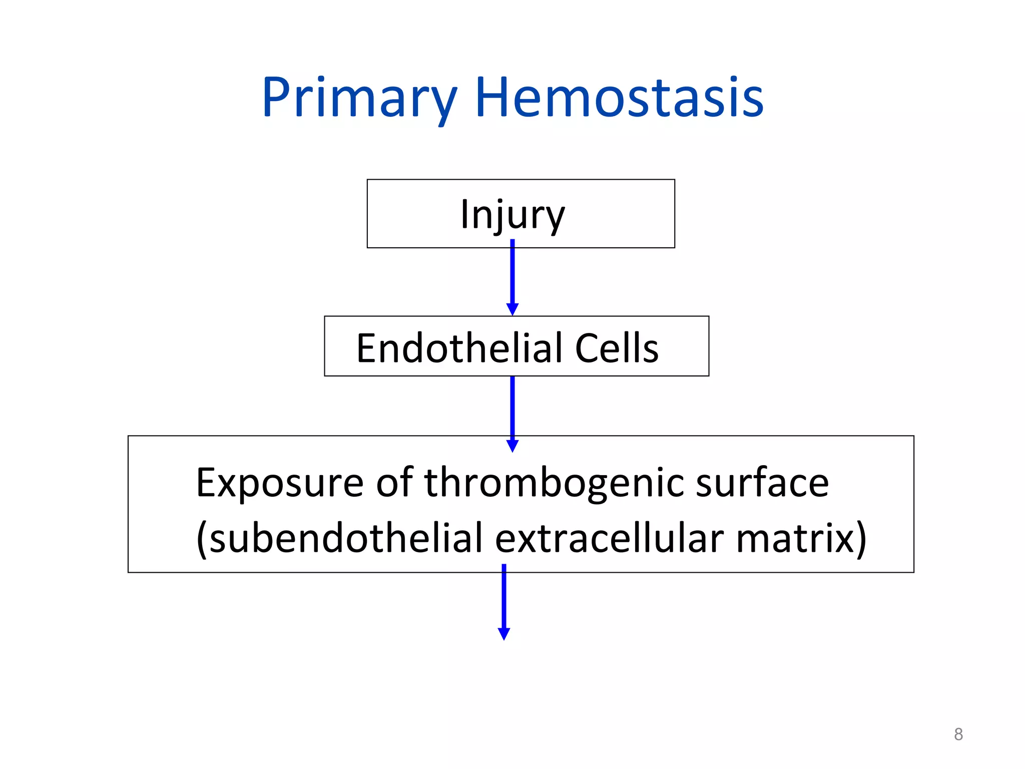
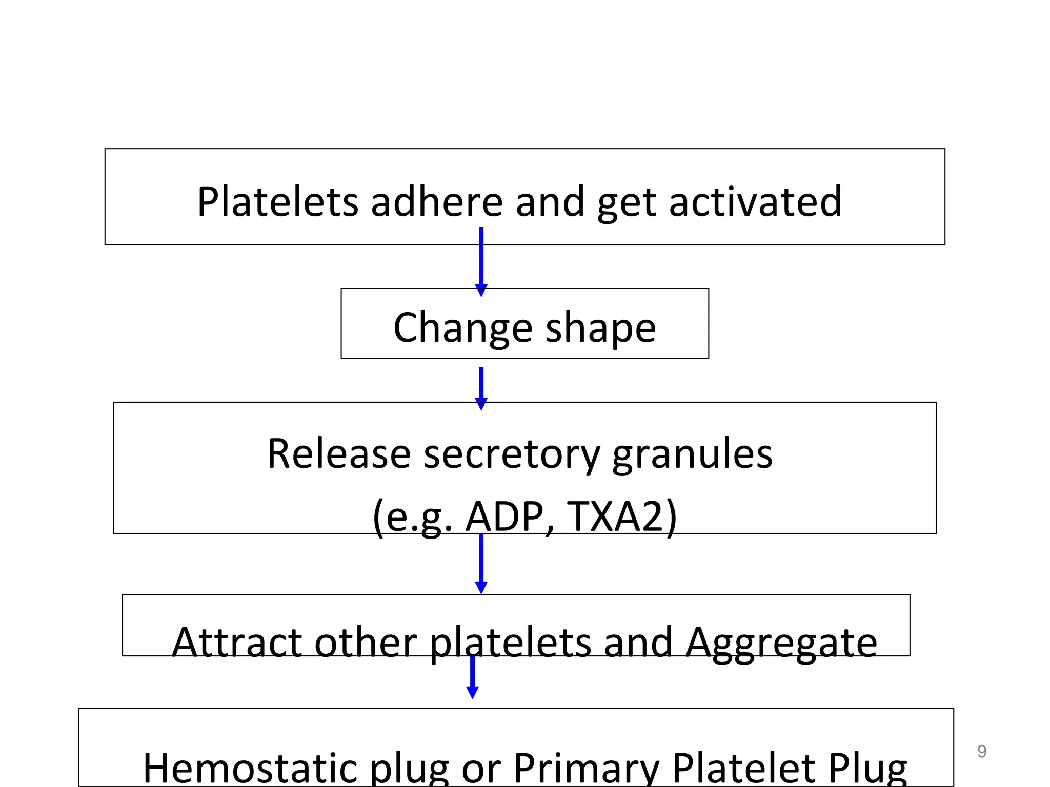
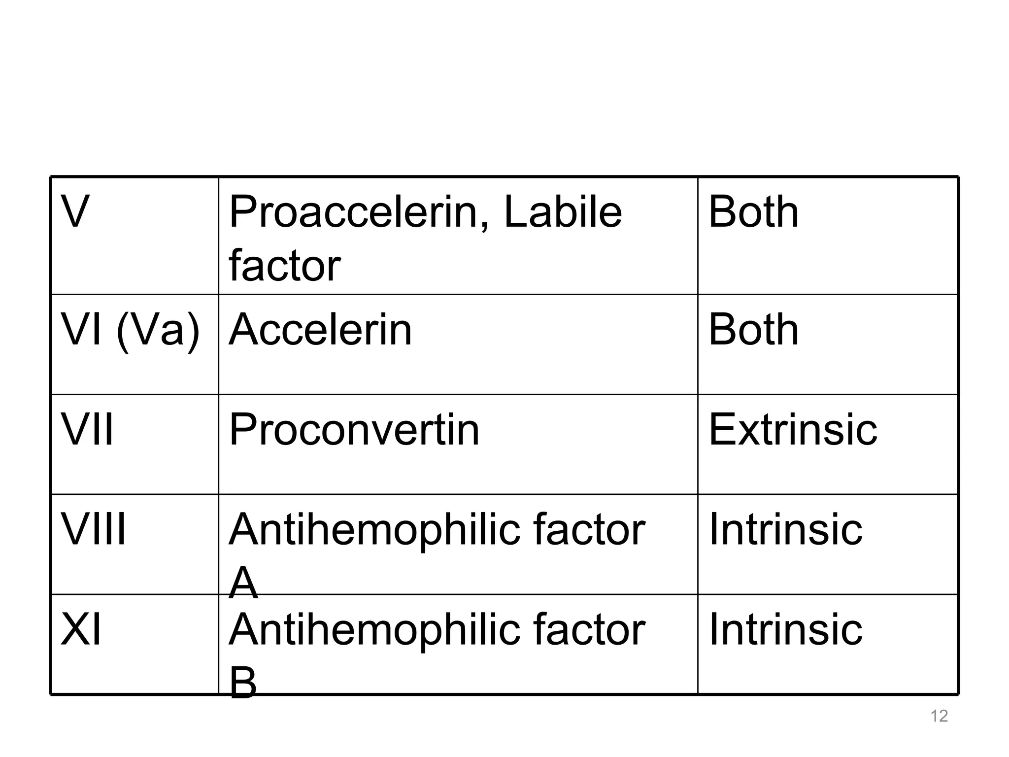
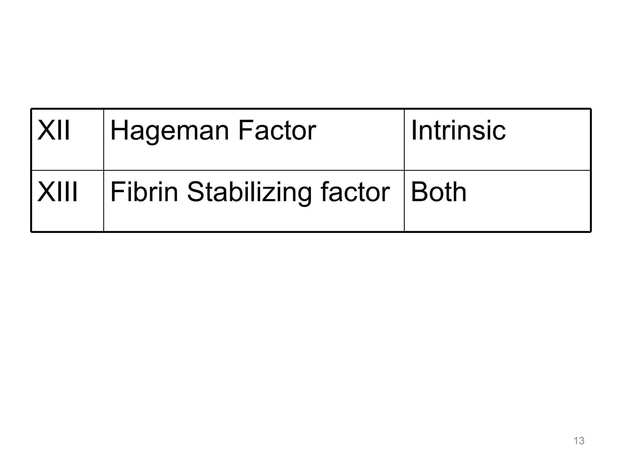
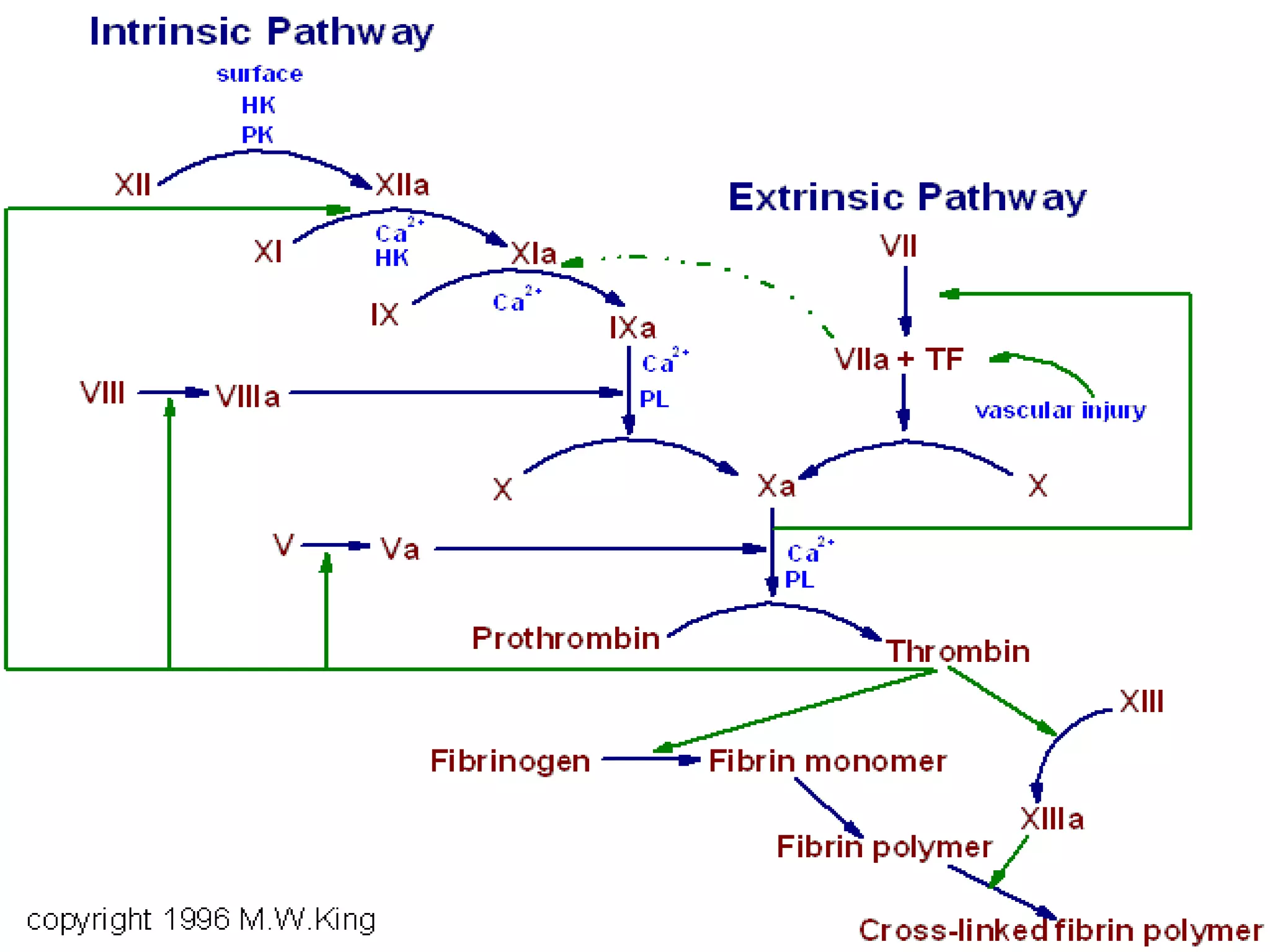
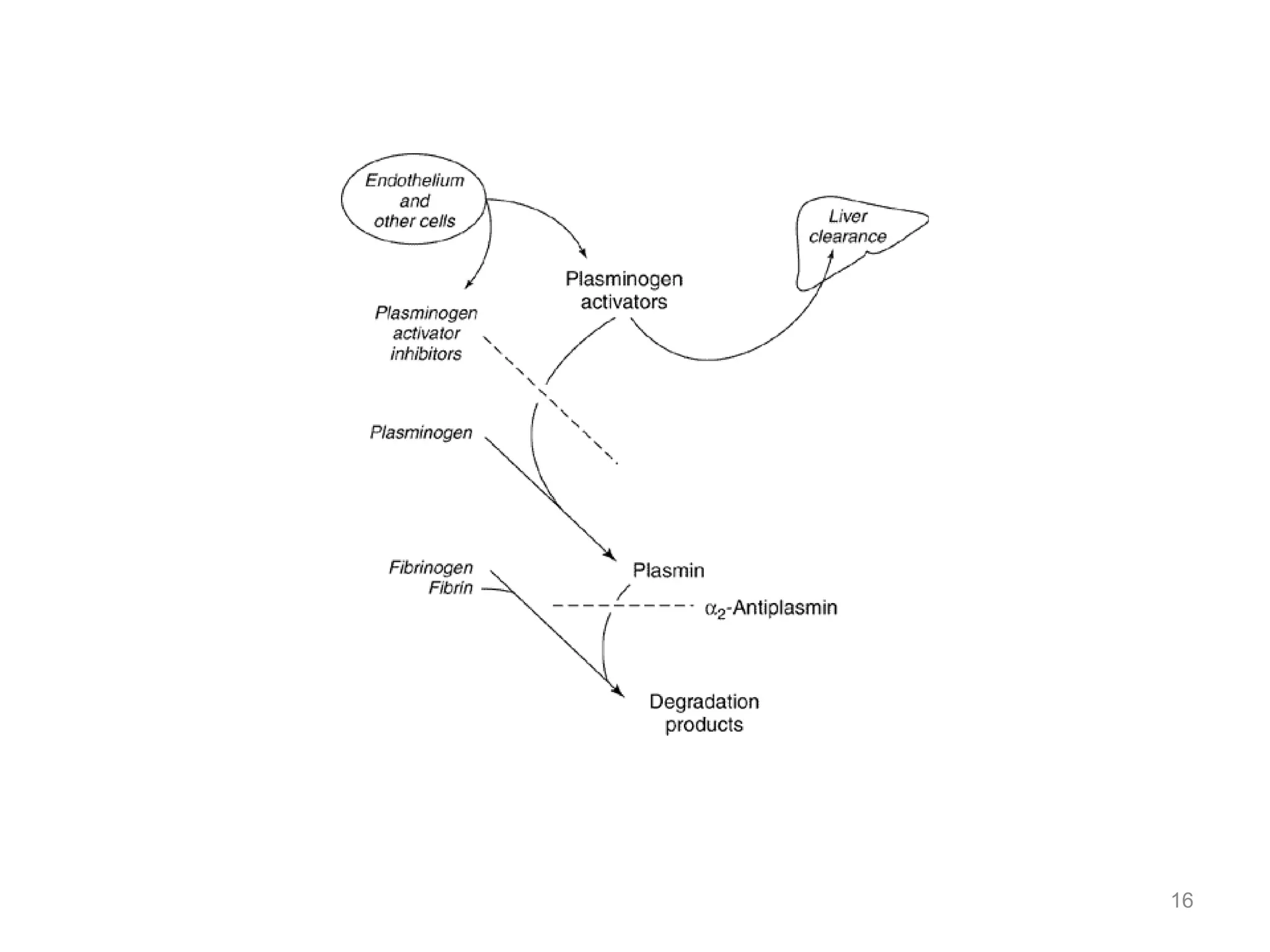
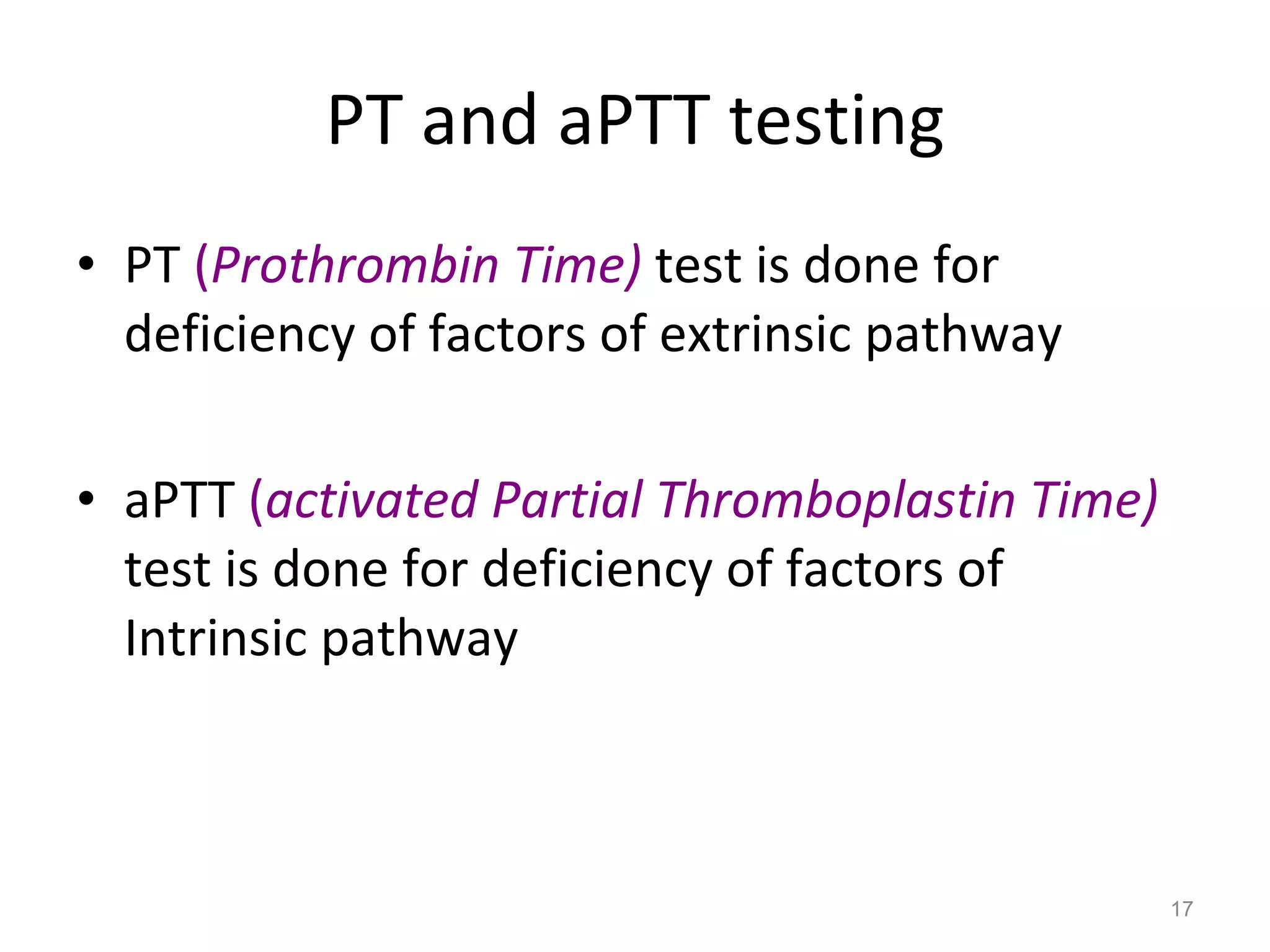
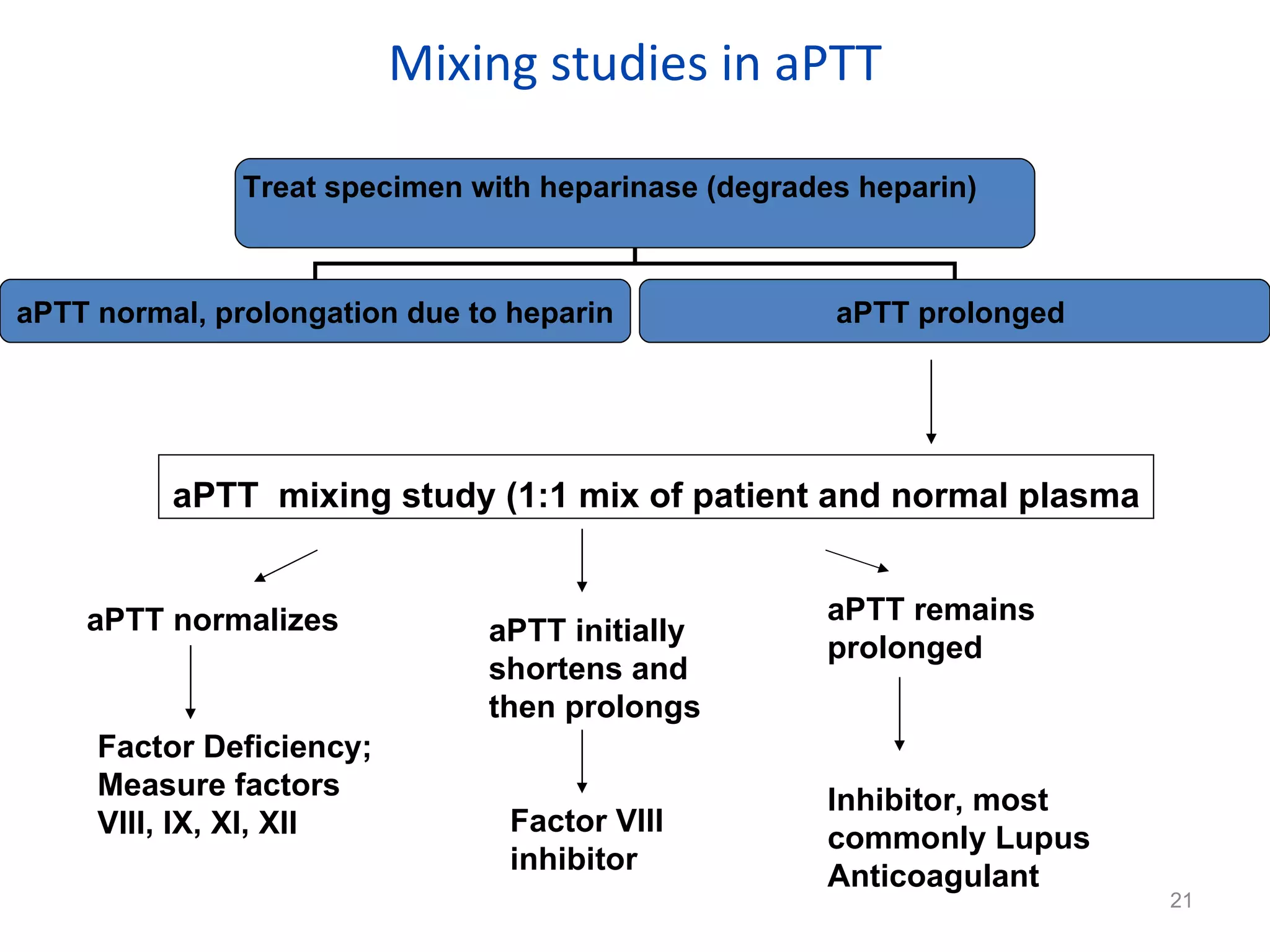
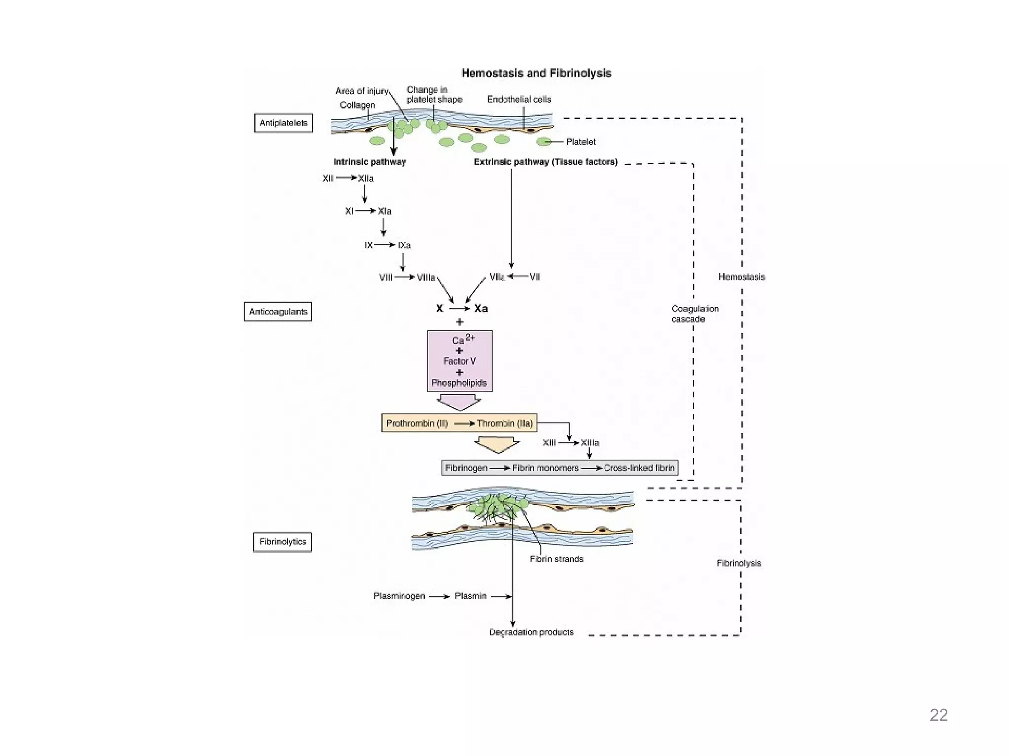
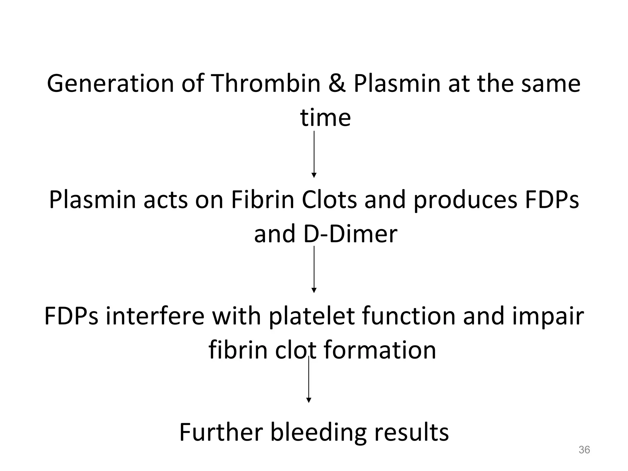
PT & aPTT PROF.DR. MOHAMMAD SOROUR CONSULTANT HAEMATOLOGY B.C.H.
Hemostasis Is a Balance Between   Clot Formation  &  Clot Dissolution.
Normal Hemostasis Clot formation (Coagulation) Clot dissolution (Fibrinolysis) PT aPTT Thrombin Time Fibrinogen  Individual factor tests FDP  D-Dimer vWF HMWK Prekallikrein
Thrombosis Abnormal Hemostasis is  Thrombosis  Formation of Blood Clot ( thrombus)  in normal blood vessels  Thrombotic occlusion of a vessel after relatively minor injury
Hemostasis involves the interaction of: Vascular Endothelium Platelets Coagulation Factors and  Fibrinolytic Proteins
Hemostasis has 2 main functions: Induce a rapid & localized hemostatic plug at the site of vascular injury  (clot formation) Maintain Blood in a fluid, clot-free state after the injury is healed  (clot dissolution)
Primary Hemostasis Injury Endothelial Cells  Exposure of thrombogenic surface (subendothelial extracellular matrix)
Platelets adhere and get activated  Change shape Release secretory granules  (e.g. ADP, TXA2) Attract other platelets and Aggregate Hemostatic plug or Primary Platelet Plug
Secondary Hemostasis Fibrin is required to stabilize the primary platelet plug Fibrin is formed by two coagulation pathways i.e. Extrinsic & Intrinsic Extrinsic Pathway is initiated when Tissue Factor (III) present in damaged organ comes in contact with Blood  Intrinsic Pathway is initiated when Factor XII binds to a negatively charged  “foreign”   surface exposed to Blood
Coagulation Factors Factor  Trivial Name  Pathway Prekallikrein Fletcher factor Intrinsic HMWK  Contact activation cofactor Intrinsic I Fibrinogen Both II Prothrombin Both III Tissue Factor Extrinsic IV Calcium Both
V Proaccelerin, Labile factor Both VI (Va) Accelerin Both VII Proconvertin Extrinsic VIII Antihemophilic factor A Intrinsic XI Antihemophilic factor B Intrinsic
XII Hageman Factor Intrinsic XIII Fibrin Stabilizing factor Both
PT and aPTT testing PT  ( Prothrombin Time)   test is done for deficiency of factors of extrinsic pathway aPTT  ( activated Partial Thromboplastin Time)   test is done for deficiency of factors of Intrinsic pathway
Effects of Hereditary or Acquired Factor Deficiency on the PT & aPTT aPTT prolonged, PT normal   Deficiencies of intrinsic pathway Factor(s) VIII, IX, XI or XII PT prolonged, aPTT normal Deficiency of extrinsic Pathway factor VII Occasionally, mild to moderate deficiency of common pathway factor(s) fibrinogen, II, V or x
Both PT and aPTT Prolonged Deficiency of common pathway factor(s) fibrinogen, II, V, or X  Multiple factor deficiencies
Mixing studies in PT PT  mixing study (1:1 mix of patient and normal plasma PT normalizes Factor Deficiency; Measure factors I, II, V, VII, X PT initially shortens and then prolongs  Factor V inhibitor (rare) aPTT remains prolonged Inhibitor (specific factor inhibitor, rare)  Treat specimen with heparinase (degrades heparin)  PT normal, prolongation due to heparin PT prolonged
Mixing studies in aPTT aPTT  mixing study (1:1 mix of patient and normal plasma aPTT normalizes Factor Deficiency; Measure factors VIII, IX, XI, XII aPTT initially shortens and then prolongs  Factor VIII inhibitor aPTT remains prolonged Inhibitor, most commonly Lupus Anticoagulant  Treat specimen with heparinase (degrades heparin)  aPTT normal, prolongation due to heparin aPTT prolonged
Fibrinolytic Mechanism Stable Fibrin Clot Activation of Protein C and Protein S Secretion of t-PA by endothelial cells Plasminogen  Plasmin Stabilized fibrin clot X, Y fragments Plasmin Degrades  D-E-D fragments E fragment + D dimer
Fibrinolysis  As soon as the injury is healed clot dissolution starts, to restore the normal flow of Blood Plasminogen is converted to the active form Plasmin by 2 distinct Plasminogen Activators (PAs): tissue plasminogen activator (t-PA)  from injured endothelial cells  Urokinase   from Kidney endothelial cells and plasma
Fibrinolysis  or  Kallikrein   from Intrinsic Pathway Plasminogen can also be activated by the bacterial product (e.g. Streptokinase) – having significance in certain Bacterial Infections Free Plasmin is neutralized by  α 2- plasmin inhibitor (PAI) t-PA activity is also blocked by PAI
Endothelial cells modulate the coagulation / anticoagulation balance by releasing PAIs PAIs block fibrinolysis by inhibiting t-PA binding to fibrin as it is most active when bound to fibrin
Fibrinolysis Three types of Natural Anti-coagulants regulate clotting: antithrombin III – inhibit thrombin activity and Factors IXa, Xa, XIa and XIIa Protein C and Protein S – Vitamin K dependent proteins, inactivate Factors Va and VIIIa Plasmin
Fibrin Degradation Products or FDP’s include: fragments X and Y – early splits and  D and E – late splits D-Dimer is the smallest cross-linked FDP
D-Dimer are specific FDP formed only by Plasmin activity on fibrin clot and not on intact fibrinogen Thus the presence of D-Dimer indicates that fibrin has been formed and so is a marker for an ongoing  in vivo  thrombotic condition
Clinical Significance of Hemostasis Hemophilia A: Caused by the deficiency of Factor VIII Hemophilia B: Caused by the deficiency of Factor IX Vitamin K deficiency (PIVKA’s)  P rotein  I nduced  V itamin  K   A ntagonism can be used in thrombotic conditions Vitamin K dependent factors are  II, V, IX, & X
Clinical Significance of Hemostasis Liver Dysfunction Fibrinogen and Factor XIII deficiency Factor XI and Contact Activation  Antithrombin Deficiency leads to DVT and PE von Willebrand Disease: Deficiency of von Willebrand Factor
DIC  (Disseminated Intravascular Coagulation) Massive Injury or Sepsis Massive release of Tissue Factor III Excessive Activation of Thrombin Coagulation becomes systemic
High consumption of Platelets, coagulation factors Over production of fibrin clot  Fibrin clot “disseminates” or spreads throughout the microcirculation Obstructing the blood flow to capillaries, smaller vessels
Lack of blood supply leads to tissue injury (decreased oxygenation, organ infarction & necrosis)  Once again release of Tissue Factor  Second time coagulation activation  More consumption of coagulation factors and platelets
Continuous thrombi formation  Production capacity of Bone Marrow and Liver reaches its maximum level  Activation of fibrinolysis at first site High consumption of Plasmin, antithrombin, Protein C and Protein S
Generation of Thrombin & Plasmin at the same time Plasmin acts on Fibrin Clots and produces FDPs and D-Dimer FDPs interfere with platelet function and impair fibrin clot formation  Further bleeding results
Thus an initial thrombotic disorder gets converted into a serious bleeding disorder  Whenever there is a widespread activation of Thrombin the chances are that it may lead to DIC
Pharmacological  Intervention Most commonly patients are on OAC (oral anti-coagulant) therapy: warfarin – over dose can lead to Vit. K deficiency heparin  Fibrinolysis:  Aspirin Streptokinase, urokinase injections t-PA injections
INR & ISI values All PT reagents are calibrated against WHO IRP (International Reference Preparation)  International Normalisation Ratio is intended to make comparison similar irrespective of the type of PT reagent used worldwide International Sensitivity Index is a measure of the sensitivity of particular PT reagent
Insensitive PT reagents will give prolonged results only when factor levels are very low Sensitive PT reagent give prolonged results even when there is a mild change in factor level  Insensitive PT reagents have higher ISI values  Sensitive PT reagents have ISI value as close to 1.0 as possible
Calculation of PT results INR = (Ratio) ISI Patient PT Time (secs)  Mean Normal PT (MNPT) MNPT = Mean PT Time of at least 20 known normal samples INR = ISI
Thanks Please send your feedbacks to: Priyank Dubey Ph: 09999990845 [email_address] Application Specialist

Pt Aptt 1218181287688715 8

  • 1.
    PT & aPTTPROF.DR. MOHAMMAD SOROUR CONSULTANT HAEMATOLOGY B.C.H.
  • 2.
    Hemostasis Is aBalance Between Clot Formation & Clot Dissolution.
  • 3.
    Normal Hemostasis Clotformation (Coagulation) Clot dissolution (Fibrinolysis) PT aPTT Thrombin Time Fibrinogen Individual factor tests FDP D-Dimer vWF HMWK Prekallikrein
  • 4.
    Thrombosis Abnormal Hemostasisis Thrombosis Formation of Blood Clot ( thrombus) in normal blood vessels Thrombotic occlusion of a vessel after relatively minor injury
  • 5.
    Hemostasis involves theinteraction of: Vascular Endothelium Platelets Coagulation Factors and Fibrinolytic Proteins
  • 6.
    Hemostasis has 2main functions: Induce a rapid & localized hemostatic plug at the site of vascular injury (clot formation) Maintain Blood in a fluid, clot-free state after the injury is healed (clot dissolution)
  • 8.
    Primary Hemostasis InjuryEndothelial Cells Exposure of thrombogenic surface (subendothelial extracellular matrix)
  • 9.
    Platelets adhere andget activated Change shape Release secretory granules (e.g. ADP, TXA2) Attract other platelets and Aggregate Hemostatic plug or Primary Platelet Plug
  • 10.
    Secondary Hemostasis Fibrinis required to stabilize the primary platelet plug Fibrin is formed by two coagulation pathways i.e. Extrinsic & Intrinsic Extrinsic Pathway is initiated when Tissue Factor (III) present in damaged organ comes in contact with Blood Intrinsic Pathway is initiated when Factor XII binds to a negatively charged “foreign” surface exposed to Blood
  • 11.
    Coagulation Factors Factor Trivial Name Pathway Prekallikrein Fletcher factor Intrinsic HMWK Contact activation cofactor Intrinsic I Fibrinogen Both II Prothrombin Both III Tissue Factor Extrinsic IV Calcium Both
  • 12.
    V Proaccelerin, Labilefactor Both VI (Va) Accelerin Both VII Proconvertin Extrinsic VIII Antihemophilic factor A Intrinsic XI Antihemophilic factor B Intrinsic
  • 13.
    XII Hageman FactorIntrinsic XIII Fibrin Stabilizing factor Both
  • 17.
    PT and aPTTtesting PT ( Prothrombin Time) test is done for deficiency of factors of extrinsic pathway aPTT ( activated Partial Thromboplastin Time) test is done for deficiency of factors of Intrinsic pathway
  • 18.
    Effects of Hereditaryor Acquired Factor Deficiency on the PT & aPTT aPTT prolonged, PT normal Deficiencies of intrinsic pathway Factor(s) VIII, IX, XI or XII PT prolonged, aPTT normal Deficiency of extrinsic Pathway factor VII Occasionally, mild to moderate deficiency of common pathway factor(s) fibrinogen, II, V or x
  • 19.
    Both PT andaPTT Prolonged Deficiency of common pathway factor(s) fibrinogen, II, V, or X Multiple factor deficiencies
  • 20.
    Mixing studies inPT PT mixing study (1:1 mix of patient and normal plasma PT normalizes Factor Deficiency; Measure factors I, II, V, VII, X PT initially shortens and then prolongs Factor V inhibitor (rare) aPTT remains prolonged Inhibitor (specific factor inhibitor, rare) Treat specimen with heparinase (degrades heparin) PT normal, prolongation due to heparin PT prolonged
  • 21.
    Mixing studies inaPTT aPTT mixing study (1:1 mix of patient and normal plasma aPTT normalizes Factor Deficiency; Measure factors VIII, IX, XI, XII aPTT initially shortens and then prolongs Factor VIII inhibitor aPTT remains prolonged Inhibitor, most commonly Lupus Anticoagulant Treat specimen with heparinase (degrades heparin) aPTT normal, prolongation due to heparin aPTT prolonged
  • 23.
    Fibrinolytic Mechanism StableFibrin Clot Activation of Protein C and Protein S Secretion of t-PA by endothelial cells Plasminogen Plasmin Stabilized fibrin clot X, Y fragments Plasmin Degrades D-E-D fragments E fragment + D dimer
  • 24.
    Fibrinolysis Assoon as the injury is healed clot dissolution starts, to restore the normal flow of Blood Plasminogen is converted to the active form Plasmin by 2 distinct Plasminogen Activators (PAs): tissue plasminogen activator (t-PA) from injured endothelial cells Urokinase from Kidney endothelial cells and plasma
  • 25.
    Fibrinolysis or Kallikrein from Intrinsic Pathway Plasminogen can also be activated by the bacterial product (e.g. Streptokinase) – having significance in certain Bacterial Infections Free Plasmin is neutralized by α 2- plasmin inhibitor (PAI) t-PA activity is also blocked by PAI
  • 26.
    Endothelial cells modulatethe coagulation / anticoagulation balance by releasing PAIs PAIs block fibrinolysis by inhibiting t-PA binding to fibrin as it is most active when bound to fibrin
  • 27.
    Fibrinolysis Three typesof Natural Anti-coagulants regulate clotting: antithrombin III – inhibit thrombin activity and Factors IXa, Xa, XIa and XIIa Protein C and Protein S – Vitamin K dependent proteins, inactivate Factors Va and VIIIa Plasmin
  • 28.
    Fibrin Degradation Productsor FDP’s include: fragments X and Y – early splits and D and E – late splits D-Dimer is the smallest cross-linked FDP
  • 29.
    D-Dimer are specificFDP formed only by Plasmin activity on fibrin clot and not on intact fibrinogen Thus the presence of D-Dimer indicates that fibrin has been formed and so is a marker for an ongoing in vivo thrombotic condition
  • 30.
    Clinical Significance ofHemostasis Hemophilia A: Caused by the deficiency of Factor VIII Hemophilia B: Caused by the deficiency of Factor IX Vitamin K deficiency (PIVKA’s) P rotein I nduced V itamin K A ntagonism can be used in thrombotic conditions Vitamin K dependent factors are II, V, IX, & X
  • 31.
    Clinical Significance ofHemostasis Liver Dysfunction Fibrinogen and Factor XIII deficiency Factor XI and Contact Activation Antithrombin Deficiency leads to DVT and PE von Willebrand Disease: Deficiency of von Willebrand Factor
  • 32.
    DIC (DisseminatedIntravascular Coagulation) Massive Injury or Sepsis Massive release of Tissue Factor III Excessive Activation of Thrombin Coagulation becomes systemic
  • 33.
    High consumption ofPlatelets, coagulation factors Over production of fibrin clot Fibrin clot “disseminates” or spreads throughout the microcirculation Obstructing the blood flow to capillaries, smaller vessels
  • 34.
    Lack of bloodsupply leads to tissue injury (decreased oxygenation, organ infarction & necrosis) Once again release of Tissue Factor Second time coagulation activation More consumption of coagulation factors and platelets
  • 35.
    Continuous thrombi formation Production capacity of Bone Marrow and Liver reaches its maximum level Activation of fibrinolysis at first site High consumption of Plasmin, antithrombin, Protein C and Protein S
  • 36.
    Generation of Thrombin& Plasmin at the same time Plasmin acts on Fibrin Clots and produces FDPs and D-Dimer FDPs interfere with platelet function and impair fibrin clot formation Further bleeding results
  • 37.
    Thus an initialthrombotic disorder gets converted into a serious bleeding disorder Whenever there is a widespread activation of Thrombin the chances are that it may lead to DIC
  • 38.
    Pharmacological InterventionMost commonly patients are on OAC (oral anti-coagulant) therapy: warfarin – over dose can lead to Vit. K deficiency heparin Fibrinolysis: Aspirin Streptokinase, urokinase injections t-PA injections
  • 39.
    INR & ISIvalues All PT reagents are calibrated against WHO IRP (International Reference Preparation) International Normalisation Ratio is intended to make comparison similar irrespective of the type of PT reagent used worldwide International Sensitivity Index is a measure of the sensitivity of particular PT reagent
  • 40.
    Insensitive PT reagentswill give prolonged results only when factor levels are very low Sensitive PT reagent give prolonged results even when there is a mild change in factor level Insensitive PT reagents have higher ISI values Sensitive PT reagents have ISI value as close to 1.0 as possible
  • 41.
    Calculation of PTresults INR = (Ratio) ISI Patient PT Time (secs) Mean Normal PT (MNPT) MNPT = Mean PT Time of at least 20 known normal samples INR = ISI
  • 42.
    Thanks Please sendyour feedbacks to: Priyank Dubey Ph: 09999990845 [email_address] Application Specialist

Editor's Notes

  • #2 J. Mitra & Co. Ltd. Annual Sales Conference 2005 (South & West)
  • #33 J. Mitra & Co. Ltd. Annual Sales Conference 2005 (South & West)