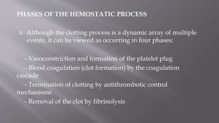
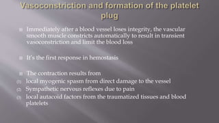
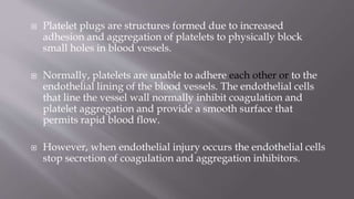
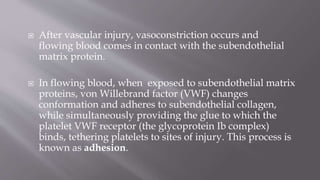
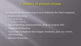
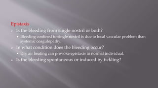
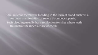
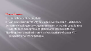
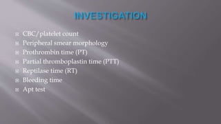
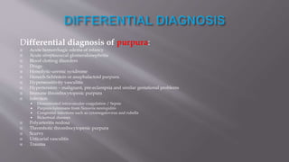
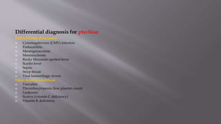
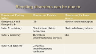
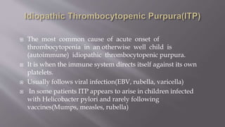
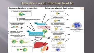
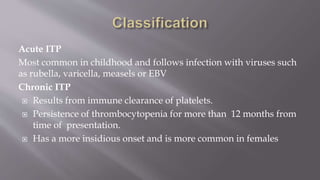
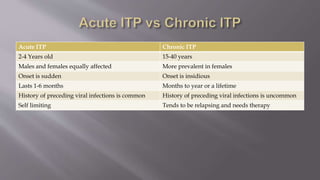
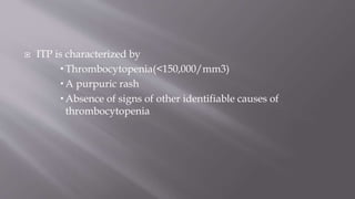
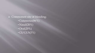
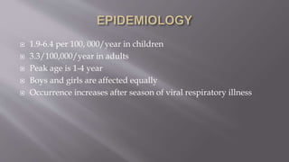
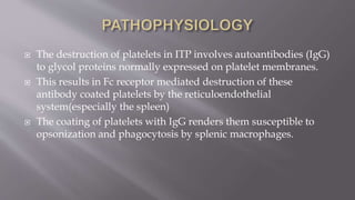
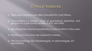
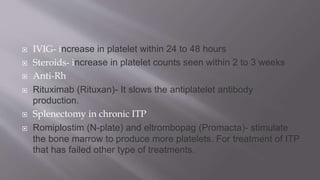
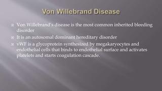
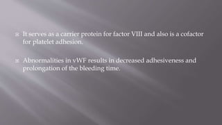
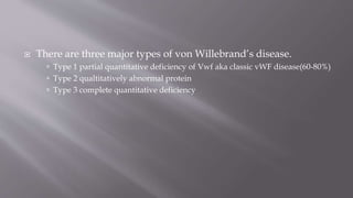
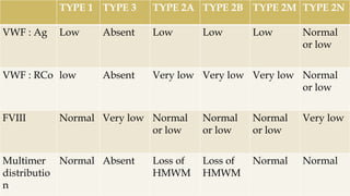
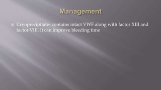
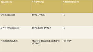
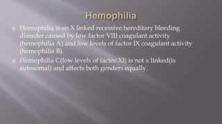
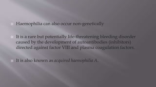
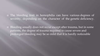
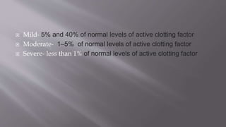
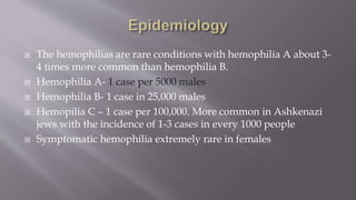
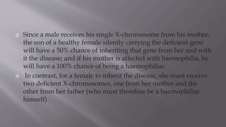
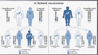
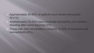
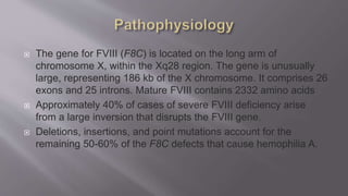
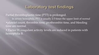
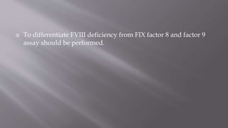
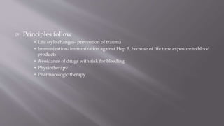
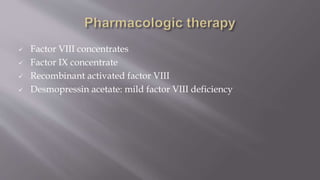
The seminar presentation covered hemostasis and approaches to bleeding disorders in pediatrics. It discussed the pathophysiology, clinical features, laboratory findings and management of idiopathic thrombocytopenic purpura, Von Willebrand's disease, and hemophilia. It provided an overview of hemostasis and the coagulation cascade, approaches to evaluating a child with bleeding, and specifics on selected bleeding disorders. The presentation included descriptions of laboratory tests used to evaluate coagulation factors and identify bleeding disorders.
UAEU - CMHS - Hematology-Oncology Course - MMH 302 - HONC 320. Education material for medical students - It cover basic principles of hematology and oncology, including CAR-T and gene editing. It can be used for study and review. It illustrates main principles of hematology and oncology.
Approach to a bleeding disorder: These presentation has the approach for a patient of bleeding disorder. it has History, physical finding, Investigations.
Hemostasis is the mechanism that leads to cessation of bleeding from a blood vessel. It is a process that involves multiple interlinked steps. This cascade culminates into the formation of a “plug” that closes up the damaged site of the blood vessel controlling the bleeding.
Title: Sense of Taste
Presenter: Dr. Faiza, Assistant Professor of Physiology
Qualifications:
MBBS (Best Graduate, AIMC Lahore)
FCPS Physiology
ICMT, CHPE, DHPE (STMU)
MPH (GC University, Faisalabad)
MBA (Virtual University of Pakistan)
Learning Objectives:
Describe the structure and function of taste buds.
Describe the relationship between the taste threshold and taste index of common substances.
Explain the chemical basis and signal transduction of taste perception for each type of primary taste sensation.
Recognize different abnormalities of taste perception and their causes.
Key Topics:
Significance of Taste Sensation:
Differentiation between pleasant and harmful food
Influence on behavior
Selection of food based on metabolic needs
Receptors of Taste:
Taste buds on the tongue
Influence of sense of smell, texture of food, and pain stimulation (e.g., by pepper)
Primary and Secondary Taste Sensations:
Primary taste sensations: Sweet, Sour, Salty, Bitter, Umami
Chemical basis and signal transduction mechanisms for each taste
Taste Threshold and Index:
Taste threshold values for Sweet (sucrose), Salty (NaCl), Sour (HCl), and Bitter (Quinine)
Taste index relationship: Inversely proportional to taste threshold
Taste Blindness:
Inability to taste certain substances, particularly thiourea compounds
Example: Phenylthiocarbamide
Structure and Function of Taste Buds:
Composition: Epithelial cells, Sustentacular/Supporting cells, Taste cells, Basal cells
Features: Taste pores, Taste hairs/microvilli, and Taste nerve fibers
Location of Taste Buds:
Found in papillae of the tongue (Fungiform, Circumvallate, Foliate)
Also present on the palate, tonsillar pillars, epiglottis, and proximal esophagus
Mechanism of Taste Stimulation:
Interaction of taste substances with receptors on microvilli
Signal transduction pathways for Umami, Sweet, Bitter, Sour, and Salty tastes
Taste Sensitivity and Adaptation:
Decrease in sensitivity with age
Rapid adaptation of taste sensation
Role of Saliva in Taste:
Dissolution of tastants to reach receptors
Washing away the stimulus
Taste Preferences and Aversions:
Mechanisms behind taste preference and aversion
Influence of receptors and neural pathways
Impact of Sensory Nerve Damage:
Degeneration of taste buds if the sensory nerve fiber is cut
Abnormalities of Taste Detection:
Conditions: Ageusia, Hypogeusia, Dysgeusia (parageusia)
Causes: Nerve damage, neurological disorders, infections, poor oral hygiene, adverse drug effects, deficiencies, aging, tobacco use, altered neurotransmitter levels
Neurotransmitters and Taste Threshold:
Effects of serotonin (5-HT) and norepinephrine (NE) on taste sensitivity
Supertasters:
25% of the population with heightened sensitivity to taste, especially bitterness
Increased number of fungiform papillae
micro teaching on communication m.sc nursing.pdfAnurag Sharma
Microteaching is a unique model of practice teaching. It is a viable instrument for the. desired change in the teaching behavior or the behavior potential which, in specified types of real. classroom situations, tends to facilitate the achievement of specified types of objectives.
Ozempic: Preoperative Management of Patients on GLP-1 Receptor Agonists Saeid Safari
Preoperative Management of Patients on GLP-1 Receptor Agonists like Ozempic and Semiglutide
ASA GUIDELINE
NYSORA Guideline
2 Case Reports of Gastric Ultrasound
Local Advanced Lung Cancer: Artificial Intelligence, Synergetics, Complex Sys...Oleg Kshivets
Overall life span (LS) was 1671.7±1721.6 days and cumulative 5YS reached 62.4%, 10 years – 50.4%, 20 years – 44.6%. 94 LCP lived more than 5 years without cancer (LS=2958.6±1723.6 days), 22 – more than 10 years (LS=5571±1841.8 days). 67 LCP died because of LC (LS=471.9±344 days). AT significantly improved 5YS (68% vs. 53.7%) (P=0.028 by log-rank test). Cox modeling displayed that 5YS of LCP significantly depended on: N0-N12, T3-4, blood cell circuit, cell ratio factors (ratio between cancer cells-CC and blood cells subpopulations), LC cell dynamics, recalcification time, heparin tolerance, prothrombin index, protein, AT, procedure type (P=0.000-0.031). Neural networks, genetic algorithm selection and bootstrap simulation revealed relationships between 5YS and N0-12 (rank=1), thrombocytes/CC (rank=2), segmented neutrophils/CC (3), eosinophils/CC (4), erythrocytes/CC (5), healthy cells/CC (6), lymphocytes/CC (7), stick neutrophils/CC (8), leucocytes/CC (9), monocytes/CC (10). Correct prediction of 5YS was 100% by neural networks computing (error=0.000; area under ROC curve=1.0).
Knee anatomy and clinical tests 2024.pdfvimalpl1234
This includes all relevant anatomy and clinical tests compiled from standard textbooks, Campbell,netter etc..It is comprehensive and best suited for orthopaedicians and orthopaedic residents.
Basavarajeeyam is a Sreshta Sangraha grantha (Compiled book ), written by Neelkanta kotturu Basavaraja Virachita. It contains 25 Prakaranas, First 24 Chapters related to Rogas& 25th to Rasadravyas.
Muktapishti is a traditional Ayurvedic preparation made from Shoditha Mukta (Purified Pearl), is believed to help regulate thyroid function and reduce symptoms of hyperthyroidism due to its cooling and balancing properties. Clinical evidence on its efficacy remains limited, necessitating further research to validate its therapeutic benefits.
Basavarajeeyam is an important text for ayurvedic physician belonging to andhra pradehs. It is a popular compendium in various parts of our country as well as in andhra pradesh. The content of the text was presented in sanskrit and telugu language (Bilingual). One of the most famous book in ayurvedic pharmaceutics and therapeutics. This book contains 25 chapters called as prakaranas. Many rasaoushadis were explained, pioneer of dhatu druti, nadi pareeksha, mutra pareeksha etc. Belongs to the period of 15-16 century. New diseases like upadamsha, phiranga rogas are explained.
Tom Selleck Health: A Comprehensive Look at the Iconic Actor’s Wellness Journeygreendigital
Tom Selleck, an enduring figure in Hollywood. has captivated audiences for decades with his rugged charm, iconic moustache. and memorable roles in television and film. From his breakout role as Thomas Magnum in Magnum P.I. to his current portrayal of Frank Reagan in Blue Bloods. Selleck's career has spanned over 50 years. But beyond his professional achievements. fans have often been curious about Tom Selleck Health. especially as he has aged in the public eye.
Follow us on: Pinterest
Introduction
Many have been interested in Tom Selleck health. not only because of his enduring presence on screen but also because of the challenges. and lifestyle choices he has faced and made over the years. This article delves into the various aspects of Tom Selleck health. exploring his fitness regimen, diet, mental health. and the challenges he has encountered as he ages. We'll look at how he maintains his well-being. the health issues he has faced, and his approach to ageing .
Early Life and Career
Childhood and Athletic Beginnings
Tom Selleck was born on January 29, 1945, in Detroit, Michigan, and grew up in Sherman Oaks, California. From an early age, he was involved in sports, particularly basketball. which played a significant role in his physical development. His athletic pursuits continued into college. where he attended the University of Southern California (USC) on a basketball scholarship. This early involvement in sports laid a strong foundation for his physical health and disciplined lifestyle.
Transition to Acting
Selleck's transition from an athlete to an actor came with its physical demands. His first significant role in "Magnum P.I." required him to perform various stunts and maintain a fit appearance. This role, which he played from 1980 to 1988. necessitated a rigorous fitness routine to meet the show's demands. setting the stage for his long-term commitment to health and wellness.
Fitness Regimen
Workout Routine
Tom Selleck health and fitness regimen has evolved. adapting to his changing roles and age. During his "Magnum, P.I." days. Selleck's workouts were intense and focused on building and maintaining muscle mass. His routine included weightlifting, cardiovascular exercises. and specific training for the stunts he performed on the show.
Selleck adjusted his fitness routine as he aged to suit his body's needs. Today, his workouts focus on maintaining flexibility, strength, and cardiovascular health. He incorporates low-impact exercises such as swimming, walking, and light weightlifting. This balanced approach helps him stay fit without putting undue strain on his joints and muscles.
Importance of Flexibility and Mobility
In recent years, Selleck has emphasized the importance of flexibility and mobility in his fitness regimen. Understanding the natural decline in muscle mass and joint flexibility with age. he includes stretching and yoga in his routine. These practices help prevent injuries, improve posture, and maintain mobilit
5. Prothrombin time (PT) – the time it takes plasma to clot after
addition of tissue factor. This measures the quality of the extrinsic
pathway (as well as the common pathway) of coagulation.
- Normal value range = 12 – 15 sec. The speed of the extrinsic
pathway is affected by levels of functional factor VII in the body.
- PT can be prolonged due to vitamin K deficiency, warfarin therapy,
malabsorption, poor factor VII synthesis (liver disease), or increased
consumption in DIC.
6. Partial thromboplastin time – measures the speed at which
blood clots form by means of the intrinsic and common
pathways. It measures factors I, II, V, VIII, X, XI and XII. It’s
commonly used in conjunction with PT test.
- The typical reference range is between 30 and 50 seconds.
- Prolonged PTT may indicate coagulation factor deficiency
(hemophilia A and B), use of heparin, sepsis (coagulation factor
consumption), presence of antibodies against coagulation factors
7. International Normalized Ration (INR) – is the ratio of a
patient’s prothrombin time to a normal (controlled sample),
raised to the value of the ISI value for the analytical system
being used.
INR = (PTtest/PTnormal)ISI
- INR is typically used to monitor patients on warfarin or
related oral anticoagulant therapy. The normal range for a
person not on warfarin is 0.8 – 1.2. For people on warfarin
therapy an INR of 2.0 – 3.0 is usually targeted.
- A high INR indicates a higher risk of bleeding, while a low
INR suggests a higher risk of developing a clot.
8. Hemostasis is the active process that clots blood in areas of blood
vessel injury to limit blood loss, yet simultaneously limits the
clot size only to the areas of injury.
It needs to be rapid and regulated such that trauma does not
trigger a systemic reaction but must initiate a rapid, localized
response.
If clotting is impaired, hemorrhage occurs and if clotting is
excessive, thrombotic complications ensue.
9. The process is carefully regulated through feedback loops and
mediators to ensure appropriate response.
The main components of the hemostatic process are the vessel
wall, platelets, coagulation proteins, anticoagulant proteins,
and fibrinolytic system.
10. PHASES OF THE HEMOSTATIC PROCESS
Although the clotting process is a dynamic array of multiple
events, it can be viewed as occurring in four phases:
- Vasoconstriction and formation of the platelet plug
- Blood coagulation (clot formation) by the coagulation
cascade
- Termination of clotting by antithrombotic control
mechanisms
- Removal of the clot by fibrinolysis
11. Immediately after a blood vessel loses integrity, the vascular
smooth muscle constricts automatically to result in transient
vasoconstriction and limit the blood loss
It’s the first response in hemostasis
The contraction results from
(1) local myogenic spasm from direct damage to the vessel
(2) Sympathetic nervous reflexes due to pain
(3) local autacoid factors from the traumatized tissues and blood
platelets
12. Platelet plugs are structures formed due to increased
adhesion and aggregation of platelets to physically block
small holes in blood vessels.
Normally, platelets are unable to adhere each other or to the
endothelial lining of the blood vessels. The endothelial cells
that line the vessel wall normally inhibit coagulation and
platelet aggregation and provide a smooth surface that
permits rapid blood flow.
However, when endothelial injury occurs the endothelial cells
stop secretion of coagulation and aggregation inhibitors.
13. After vascular injury, vasoconstriction occurs and
flowing blood comes in contact with the subendothelial
matrix protein.
In flowing blood, when exposed to subendothelial matrix
proteins, von Willebrand factor (VWF) changes
conformation and adheres to subendothelial collagen,
while simultaneously providing the glue to which the
platelet VWF receptor (the glycoprotein Ib complex)
binds, tethering platelets to sites of injury. This process is
known as adhesion.
14. In addition, this interaction between VWF and platelets
causes activation of the platelet, triggering secretion of
storage granules containing adenosine diphosphate (ADP),
serotonin, and thromboxane A2. This process is known as
secretion.
Those secretions result in further vasoconstriction, as well the
expression of fibrinogen receptors on platelets.
Fibrinogen acts as the ligand that connects two platelets, and
its activation results in the recruitment and aggregation of
other platelets to form the platelet plug. This process is
known as aggregation.
15. The final process is procoagulant activity. Platelet
procoagulant activity is an important aspect of platelet
activation and involves
- exposure of procoagulant phospholipids (primarily
phosphatidylserine), and
- the subsequent assembly of the enzyme complexes in the
clotting cascade on the platelet surface
These complexes are an important example of the close
interrelationship between platelet activation and activation of
the clotting cascade
16.
17. Blood coagulation is the process through which the liquid blood
is converted to gel (clot).
It’s a slow but long lasting mechanism by which bleeding is
stopped and the weak platelet plug is stabilized.
The process is mediated through the sequential activation of a
series of proenzymes or inactive precursor proteins ,called blood
clotting factors, to their active form.
18. There are 12
blood clotting
factors in the
plasma:
19. These proteins are synthesized in the liver in their inactive form.
Activation of one of these factors results in activation of the next
one in the cascade, ending in fibrin clot formation.
Stages of Blood Coagulation
1. Formation of prothrombin activators through
The intrinsic pathway
The extrinsic pathway
2. Conversion of prothrombin into thrombin by the action of
prothrombin activators
3. Conversion of fibrinogen into fibrin thread by the action of
thrombin
20. Activated factor X, along with Ca++ ion, Labile factor (V), and platelet
factor 3 (PF3 ) are collectively referred to as the
prothrombin activator
Prothrombin → thrombin
fibrinogen → fibrin
fibrin threads form a meshwork that traps blood cells, platelets, and plasma
to form the blood clot.
21. The intrinsic pathway is initiated by the exposure of factor XII
and platelets to collagen
The extrinsic pathway is activated by tissue factor exposed at
the site of injury or tissue factor-like material
(thromboplastin)
Both pathways converge on the activation of factor X which,
as a component of prothrombin activators, converts
prothrombin to thrombin, the final enzyme of the clotting
cascade. Thrombin converts fibrinogen from a soluble plasma
protein into an insoluble fibrin clot.
22.
23. Clot retraction - once the clot is formed, the platelets trapped
within it contract, shrinking the fibrin meshwork. This clot
retraction pulls the edges of the damaged vessel closer
together.
- The clot is also stabilized by squeezing serum from the fibrin
strands.
Clot repair - PDGF stimulates rebuilding of blood vessel wall.
Fibroblasts form a tissue patch stimulated by VEGF, causing
endothelial cells to multiply and restore the endothelial lining.
24. Virtually all procoagulant proteins are balanced by an
anticoagulant protein that regulates or inhibits procoagulant
function.
Four clinically important, naturally occurring anticoagulants
regulate the extension of the clotting process: antithrombin III
(AT-III), protein C, protein S, and tissue factor pathway
inhibitor.
AT-III is a protease inhibitor that regulates factor X and
thrombin
25. When thrombin in flowing blood encounters intact
endothelium, thrombin binds to thrombomodulin, its
endothelial receptor. The thrombin–thrombomodulin
complex reduces the availability of thrombin and activates
protein C.
In the presence of the cofactor protein S, activated protein C
proteolyses and inactivates factor V and factor VIII.
Tissue factor pathway inhibitor limits activation of factor X.
26. Once a stable fibrin-platelet plug is formed, the fibrinolytic
system limits its extension and lyses the clot to reestablish
vascular integrity and prevent permanent obstruction.
This is accomplished by the enzyme plasmin, a proteolytic
enzyme that digests fibrin.
Plasmin is found in plasma as plasminogen and is activated
by either urokinase-like or tissue-type plasminogen activator.
27. The conversion of plasminogen into plasmin involves several
substances, including factor XII.
The process is also regulated by plasminogen activator
inhibitors and α2-antiplasmin, as well as by the thrombin-
activatable fibrinolysis inhibitor.
Within a few days after the blood has clotted, enough plasmin
has been formed to dissolve the clot.
Finally, the flow of blood in and around the clot is crucial,
because flowing blood returns to the liver, where activated
clotting factor complexes are removed.
30. Children with bleeding disorder may present with
Epistaxis
Gum bleeding
Gross haematuria
Joint pain or swelling due to Intra-articular hemorrhage, commonly
affected joints include the knees, ankles and elbows.
Excessive bruising, ptechiae, purpura
contusions or spontaneous haemorrhage during childhood play.
Prolonged bleeding after after certain procedure (e.g. tooth extraction,
circumcision)
Menorrhagia in adolescent girls
31. Neurological symptoms due to intracranial haemorrhage
Haematemesis,
Melaena or frank rectal bleeding (from gastrointestinal
bleeding).
Haemoptysis
32. We should ask the following so as to elaborate the chief complaint
Onset of bleeding-
age at onset,
acute vs chronic
Site of bleeding (mucocutanous, deep or surgical site)
Duration of bleeding
Associated symptoms like fatigue, headache, pain any where,
easy bruising
History of trauma
33. Epistaxis
Is the bleeding from single nostril or both?
Bleeding confined to single nostril is due to local vascular problem than
systemic coagulopathy.
In what condition does the bleeding occur?
Dry air heating can provoke epistaxis in normal individual.
Is the bleeding spontaneous or induced by tickling?
34. Oral mucous membrane bleeding in the form of blood blister is a
common manifestation of severe thrombocytopenia.
Such bleeding usually has predilection for sites where teeth
traumatize the inner surface of cheek.
35. Hemarthroses
It is hallmark of hemophilia
Can also occur in vWD type 3 and severe factor VII deficiency
Excessive bleeding following circumcision in male is usually first
symptom of hemophilia or glanzmann thromboasthenia.
Bleeding from umbilical stump is characteristic of factor VIII
deficiency or afibrinogenemia.
36. history of infection
history of RVI
history of birth asyphyxia
Hx of previous hospital visit for bleeding symptoms
Hx of blood transfussion
Results of previous laboratory evaluation
Hx of anemia
37. Family hx is particularly important when hereditary diseases are
considered.
Ask for hx of chronic maternal diseases
Hx of bleeding disorder among family members or close
relatives.
Common inherited bleeding disorders are
von Willebrand disease.
Hemophilia A (factor VIII deficiency)
Hemophilia B (factor IX deficiency)
38. Social history
consider the possibility of violence as the cause of excessive
bruising or bleeding (e.g. domestic violence).
39. Hx of vitamin K administration at birth or during pregnancy period
several different medications can alter the ability of the clotting
cascade to function effectively.
e.g.
warfarin,
Heparin
NSAIDs
NOACs (rivaroxaban, dabigatran, apixaban, edoxaban)
prolonged use of wide spectrum antibiotics
Patient or maternal use of anticonvulsants
40. Ask hx of herbal use
Commonly used herbs that can cause platelet dysfuction
o Ginkgo biloba
o Ginseng
44. Prenatal hx:
hx of maternal infection during pregnancy
condition of baby right after birth, admitted to NICU or not
Umbilical stump bleeding
45. Detailed nutritional hx should be asked to assess for
Vitamin K deficiency
Vitamin C deficiency, b/c patient may have skin bleeding cosistent with
scurvy (perifollicular purpura)
General malnutrition
46. Hemophilia is associated with
substantial brain dysfunction,
problems with coordination and motor function,
lower intelligence, academic and adaptive skills, and
more behavioral/emotional problems
47. 1. General appearance
Sick looking – possible causes
DIC, infection, liver diseases
NEC
Well looking – possible causes
VKDB, clotting factor deficiency
Inherited bleeding disorders
Immune thrombocytopenia
Bleeding due to local trauma
2. Vital sign
BP -
hemophilia patients suffer from higher blood pressure levels than the general population at
all age
Patient may be hypotensive in case of excessive overt or occult bleeding
48. 3. Systemic Inquiry
HEENT
Head – look for
Contusion, laceration, caputsucedanum
Eye
conjunctiva, intra ocular hemorrhage, jaundice
Ear
Asses for hemotympanema which may be caused by haemophilia or anticoagulant medication
Ear bleeding
Nose
Position of septum, nasal polyp, intranasal laceration
Throat and mouth
Extracted tooth, bleeding of gum, sharp material in mouth or throat, laceration of oral mucosa
LGS
Look for lymphadenopathy
49. Respiratory system
Haemoptysis
Breathing difficulty
May caused by diffused alveolar haemorrhage or pulmonary
haemorrhage
Dullness of thorax on percussion may indicate hemothorax.
51. GI system
Intraperitoneal bleeding appear as
Abdominal tenderness
Tense and rigid abdomen
Rectal bleeding
Stool color
palpate liver and spleen
55. CNS
Neurologic manifestations of intracranial hemorrhage
Weakness or loss of muscle strength
Loss of sight or double vision
Memory loss
Impaired mental ability
Lack of coordination
56. CBC/platelet count
Peripheral smear morphology
Prothrombin time (PT)
Partial thromboplastin time (PTT)
Reptilase time (RT)
Bleeding time
Apt test
57. Differential diagnosis of purpura:
Acute hemorrhagic edema of infancy
Acute streptococcal glomerulonephritis
Blood clotting disorders
Drugs
Hemolytic-uremic syndrome
Henoch-Schönlein or anaphalactoid purpura.
Hypersensitivity vasculitis
Hypertension – malignant, pre-eclampsia and similar gestational problems
Immune thrombocytopenic purpura
Infection
Disseminated intravascular coagulation / Sepsis
Purpura fulminans from Neisseria meningiditis
Congenital infections such as cytomegalovirus and rubella
Rickettsial diseases
Polyarteritis nodosa
Thrombotic thrombocytopenic purpura
Scurvy
Urticarial vasculitis
Trauma
59. Differential diagnosis for ecchymosis
Trauma
Melanosis
Cutaneous malignant melanoma
Addison disease
Hemachromatosis
Wilson disease
Drug-induced pigmentation (E.g. Hydroxychoroquine, chloroquine,
quinidine, and quinacrine can cause a blue-black pigmentation of the
extremities, face, oral mucosa, nails, and ear cartilage )
60. Differential Diagnoses for Epistaxis
Allergic Rhinitis
Barotrauma
Drug toxicity
Disseminated Intravascular Coagulation
Nasal Foreign Bodies
Pediatric Osler-Weber-Rendu Syndrome
Sinusitis (Rhinosinusitis)
Type A Hemophilia
Type B Hemophilia
von Willebrand Disease
61. DDX for gum bleeding
Gingivitis
Periodontitis
Vitamin C deficiencies (scurvy)
vitamin K deficiencies
Leukemia
Thrombocytopenia
62.
63. Disorders of Clotting
Factors
Disorders of Platelets Disorders of the blood
vessels
Hemophila A and
Hemophilia B
ITP Henoch schonlein purpura
Factor XI deficiency Non immune platelet
destruction
Eholers danlons syndrome
Factor 2 deficiency Thrombotic
thrombocytopenic purpura
SLE
Factor XIII deficieny Congenital
thrombocytopenic
syndromes
64.
65.
66. The most common cause of acute onset of
thrombocytopenia in an otherwise well child is
(autoimmune) idiopathic thrombocytopenic purpura.
It is when the immune system directs itself against its own
platelets.
Usually follows viral infection(EBV, rubella, varicella)
In some patients ITP appears to arise in children infected
with Helicobacter pylori and rarely following
vaccines(Mumps, measles, rubella)
67.
68. Acute ITP
Most common in childhood and follows infection with viruses such
as rubella, varicella, measels or EBV
Chronic ITP
Results from immune clearance of platelets.
Persistence of thrombocytopenia for more than 12 months from
time of presentation.
Has a more insidious onset and is more common in females
69. Acute ITP Chronic ITP
2-4 Years old 15-40 years
Males and females equally affected More prevalent in females
Onset is sudden Onset is insidious
Lasts 1-6 months Months to year or a lifetime
History of preceding viral infections is common History of preceding viral infections is uncommon
Self limiting Tends to be relapsing and needs therapy
70. ITP is characterized by
Thrombocytopenia(<150,000/mm3)
A purpuric rash
Absence of signs of other identifiable causes of
thrombocytopenia
71. Commonest site of bleeding;
Cutaneous(86%)
Nasal(20%)
Oral(20%)
GI/GUS(3%)
72. 1.9-6.4 per 100, 000/year in children
3.3/100,000/year in adults
Peak age is 1-4 year
Boys and girls are affected equally
Occurrence increases after season of viral respiratory illness
73. The destruction of platelets in ITP involves autoantibodies (IgG)
to glycol proteins normally expressed on platelet membranes.
This results in Fc receptor mediated destruction of these
antibody coated platelets by the reticuloendothelial
system(especially the spleen)
The coating of platelets with IgG renders them susceptible to
opsonization and phagocytosis by splenic macrophages.
74. Sign and symptoms are often preceded by viral illness
Presentation is a sudden onset of generalized petechiae and
purpura in a previously healthy 1-4 yr old child
Bleeding from mucous membranes is seen in third of the cases.
Bruises occur in areas not exposed to trauma.
Physical findings like hepatomegaly or splenomegaly are
uncommon
75. Classsification to characterize severity of bleeding :
1.No symptoms
2. Mild symptoms: bruising and petechiae, occasional minor
epistaxis, very little interference with daily living
3. Moderate: more severe skin and mucosal lesions, more
troublesome epistaxis and menorrhagia
4. Severe: bleeding episodes—menorrhagia, epistaxis, melena
76. Therapy does not appear to affect the natural history of the
illness.
Spontaneous resolution occurs within 6 months
1% of patients may develop an intracranial hemorrhage
20% of children who present with acute ITP go on to have
chronic ITP. The older the child presents with ITP the more
likely it will develop to chronic ITP.
77. Low platelet count (<50,000)
Bleeding time is prolonged.
PT and PTT are normal.
Mild anemia may be present in some patients due to the
bleeding
WBC and differential count is normal
Bone marrow examination reveals increased or normal numbers
of megakaryocytes and normal myeloid & erythroid cells
78. Many children don’t need therapy.
Avoiding trauma and physical activity, drugs that alter platelet
function.
Platelet transfusion may be indicated in emergency situations
but they are destroyed rapidly.
79. IVIG- increase in platelet within 24 to 48 hours
Steroids- increase in platelet counts seen within 2 to 3 weeks
Anti-Rh
Rituximab (Rituxan)- It slows the antiplatelet antibody
production.
Splenectomy in chronic ITP
Romiplostim (N-plate) and eltrombopag (Promacta)- stimulate
the bone marrow to produce more platelets. For treatment of ITP
that has failed other type of treatments.
80. Von Willebrand’s disease is the most common inherited bleeding
disorder
It is an autosomal dominant hereditary disorder
vWF is a glycoprotein synthesized by megakaryocytes and
endothelial cells that binds to endothelial surface and activates
platelets and starts coagulation cascade.
81. It serves as a carrier protein for factor VIII and also is a cofactor
for platelet adhesion.
Abnormalities in vWF results in decreased adhesiveness and
prolongation of the bleeding time.
82. There are three major types of von Willebrand’s disease.
Type 1 partial quantitative deficiency of Vwf aka classic vWF disease(60-80%)
Type 2 qualtitatively abnormal protein
Type 3 complete quantitative deficiency
83. Typical presentations of type 1 VWD include mucosal bleeding,
epistaxis, menorrhagia and easy bruising.
In Type 3 VWD in addition to mucosal bleeding patients may
present with joint bleeding or CNS hemorrhage.
84. The commonest hereditary bleeding disorder (3-4 out of I
00.000). And the mild type is the commonest type
It is more common in women
It is more severe with blood type O
85. Bleeding in mucocutaneous surfaces is very common.
There maybe
epistaxis, gum bleeding and menorrhagia
Post operative bleeding
Very rarely purpura or hemarthrosis
86. Unfortunately there is no single test that can reliably diagnose
VWD.
Instead a panel of tests is usually required
Bleeding time is prolonged but platelet count is normal in most
cases.
PT is normal but PTT may be prolonged
Anemia in significant bleeding
87. TYPE 1 TYPE 3 TYPE 2A TYPE 2B TYPE 2M TYPE 2N
VWF : Ag Low Absent Low Low Low Normal
or low
VWF : RCo low Absent Very low Very low Very low Normal
or low
FVIII Normal Very low Normal
or low
Normal
or low
Normal
or low
Very low
Multimer
distributio
n
Normal Absent Loss of
HMWM
Loss of
HMWM
Normal Normal
89. Treatment VWD types Administration
Desmopressin Type 1 VWD IV
VWF concentrates Type 2 and Type 3 IV
Antifibrinolytics Mucosal bleeding, all types
of VWD
PO or IV
90. Hemophilia is an X linked recessive hereditary bleeding
disorder caused by low factor VIII coagulant activity
(hemophilia A) and low levels of factor IX coagulant activity
(hemophilia B).
Hemophilia C(low levels of factor XI) is not x linked(is
autosomal) and affects both genders equally.
91. Haemophilia can also occur non-genetically
It is a rare but potentially life-threatening bleeding disorder
caused by the development of autoantibodies (inhibitors)
directed against factor VIII and plasma coagulation factors.
It is also known as acquired haemophilia A.
92. The bleeding trait in hemophilia can have various degrees of
severity, depending on the character of the genetic deficiency.
Bleeding usually does not occur except after trauma, but in some
patients, the degree of trauma required to cause severe and
prolonged bleeding may be so mild that it is hardly noticeable
93. Mild- 5% and 40% of normal levels of active clotting factor
Moderate- 1–5% of normal levels of active clotting factor
Severe- less than 1% of normal levels of active clotting factor
94. The hemophilias are rare conditions with hemophilia A about 3-
4 times more common than hemophilia B.
Hemophilia A- 1 case per 5000 males
Hemophilia B- 1 case in 25,000 males
Hemopilia C – 1 case per 100,000. More common in Ashkenazi
jews with the incidence of 1-3 cases in every 1000 people
Symptomatic hemophilia extremely rare in females
95. Since a male receives his single X-chromosome from his mother,
the son of a healthy female silently carrying the deficient gene
will have a 50% chance of inheriting that gene from her and with
it the disease; and if his mother is affected with haemophilia, he
will have a 100% chance of being a haemophiliac
In contrast, for a female to inherit the disease, she must receive
two deficient X-chromosomes, one from her mother and the
other from her father (who must therefore be a haemophiliac
himself)
96.
97. Approximately 50-60% of patients have severe hemophilia
A(<1%)
Approximately 25-30% have moderate hemophilia and manifest
bleeding after minor trauma(2-5%)
Those with mild hemophilia A comprise 15-20% of all people with
hemophilia(5-40%)
98. The gene for FVIII (F8C) is located on the long arm of
chromosome X, within the Xq28 region. The gene is unusually
large, representing 186 kb of the X chromosome. It comprises 26
exons and 25 introns. Mature FVIII contains 2332 amino acids
Approximately 40% of cases of severe FVIII deficiency arise
from a large inversion that disrupts the FVIII gene.
Deletions, insertions, and point mutations account for the
remaining 50-60% of the F8C defects that cause hemophilia A.
99. FVIII deficiency leads to the disruption of the normal intrinsic
coagulation cascade, resulting in excessive hemorrhage in
response to trauma and, in severe cases, spontaneous
hemorrhage.
100. Injury
Formation of the platelet plug
Generation of the fibrin clot that prevents further hemorrhage
Inadequate thrombin generation
Failure to form a tightly cross-linked fibrin clot to support the
platelet plug
Formation of a soft, friable clot
Bleeding
101. Hemorrhage sites include
joints (the hallmark);
muscles;
the central nervous system (CNS);
gastrointestinal,
genitourinary,
pulmonary, and
cardiovascular systems.
102. The clinical features of hemophilia A and B are generally
indistinguishable from each other
Symptoms include easy bruising, intramuscular hematomas, and
hemarthroses
Minor traumatic lacerations of the mouth (a torn frenulum) may persist
for hours or days
Bleeding into the joints leads to synovial inflammation which leads to
further bleeding.
This joint can become a “target joint” i.e a joint with 4 or more bleeds
within 6 months.
Repeated bleeding leads to synovial hypertrophy,, fibrosis, and damage
to cartilage, This results in permanent deformities, misalignment, loss of
mobility, and extremities of unequal lengths.
103. General- weakness, acutely sick looking, orthostasis
• HEENT- Pale conjutiva, frequent nosebleeds, bleeding gums,
tounge and mouth lacerations
Chest- tachycardia
Respiratory- tachypnea, dyspnea
• GIS- blood in the stool
• GUS- Hematuria, renal colic, and post circumcision bleeding
Integumentery- large unexplained bruises,
Musculoskeletal- warmth, pain, stiffness, and refusal to use joint,
limited ROM, flexed, internally rotated position of the
hips(irritation of the iliopsoas)
CNS- lethargy, irritability or even seizures
104. Partial thromboplastin time (PTT) is prolonged.
- In severe hemophila, PTT is usually 2-3 times the upper limit of normal
•platelet count, thrombin time, prothrombin time, and bleeding
time are normal
• Factor IX coagulant activity levels are reduced in patients with
hemophilia B.
105. To differentiate FVIII deficiency from FIX factor 8 and factor 9
assay should be performed.
106. Early, appropriate therapy is the hallmark of excellent
hemophilia care
Prophylaxis is the standard of care for most children with severe
hemophilia, to prevent spontaneous bleeding and early joint
deformities.
107. Principles follow
Life style changes- prevention of trauma
Immunization- immunization against Hep B, because of life time exposure to blood
products
Avoidance of drugs with risk for bleeding
Physiotherapy
Pharmacologic therapy
108. Factor VIII concentrates
Factor IX concentrate
Recombinant activated factor VIII
Desmopressin acetate: mild factor VIII deficiency
109. Prognosis is good with early detection and intervention.
Editor's Notes
The speed of the extrinsic pathway is affected by levels of functional factor VII in the body. It has a short half life, and vitamin K is required to carboxylate its glutamate residues.
ISI is international sensitivity index used to indicate ow a particular batch of tissue factors compares to an international reference tissue factor. This is due to the variations between different types of manufacturer’s tissue factors used in the reagent to perform the PT test. The INR was devised to standardize the results.
The endothelial cells that line the vessel wall normally inhibit coagulation with a heparin-like molecule and thrombomodulin and platelet aggregation with nitric oxide and prostacyclin and provide a smooth surface that permits rapid blood flow.
The clot begins to develop in 15 to 20 seconds if the trauma to the vascular wall has been severe, and in 1 to 2 minutes if the trauma has been minor.
Prothrombin activators are chemicals that lyse prothrombin to form thrombin molecules.
Prothrombin activator is generally considered to be formed in two ways, although, in reality, the two ways interact constantly with each other: (1) by the extrinsic pathway that begins with trauma to the vascular wall and surrounding tissues and (2) by the intrinsic pathway that begins in the blood itself.
In both the extrinsic and the intrinsic pathways, a series of different plasma proteins called blood-clotting factors plays a major role. Most of these proteins are inactive
forms of proteolytic enzymes. When converted to the active forms, their enzymatic actions cause the successive, cascading reactions of the clotting process.
The extrinsic pathway for initiating the formation of prothrombin activator begins with a traumatized vascular wall or traumatized extravascular tissues that come in
contact with the blood.
1. Release of tissue factor. Traumatized tissue releases a complex of several factors called tissue factor or tissue thromboplastin
thrombin has a direct proteolytic effect on prothrombin itself, tending to convert this into still more thrombin
Within 3 to 6 minutes after rupture of a vessel, if the vessel opening is not too large, the entire opening or broken end of the vessel is filled with clot. After 20 minutes to an hour, the clot retracts; this closes the vessel still further.
Platelets also play an important role in this clot retraction,
Vascular Endothelial GF
Probably the most important factor for preventing clotting in the normal vascular system is the smoothness of the endothelial cell surface, which prevents contact activation of the intrinsic clotting system;
Purpura, also called blood spots or skin hemorrhages, refers to purple-colored spots that are most recognizable on the skin. The spots may also appear on organs or mucous membranes, including the membranes on the inside of the mouth. Purpura occurs when small blood vessels burst, causing blood to pool under the skin.
https://www.ncbi.nlm.nih.gov/pmc/articles/PMC4270245/
Viruses can trigger a decrease in platelet production by (I) infection of megakaryocytes, which can lead to (A) apoptosis of megakaryocytes, (B) decreased maturation and ploidy of megakaryocytes, or (C) decreased expression of thrombopoietin receptor c-Mpl. Viruses can also infect hematopoietic stem cells (II), which results (A) in a decrease of progenitor cells and (B) the induction of growth deficient megakaryocyte colony forming units, due to disturbed cytokine production by the infected cells in the bone marrow. Viruses can further indirectly influence platelet production (III) via induction of IFNα/β, which suppresses proplatelet formation or (IV) by targeting and modulating liver functions, which are important for the production of megakaryocyte growth and development factor thrombopoietin. Another mechanism for how viruses cause thrombocytopenia is by favoring platelet destruction, which frequently occurs during viremia. Viruses can either (V) directly interact with platelets or (VI) platelets recognize immunocomplexes of IgGs and viral antigens. Antiviral antigens often show cross-reactivity with platelet surface integrins (VII), which provides another mechanism of virus-induced destruction of platelets. The virus-induced pro-inflammatory environment itself often leads to further platelet activation in viremic patients (VIII). Additionally, changes in the portal vein pressure (IX) and an enlarged spleen (X) can serve as trigger for platelet activation. Once activated, platelets are recognized by circulating leukocytes or by cells in spleen and liver and rapidly cleared from the circulation. IFN, interferon, TPO, thrombopoietin; FcγRII, Fc receptor γ R
IgG autoantibodies are also thought to cause damage on megakaryocytes. Some studies show impaired production of thrombopoetin may be a contributing factor for low circulating platelets
Those who favor interventional therapy argue that the objective of early therapy is to raise the platelet count to >20 ×109/L and prevent the rare development of intracranial hemorrhage. There is no evidence that therapy prevents serious bleeding
The diagnosis of ITP is established by the exclusion of other causes of thrombocythemia
Increased number of megakaryotes shows adaptation to the increased turnover of the platelets
normal myeloid & erythroid cells rules out aplastic anemia
drugs that alter platelet function(asprin, heparin, chloramphenicol, sulfonamides, carbamazepine, valproic acid and digoxin)
Steroids include prednisolone and methylprednisolone.
https://www.hopkinsmedicine.org/health/conditions-and-diseases/idiopathic-thrombocytopenic-purpura
VWF is not an enzyme and, thus, has no catalytic activity.
Type 2- a defect in multimerization
VWF antigen assay (VWF : Ag), of <30 IU/dL for diagnosis of VWD
Type 2N misdiagnosed as hemophilia a
Type 2B ass with thrombocytopenia.
Desmopressin increases VWF by inducing synthesis of VWF by endothelial cells. By stimulating release of VWF from endothelial storage sites.
Desmopressin contraindicated in type 2b because it increases platelet VWF binding which is already high leading to increased removal.
Some females with a nonfunctional gene on one of the X chromosomes may be mildly symptomatic
Incidence in females is now increasing due to the better treatment that allows affected males to survive childhood
Females may be symptomatic in case of lyonization
Those with turner syndrome are hemophilia phenotype