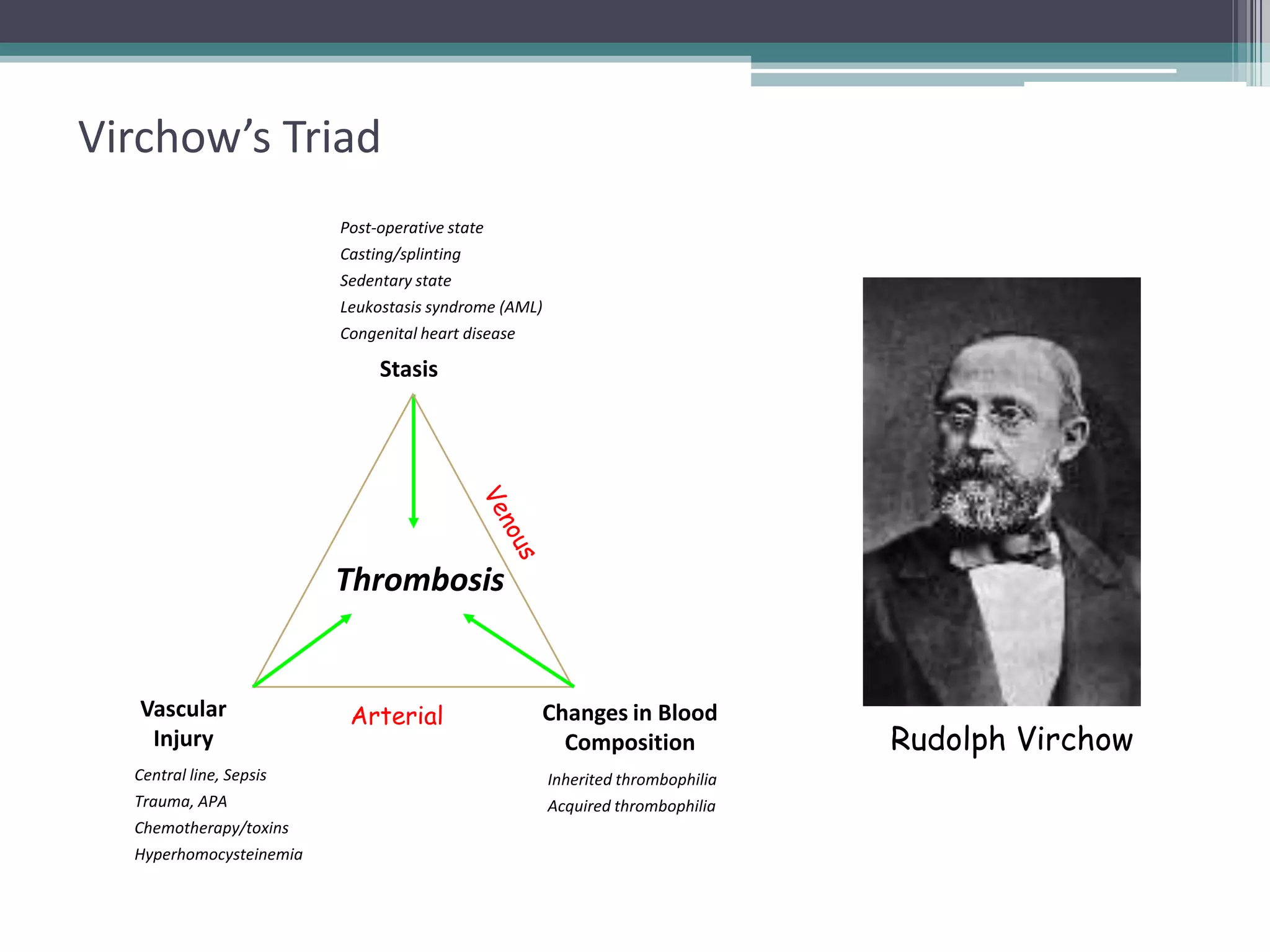
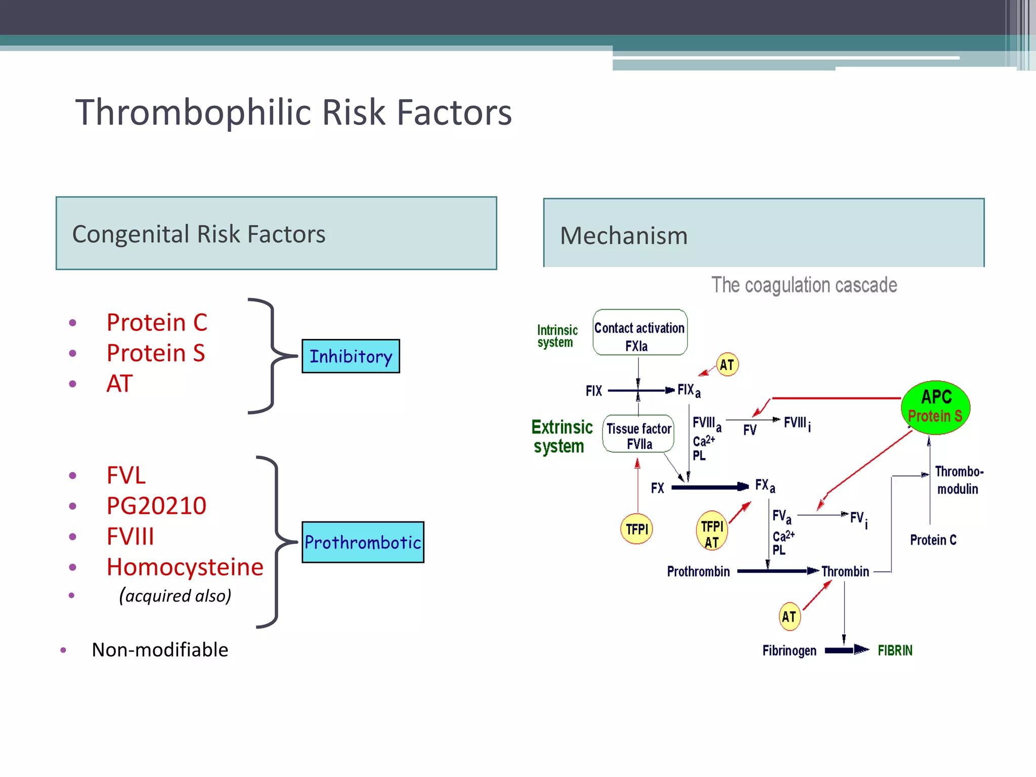
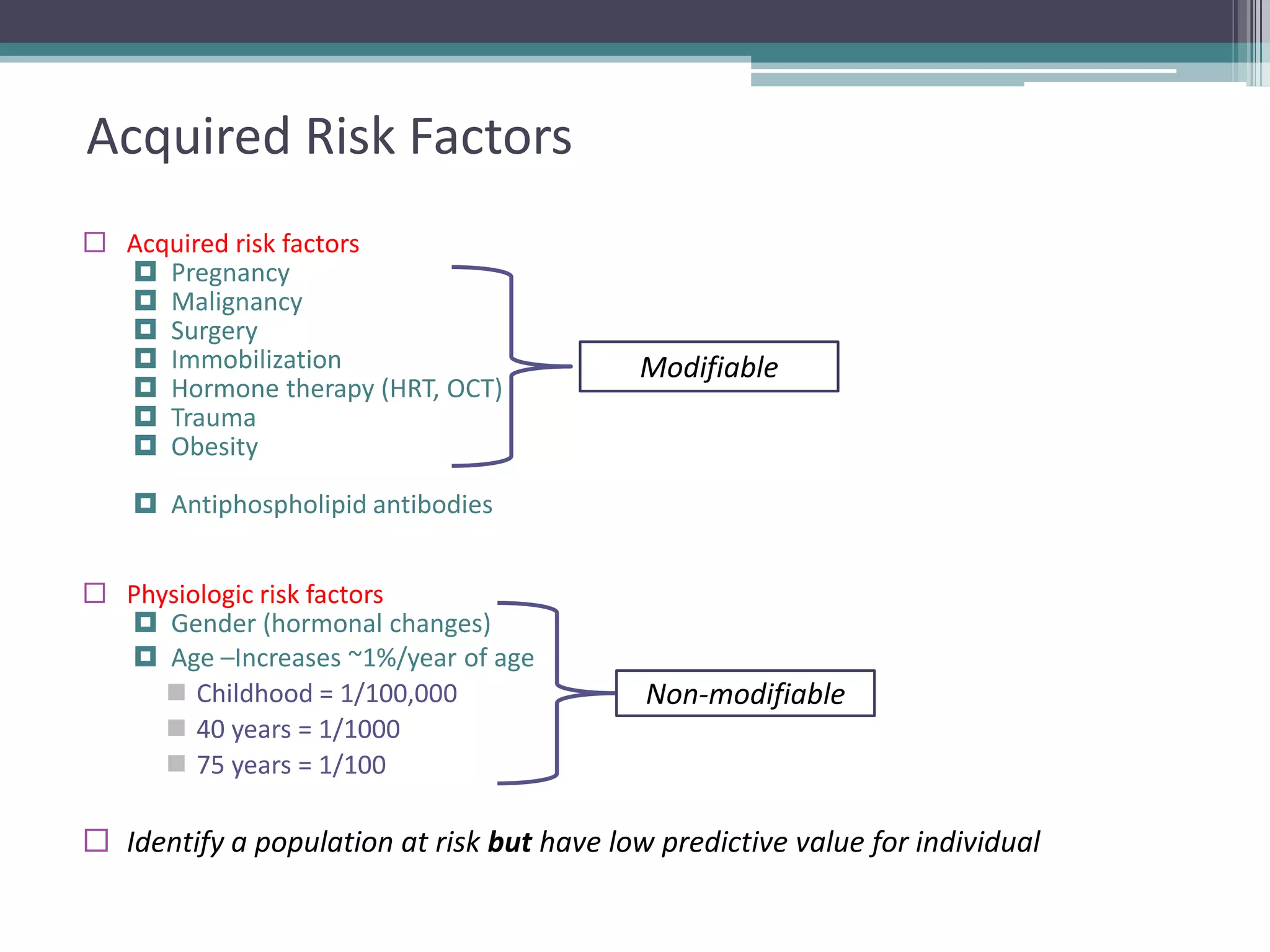
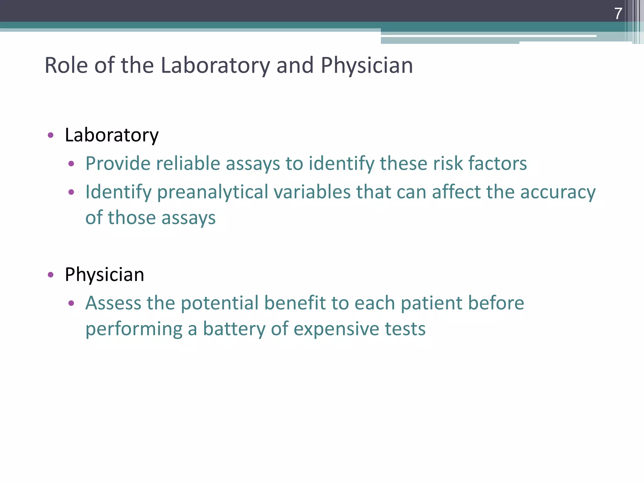
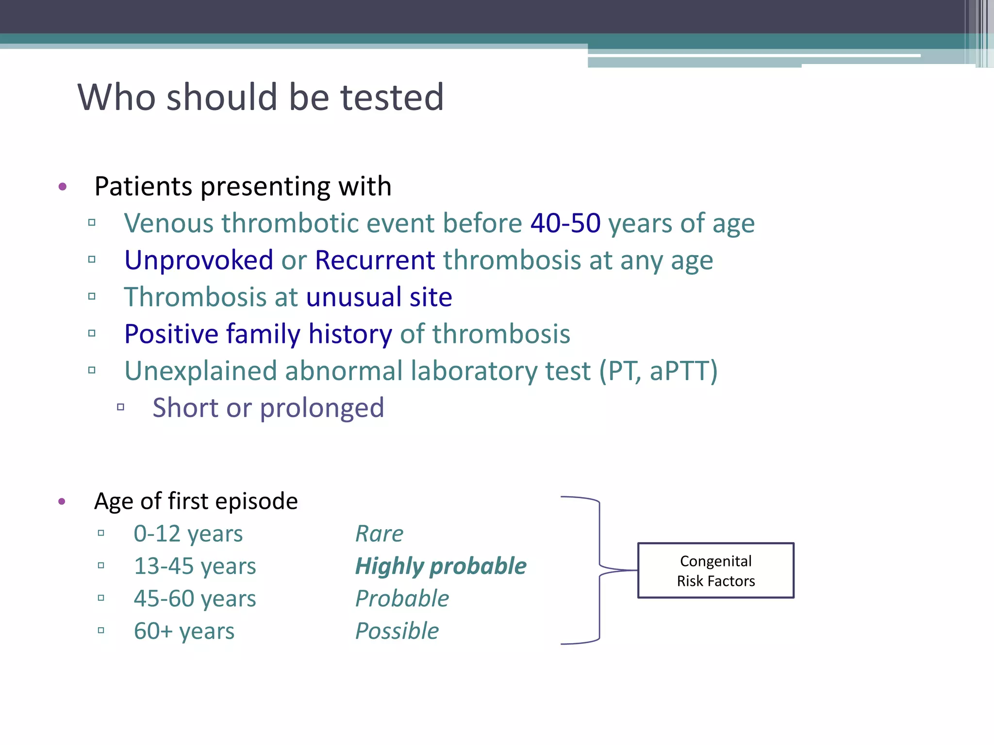
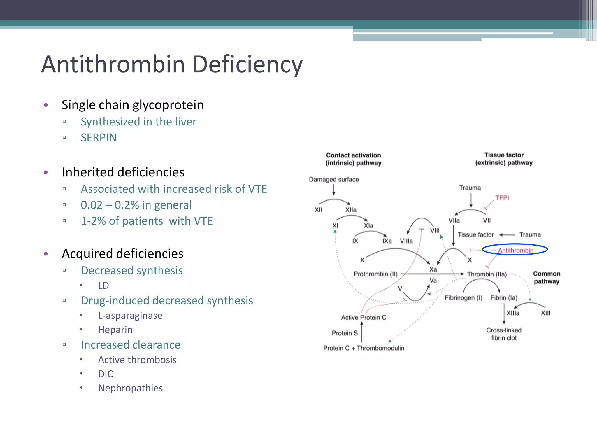
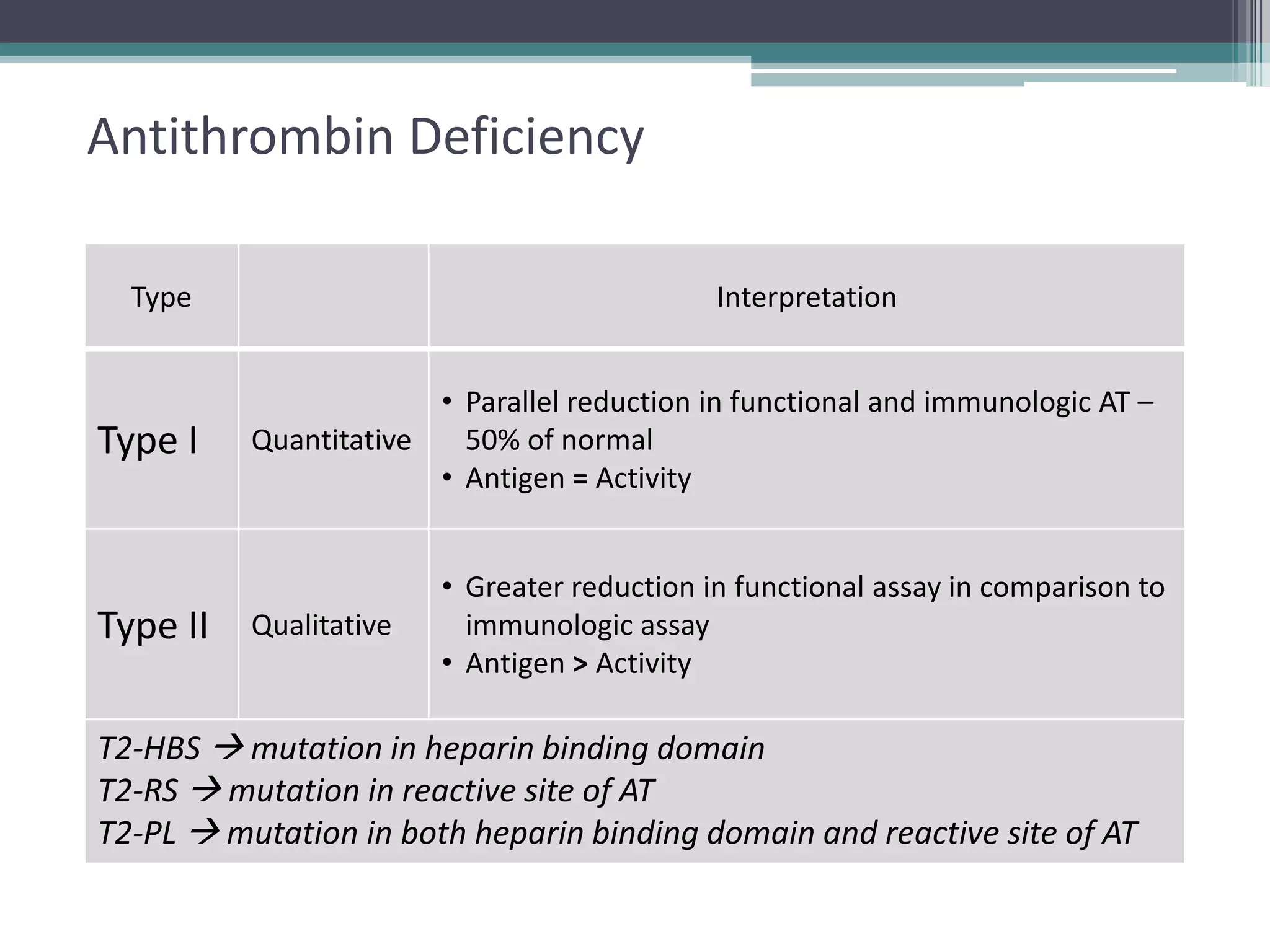
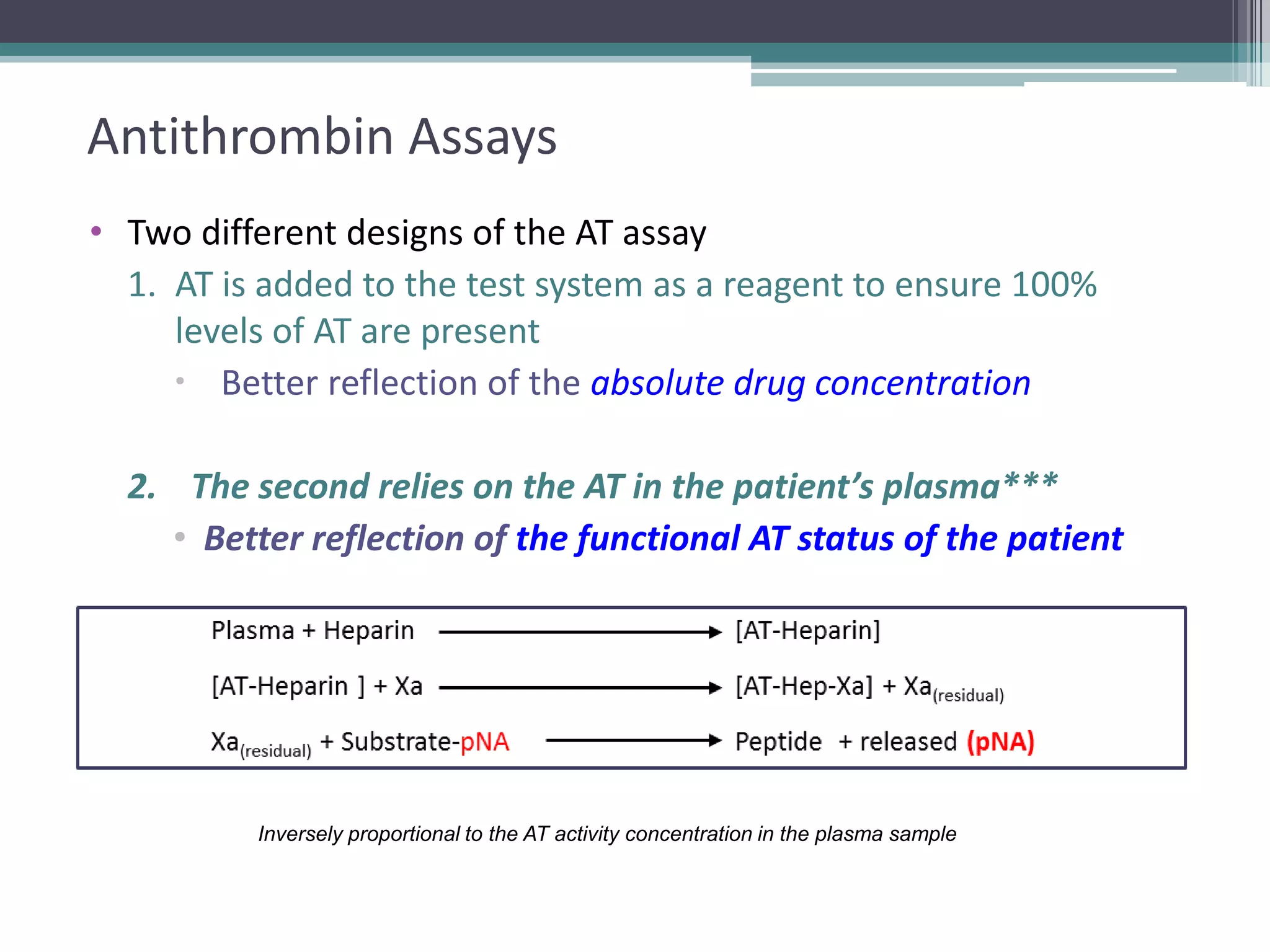
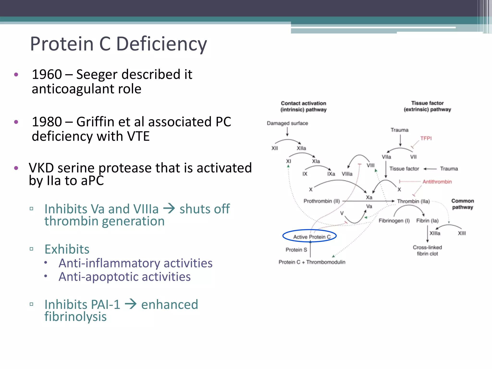
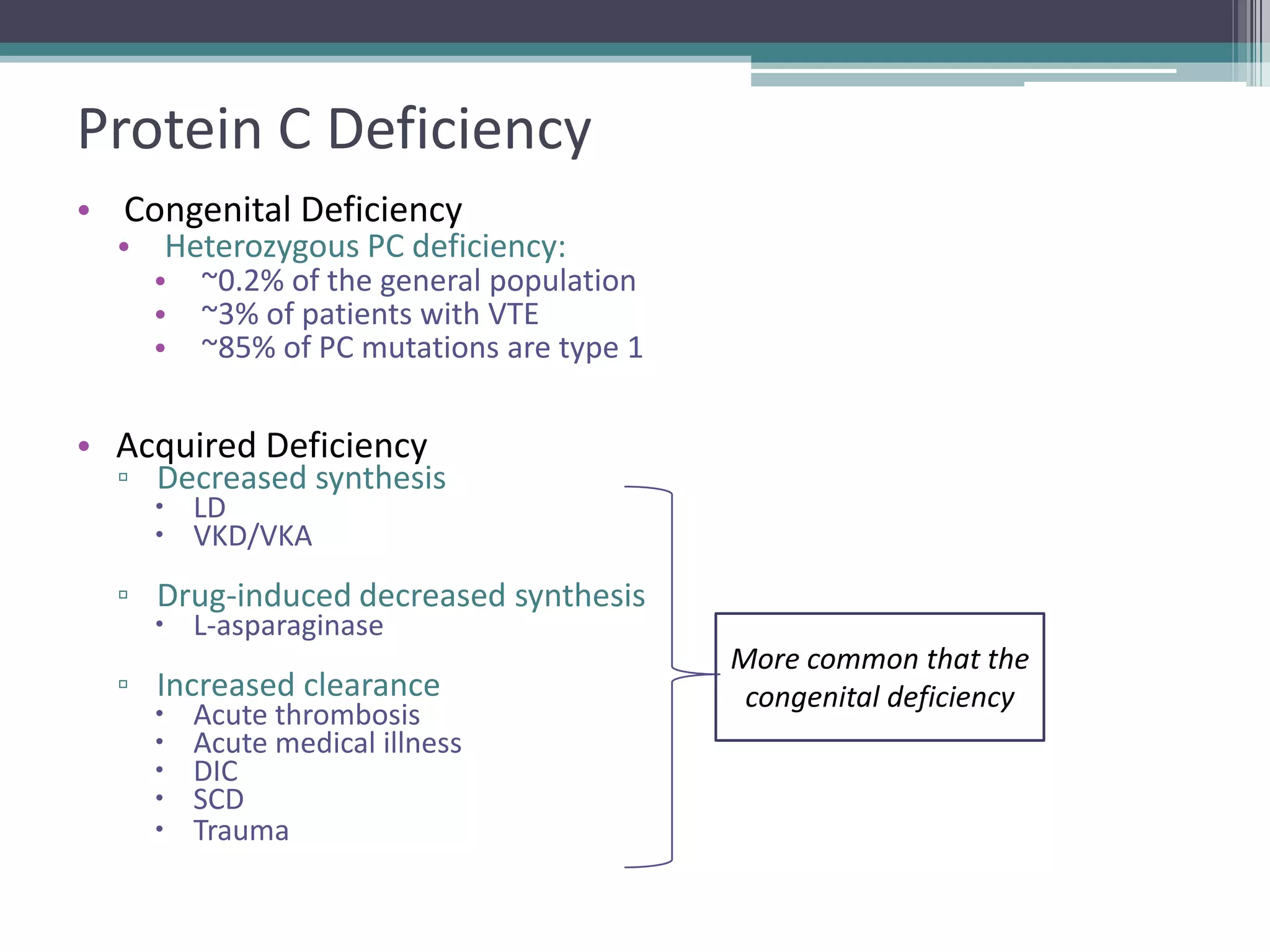
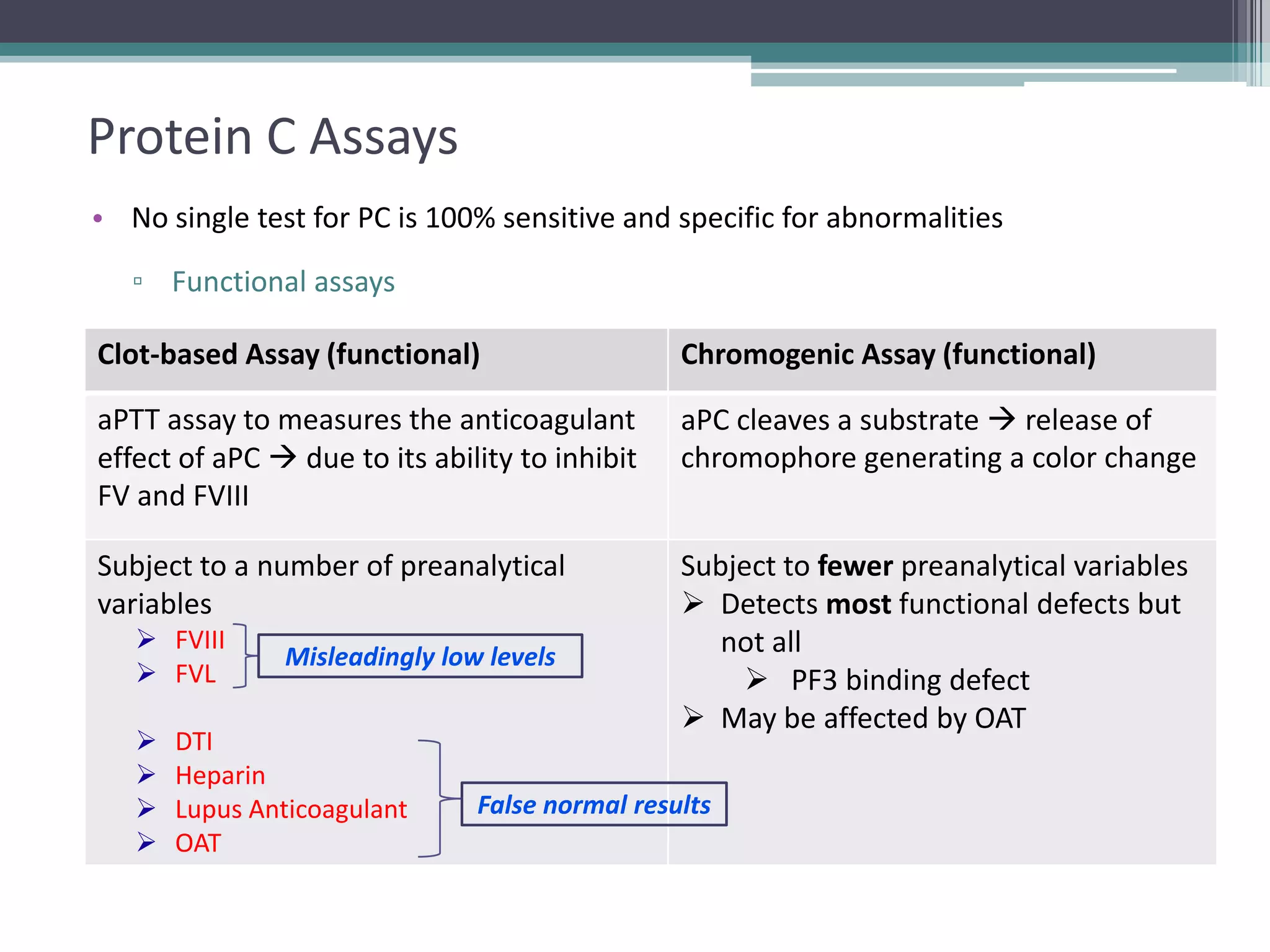
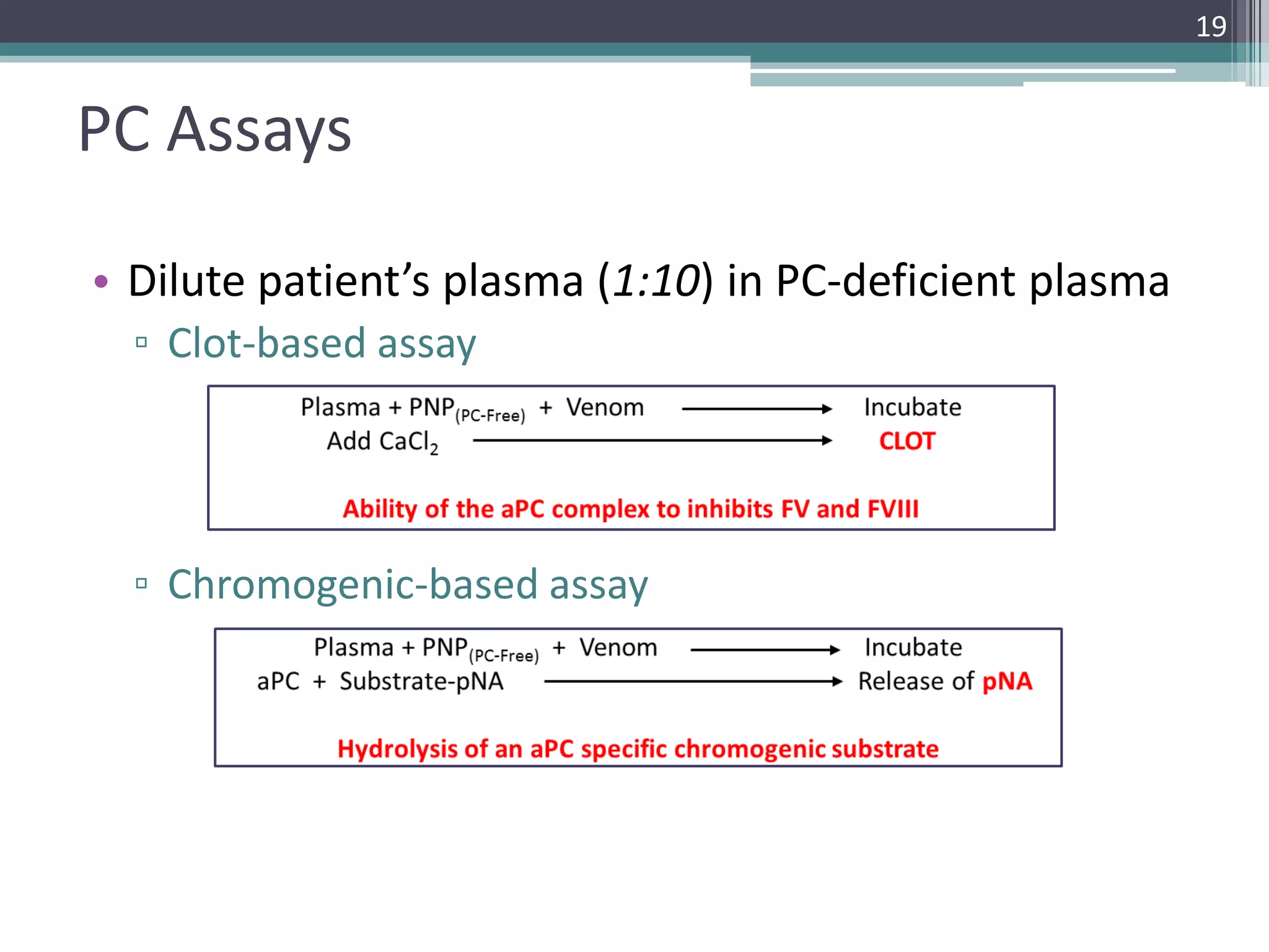
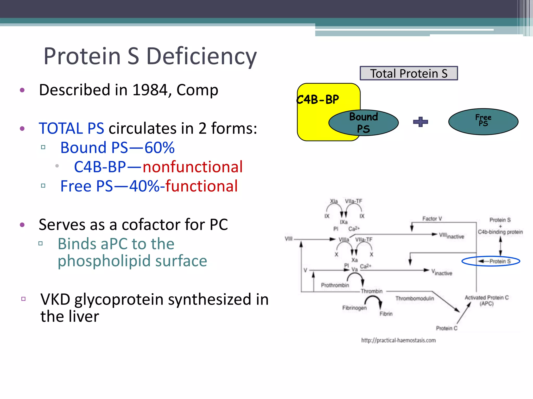
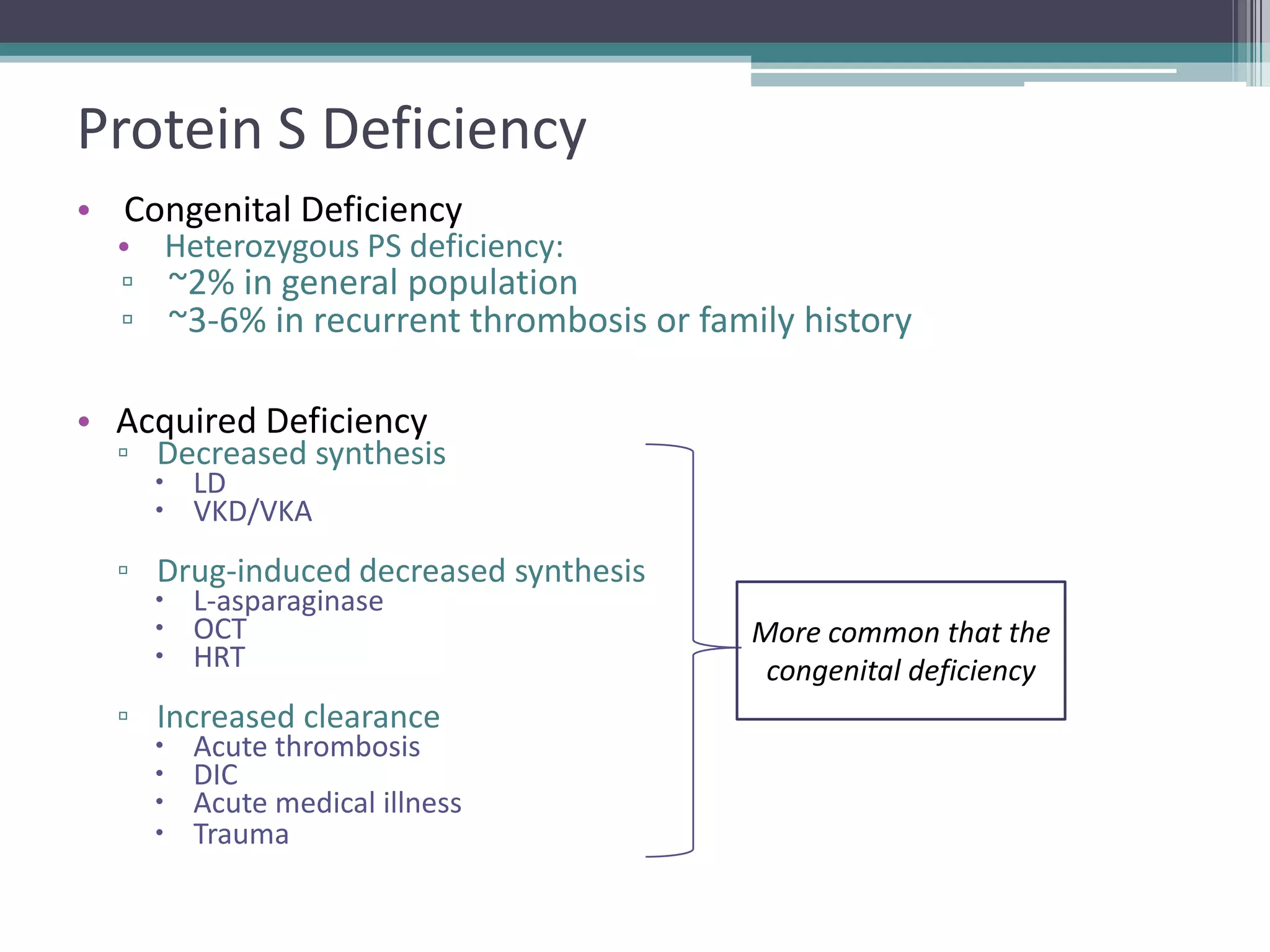
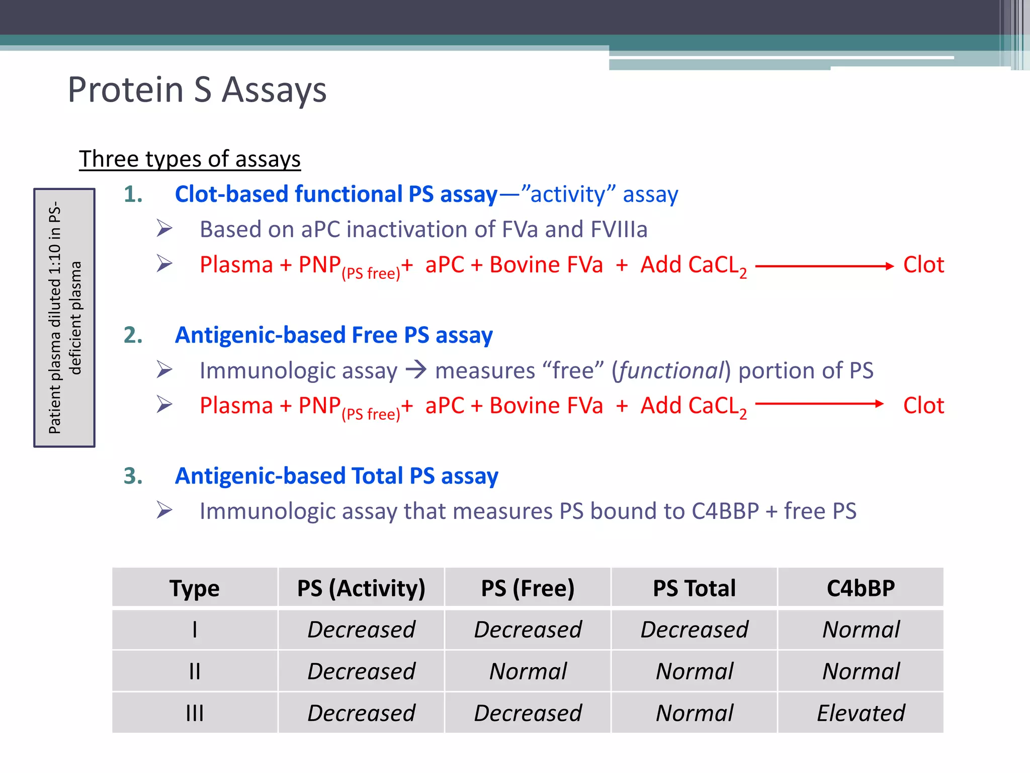
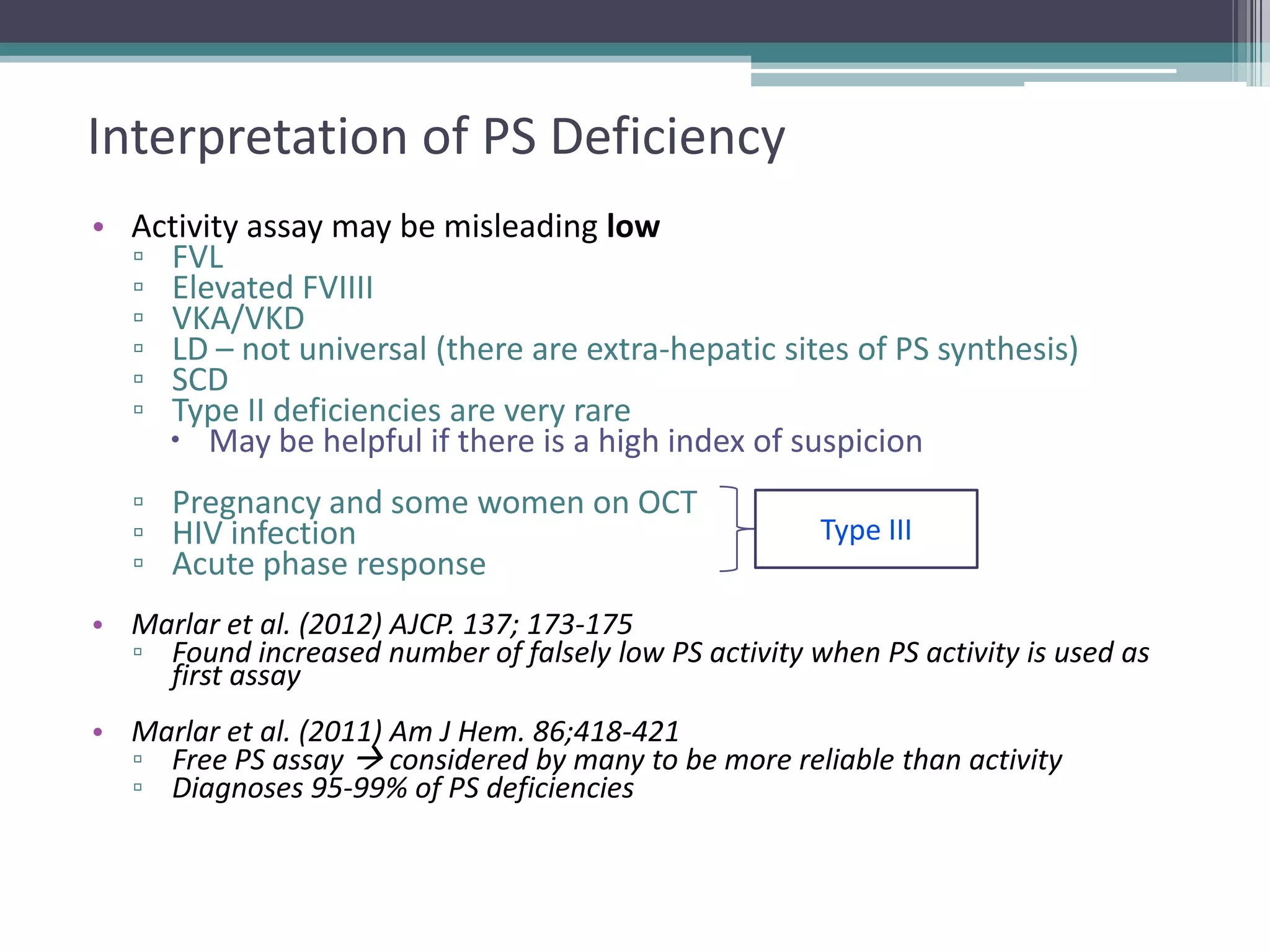
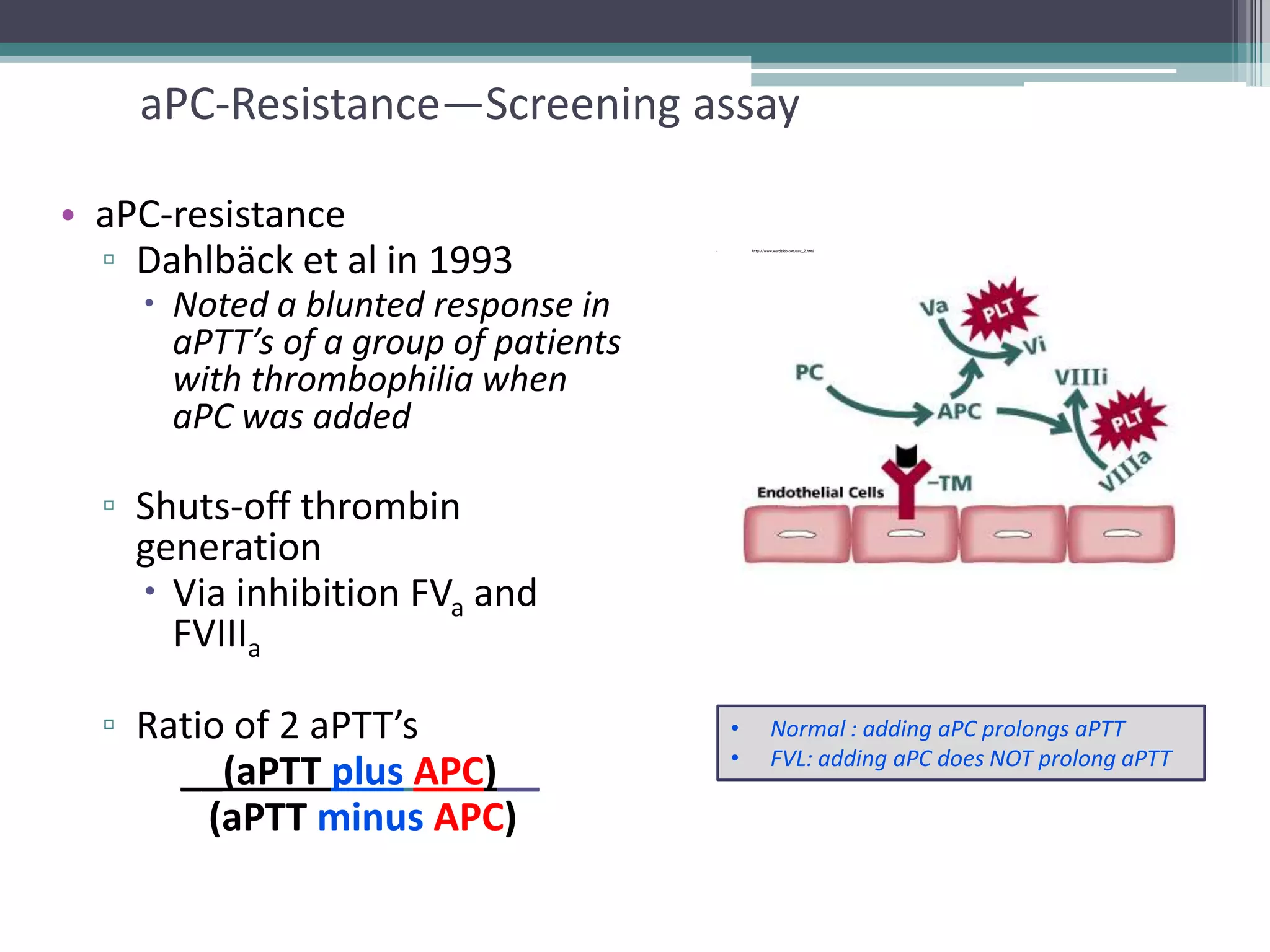
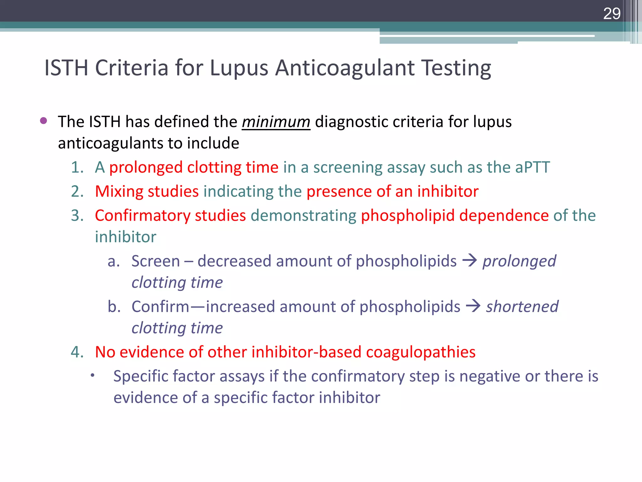
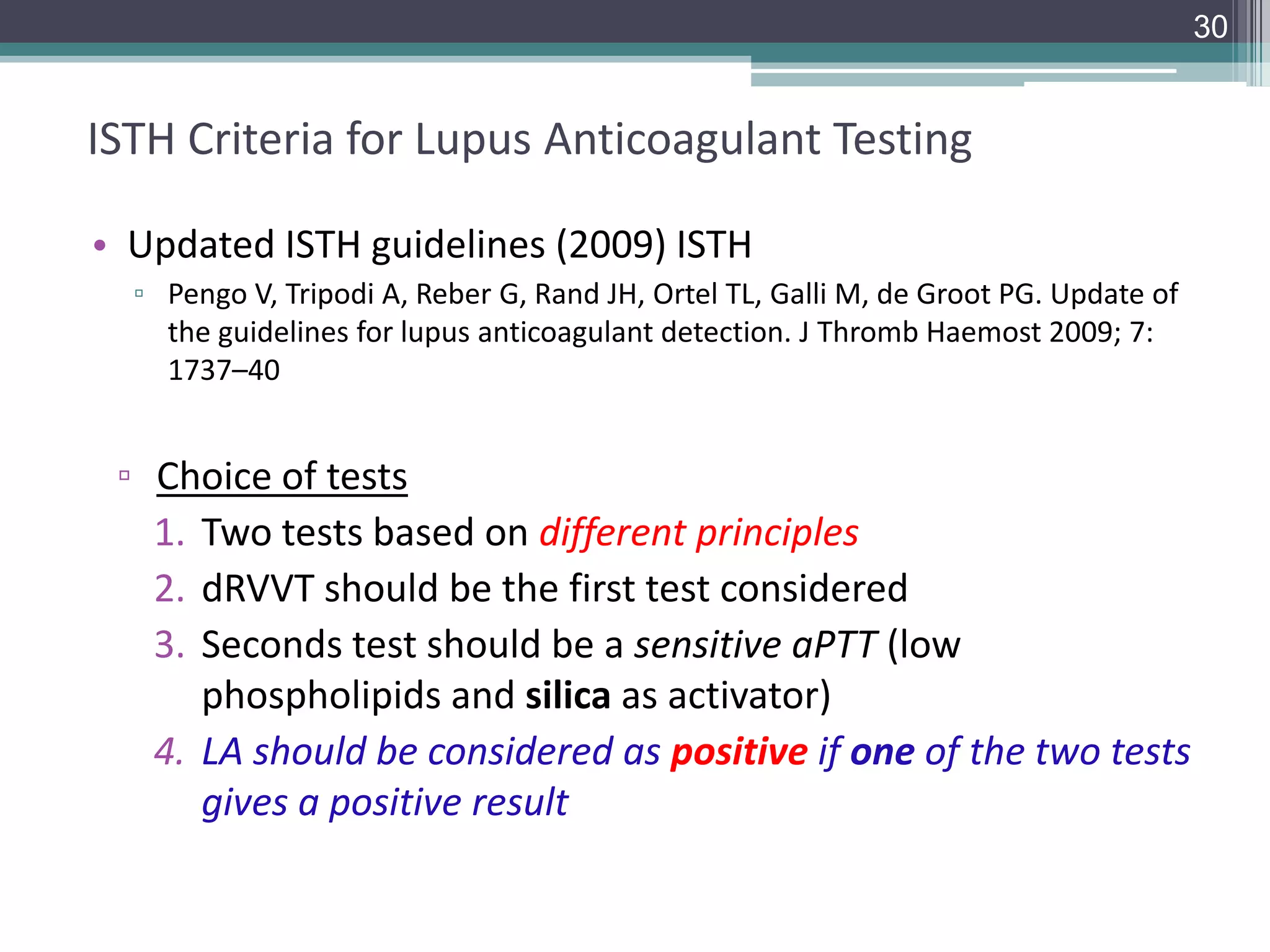
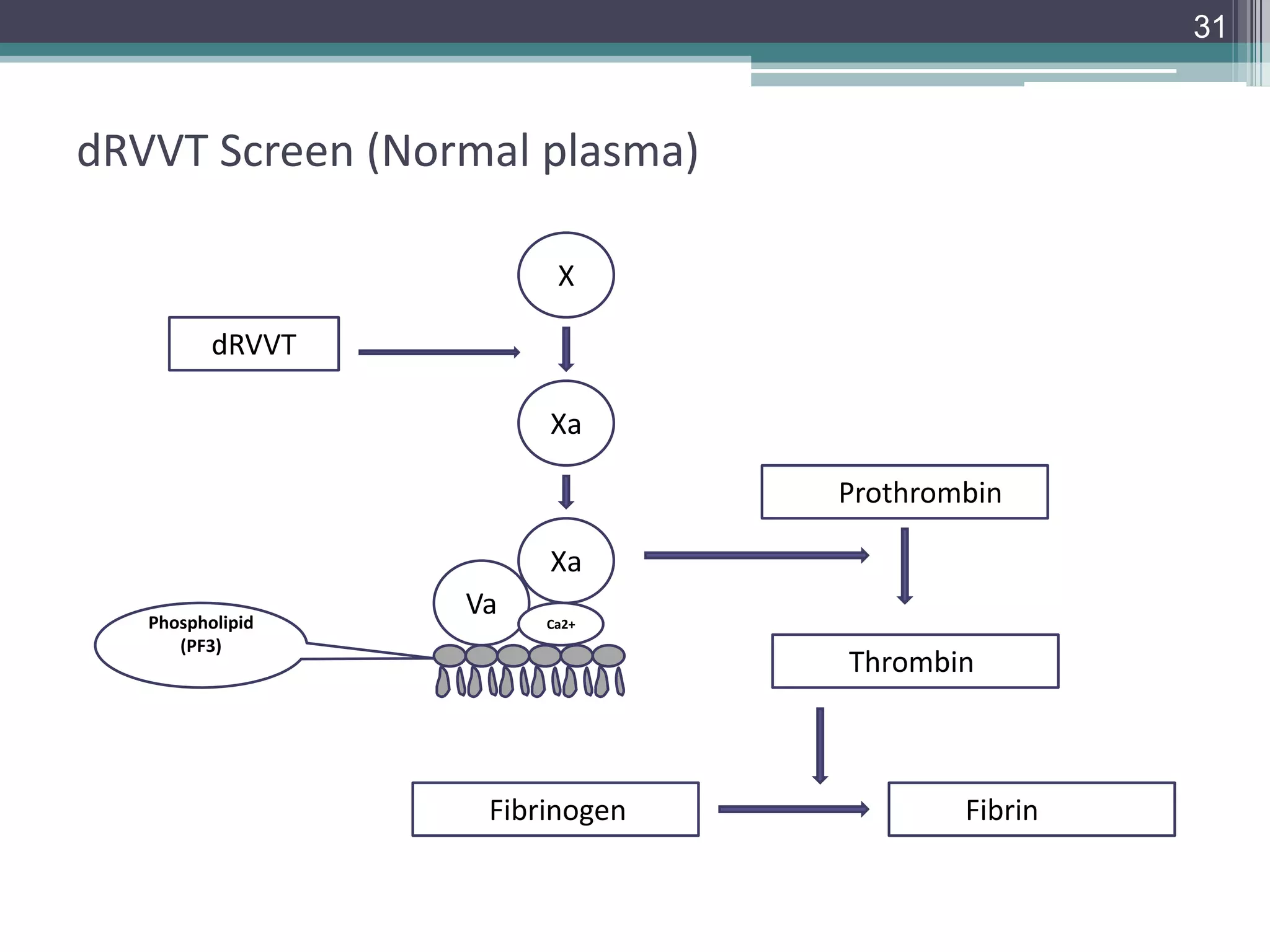
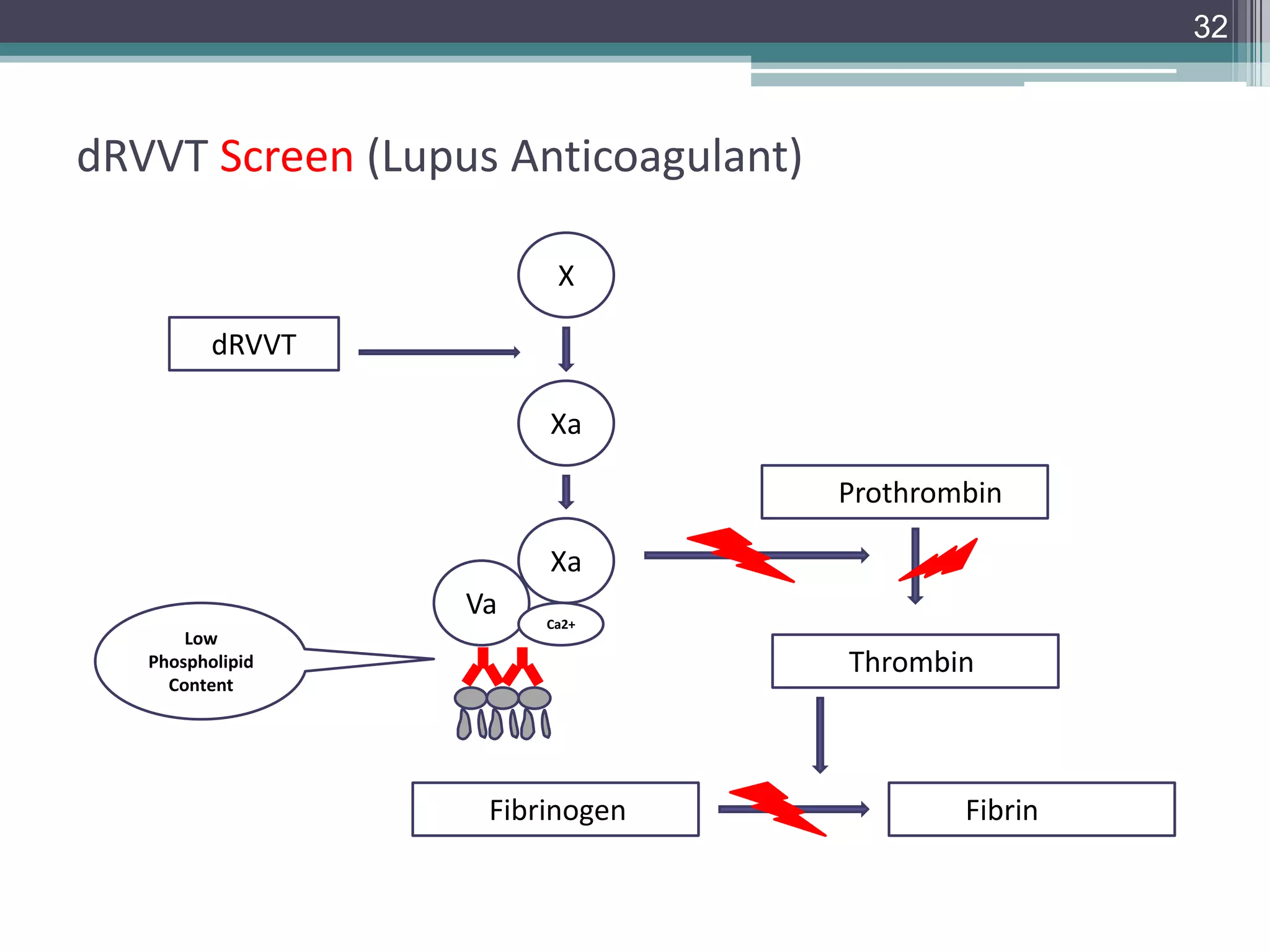
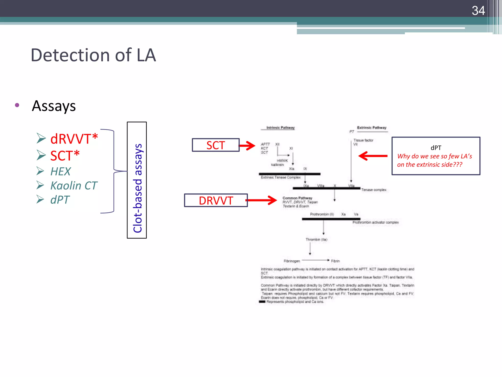
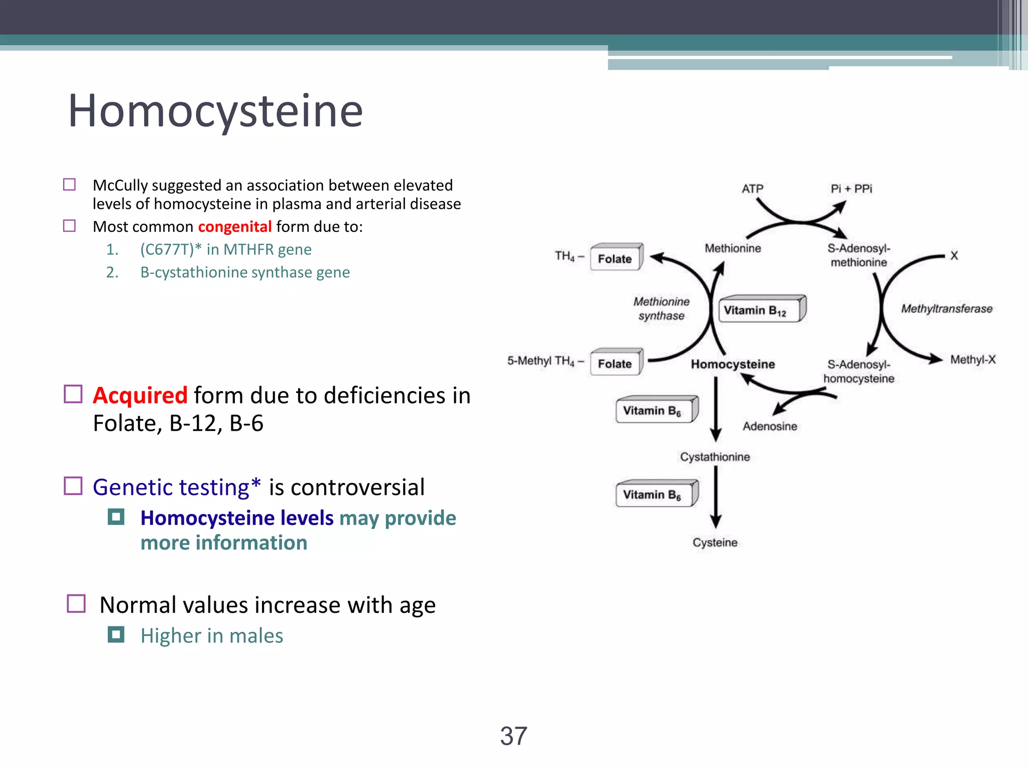
Hemostasis is the arrest of bleeding, whether it be by normal vasoconstriction (the vessel walls closing temporarily), by an abnormal obstruction (such as a plaque) or by coagulation or surgical means (such as ligation)
Autologous Blood Transfusion (ABT) means reinfusion of blood or blood products taken from the same patient
ABT is not a new concept, fear of transfusion- transmitted diseases stimulated the growth of autologous programme
Autologous Blood Transfusion (ABT) means reinfusion of blood or blood products taken from the same patient
ABT is not a new concept, fear of transfusion- transmitted diseases stimulated the growth of autologous programme
Thrombophilia test- when and what to do?
Thrombophilia is not a disease itself but a manifestation of several conditions leading to increased tendency for thrombosis. Since long it has been a grey area in thrombophilia with a lot of who, when, why and what regarding testing for this condition. As a result, the tests are being done very indiscriminately without knowing what to do with the result, whether positive or negative. Many family-screening are also commonly done simultaneously in many cases unnecessarily.
The inherited thrombophilias are mutation of factor V Leiden (FVL) or prothrombin gene and deficiency of protein C (PC), protein S (PS) or antithrombin (AT). The acquired conditions are anti-phospholipid syndrome (APS), paroxysmal nocturnal hemoglobinuria (PNH) and myeloproliferative neoplasms (MPN). Various malignancies, drugs (e.g., oral contraceptives, chemotherapy), pregnancy, trauma and surgery also increase the risk of thrombosis. In clinical point of view immobility and central venous access devises (CVAD) also add fuel to the flame (Virchow’s triad).
The excerpts from the compilation of various literatures including recent guidelines are as follows. Testing for “heritable thrombophilia” should be done if thrombosis occurs at a younger age (<50yrs), with no/weak provocation and preferably with family history of thrombosis in first degree relatives in young age. Tests if at all needed should be done after at least 3 months of anticoagulation, only after stopping of anticoagulation for specified duration for respective drugs and tests should not be done during an acute event. Genetic testing to predict a first episode of venous thrombosis is not recommended. For thrombosis at unusual venous sites (splanchnic veins or cortical venous sinuses) or arteries, PNH and MPN should be ruled out as per CBC and other ancillary features. JAK2 mutation may also present with thrombosis even before MPN develops. Apart from this, tests for APS should also be done in above cases and additionally in retinal vein occlusion, stroke, or myocardial infarction in young. Neonatal purpura fulminans is a matter of special concern; protein C/S should be tested at urgent basis as specific treatment can be offered. Women contemplating pregnancy or willing for oral contraceptives should be addressed as per personal or family history of thrombosis/thrombophilia.
Finally, an algorithmic approach should be adopted for individualizing each case of thrombosis to avoid indiscriminate testing and to avoid unnecessary anxiety of patients and family members with a proper vision to what to do with the result.
Current Component Therapy by Diane Eklund, MDbloodbankhawaii
Lorem ipsum dolor sit amet, voluptaria percipitur has eu. Nibh iriure nostrud ei mea. Vel dicta voluptua convenire ei, id pro libris viderer. Pri et legendos atomorum, vel eu noster probatus menandri. Omnes possim ut eam, sed ea labore maiorum.
New Directions in Targeted Therapeutic Approaches for Older Adults With Mantl...i3 Health
i3 Health is pleased to make the speaker slides from this activity available for use as a non-accredited self-study or teaching resource.
This slide deck presented by Dr. Kami Maddocks, Professor-Clinical in the Division of Hematology and
Associate Division Director for Ambulatory Operations
The Ohio State University Comprehensive Cancer Center, will provide insight into new directions in targeted therapeutic approaches for older adults with mantle cell lymphoma.
STATEMENT OF NEED
Mantle cell lymphoma (MCL) is a rare, aggressive B-cell non-Hodgkin lymphoma (NHL) accounting for 5% to 7% of all lymphomas. Its prognosis ranges from indolent disease that does not require treatment for years to very aggressive disease, which is associated with poor survival (Silkenstedt et al, 2021). Typically, MCL is diagnosed at advanced stage and in older patients who cannot tolerate intensive therapy (NCCN, 2022). Although recent advances have slightly increased remission rates, recurrence and relapse remain very common, leading to a median overall survival between 3 and 6 years (LLS, 2021). Though there are several effective options, progress is still needed towards establishing an accepted frontline approach for MCL (Castellino et al, 2022). Treatment selection and management of MCL are complicated by the heterogeneity of prognosis, advanced age and comorbidities of patients, and lack of an established standard approach for treatment, making it vital that clinicians be familiar with the latest research and advances in this area. In this activity chaired by Michael Wang, MD, Professor in the Department of Lymphoma & Myeloma at MD Anderson Cancer Center, expert faculty will discuss prognostic factors informing treatment, the promising results of recent trials in new therapeutic approaches, and the implications of treatment resistance in therapeutic selection for MCL.
Target Audience
Hematology/oncology fellows, attending faculty, and other health care professionals involved in the treatment of patients with mantle cell lymphoma (MCL).
Learning Objectives
1.) Identify clinical and biological prognostic factors that can guide treatment decision making for older adults with MCL
2.) Evaluate emerging data on targeted therapeutic approaches for treatment-naive and relapsed/refractory MCL and their applicability to older adults
3.) Assess mechanisms of resistance to targeted therapies for MCL and their implications for treatment selection
MANAGEMENT OF ATRIOVENTRICULAR CONDUCTION BLOCK.pdfJim Jacob Roy
Cardiac conduction defects can occur due to various causes.
Atrioventricular conduction blocks ( AV blocks ) are classified into 3 types.
This document describes the acute management of AV block.
micro teaching on communication m.sc nursing.pdfAnurag Sharma
Microteaching is a unique model of practice teaching. It is a viable instrument for the. desired change in the teaching behavior or the behavior potential which, in specified types of real. classroom situations, tends to facilitate the achievement of specified types of objectives.
Report Back from SGO 2024: What’s the Latest in Cervical Cancer?bkling
Are you curious about what’s new in cervical cancer research or unsure what the findings mean? Join Dr. Emily Ko, a gynecologic oncologist at Penn Medicine, to learn about the latest updates from the Society of Gynecologic Oncology (SGO) 2024 Annual Meeting on Women’s Cancer. Dr. Ko will discuss what the research presented at the conference means for you and answer your questions about the new developments.
Tom Selleck Health: A Comprehensive Look at the Iconic Actor’s Wellness Journeygreendigital
Tom Selleck, an enduring figure in Hollywood. has captivated audiences for decades with his rugged charm, iconic moustache. and memorable roles in television and film. From his breakout role as Thomas Magnum in Magnum P.I. to his current portrayal of Frank Reagan in Blue Bloods. Selleck's career has spanned over 50 years. But beyond his professional achievements. fans have often been curious about Tom Selleck Health. especially as he has aged in the public eye.
Follow us on: Pinterest
Introduction
Many have been interested in Tom Selleck health. not only because of his enduring presence on screen but also because of the challenges. and lifestyle choices he has faced and made over the years. This article delves into the various aspects of Tom Selleck health. exploring his fitness regimen, diet, mental health. and the challenges he has encountered as he ages. We'll look at how he maintains his well-being. the health issues he has faced, and his approach to ageing .
Early Life and Career
Childhood and Athletic Beginnings
Tom Selleck was born on January 29, 1945, in Detroit, Michigan, and grew up in Sherman Oaks, California. From an early age, he was involved in sports, particularly basketball. which played a significant role in his physical development. His athletic pursuits continued into college. where he attended the University of Southern California (USC) on a basketball scholarship. This early involvement in sports laid a strong foundation for his physical health and disciplined lifestyle.
Transition to Acting
Selleck's transition from an athlete to an actor came with its physical demands. His first significant role in "Magnum P.I." required him to perform various stunts and maintain a fit appearance. This role, which he played from 1980 to 1988. necessitated a rigorous fitness routine to meet the show's demands. setting the stage for his long-term commitment to health and wellness.
Fitness Regimen
Workout Routine
Tom Selleck health and fitness regimen has evolved. adapting to his changing roles and age. During his "Magnum, P.I." days. Selleck's workouts were intense and focused on building and maintaining muscle mass. His routine included weightlifting, cardiovascular exercises. and specific training for the stunts he performed on the show.
Selleck adjusted his fitness routine as he aged to suit his body's needs. Today, his workouts focus on maintaining flexibility, strength, and cardiovascular health. He incorporates low-impact exercises such as swimming, walking, and light weightlifting. This balanced approach helps him stay fit without putting undue strain on his joints and muscles.
Importance of Flexibility and Mobility
In recent years, Selleck has emphasized the importance of flexibility and mobility in his fitness regimen. Understanding the natural decline in muscle mass and joint flexibility with age. he includes stretching and yoga in his routine. These practices help prevent injuries, improve posture, and maintain mobilit
ARTIFICIAL INTELLIGENCE IN HEALTHCARE.pdfAnujkumaranit
Artificial intelligence (AI) refers to the simulation of human intelligence processes by machines, especially computer systems. It encompasses tasks such as learning, reasoning, problem-solving, perception, and language understanding. AI technologies are revolutionizing various fields, from healthcare to finance, by enabling machines to perform tasks that typically require human intelligence.
Ethanol (CH3CH2OH), or beverage alcohol, is a two-carbon alcohol
that is rapidly distributed in the body and brain. Ethanol alters many
neurochemical systems and has rewarding and addictive properties. It
is the oldest recreational drug and likely contributes to more morbidity,
mortality, and public health costs than all illicit drugs combined. The
5th edition of the Diagnostic and Statistical Manual of Mental Disorders
(DSM-5) integrates alcohol abuse and alcohol dependence into a single
disorder called alcohol use disorder (AUD), with mild, moderate,
and severe subclassifications (American Psychiatric Association, 2013).
In the DSM-5, all types of substance abuse and dependence have been
combined into a single substance use disorder (SUD) on a continuum
from mild to severe. A diagnosis of AUD requires that at least two of
the 11 DSM-5 behaviors be present within a 12-month period (mild
AUD: 2–3 criteria; moderate AUD: 4–5 criteria; severe AUD: 6–11 criteria).
The four main behavioral effects of AUD are impaired control over
drinking, negative social consequences, risky use, and altered physiological
effects (tolerance, withdrawal). This chapter presents an overview
of the prevalence and harmful consequences of AUD in the U.S.,
the systemic nature of the disease, neurocircuitry and stages of AUD,
comorbidities, fetal alcohol spectrum disorders, genetic risk factors, and
pharmacotherapies for AUD.
Lung Cancer: Artificial Intelligence, Synergetics, Complex System Analysis, S...Oleg Kshivets
RESULTS: Overall life span (LS) was 2252.1±1742.5 days and cumulative 5-year survival (5YS) reached 73.2%, 10 years – 64.8%, 20 years – 42.5%. 513 LCP lived more than 5 years (LS=3124.6±1525.6 days), 148 LCP – more than 10 years (LS=5054.4±1504.1 days).199 LCP died because of LC (LS=562.7±374.5 days). 5YS of LCP after bi/lobectomies was significantly superior in comparison with LCP after pneumonectomies (78.1% vs.63.7%, P=0.00001 by log-rank test). AT significantly improved 5YS (66.3% vs. 34.8%) (P=0.00000 by log-rank test) only for LCP with N1-2. Cox modeling displayed that 5YS of LCP significantly depended on: phase transition (PT) early-invasive LC in terms of synergetics, PT N0—N12, cell ratio factors (ratio between cancer cells- CC and blood cells subpopulations), G1-3, histology, glucose, AT, blood cell circuit, prothrombin index, heparin tolerance, recalcification time (P=0.000-0.038). Neural networks, genetic algorithm selection and bootstrap simulation revealed relationships between 5YS and PT early-invasive LC (rank=1), PT N0—N12 (rank=2), thrombocytes/CC (3), erythrocytes/CC (4), eosinophils/CC (5), healthy cells/CC (6), lymphocytes/CC (7), segmented neutrophils/CC (8), stick neutrophils/CC (9), monocytes/CC (10); leucocytes/CC (11). Correct prediction of 5YS was 100% by neural networks computing (area under ROC curve=1.0; error=0.0).
CONCLUSIONS: 5YS of LCP after radical procedures significantly depended on: 1) PT early-invasive cancer; 2) PT N0--N12; 3) cell ratio factors; 4) blood cell circuit; 5) biochemical factors; 6) hemostasis system; 7) AT; 8) LC characteristics; 9) LC cell dynamics; 10) surgery type: lobectomy/pneumonectomy; 11) anthropometric data. Optimal diagnosis and treatment strategies for LC are: 1) screening and early detection of LC; 2) availability of experienced thoracic surgeons because of complexity of radical procedures; 3) aggressive en block surgery and adequate lymph node dissection for completeness; 4) precise prediction; 5) adjuvant chemoimmunoradiotherapy for LCP with unfavorable prognosis.
Title: Sense of Taste
Presenter: Dr. Faiza, Assistant Professor of Physiology
Qualifications:
MBBS (Best Graduate, AIMC Lahore)
FCPS Physiology
ICMT, CHPE, DHPE (STMU)
MPH (GC University, Faisalabad)
MBA (Virtual University of Pakistan)
Learning Objectives:
Describe the structure and function of taste buds.
Describe the relationship between the taste threshold and taste index of common substances.
Explain the chemical basis and signal transduction of taste perception for each type of primary taste sensation.
Recognize different abnormalities of taste perception and their causes.
Key Topics:
Significance of Taste Sensation:
Differentiation between pleasant and harmful food
Influence on behavior
Selection of food based on metabolic needs
Receptors of Taste:
Taste buds on the tongue
Influence of sense of smell, texture of food, and pain stimulation (e.g., by pepper)
Primary and Secondary Taste Sensations:
Primary taste sensations: Sweet, Sour, Salty, Bitter, Umami
Chemical basis and signal transduction mechanisms for each taste
Taste Threshold and Index:
Taste threshold values for Sweet (sucrose), Salty (NaCl), Sour (HCl), and Bitter (Quinine)
Taste index relationship: Inversely proportional to taste threshold
Taste Blindness:
Inability to taste certain substances, particularly thiourea compounds
Example: Phenylthiocarbamide
Structure and Function of Taste Buds:
Composition: Epithelial cells, Sustentacular/Supporting cells, Taste cells, Basal cells
Features: Taste pores, Taste hairs/microvilli, and Taste nerve fibers
Location of Taste Buds:
Found in papillae of the tongue (Fungiform, Circumvallate, Foliate)
Also present on the palate, tonsillar pillars, epiglottis, and proximal esophagus
Mechanism of Taste Stimulation:
Interaction of taste substances with receptors on microvilli
Signal transduction pathways for Umami, Sweet, Bitter, Sour, and Salty tastes
Taste Sensitivity and Adaptation:
Decrease in sensitivity with age
Rapid adaptation of taste sensation
Role of Saliva in Taste:
Dissolution of tastants to reach receptors
Washing away the stimulus
Taste Preferences and Aversions:
Mechanisms behind taste preference and aversion
Influence of receptors and neural pathways
Impact of Sensory Nerve Damage:
Degeneration of taste buds if the sensory nerve fiber is cut
Abnormalities of Taste Detection:
Conditions: Ageusia, Hypogeusia, Dysgeusia (parageusia)
Causes: Nerve damage, neurological disorders, infections, poor oral hygiene, adverse drug effects, deficiencies, aging, tobacco use, altered neurotransmitter levels
Neurotransmitters and Taste Threshold:
Effects of serotonin (5-HT) and norepinephrine (NE) on taste sensitivity
Supertasters:
25% of the population with heightened sensitivity to taste, especially bitterness
Increased number of fungiform papillae
Prix Galien International 2024 Forum ProgramLevi Shapiro
June 20, 2024, Prix Galien International and Jerusalem Ethics Forum in ROME. Detailed agenda including panels:
- ADVANCES IN CARDIOLOGY: A NEW PARADIGM IS COMING
- WOMEN’S HEALTH: FERTILITY PRESERVATION
- WHAT’S NEW IN THE TREATMENT OF INFECTIOUS,
ONCOLOGICAL AND INFLAMMATORY SKIN DISEASES?
- ARTIFICIAL INTELLIGENCE AND ETHICS
- GENE THERAPY
- BEYOND BORDERS: GLOBAL INITIATIVES FOR DEMOCRATIZING LIFE SCIENCE TECHNOLOGIES AND PROMOTING ACCESS TO HEALTHCARE
- ETHICAL CHALLENGES IN LIFE SCIENCES
- Prix Galien International Awards Ceremony
Pulmonary Thromboembolism - etilogy, types, medical- Surgical and nursing man...VarunMahajani
Disruption of blood supply to lung alveoli due to blockage of one or more pulmonary blood vessels is called as Pulmonary thromboembolism. In this presentation we will discuss its causes, types and its management in depth.
How to Give Better Lectures: Some Tips for Doctors
Laboratory tests of hemostasis and coagulation system
1. Coagulation Assays – Part 2
Larry Smith, PhD
Director, Coagulation/Hemostasis
Assistant Attending
2. 2
Thrombophilia Testing
• Increased tendency to VTE
• Not a disease per se but may be associated with
▫ Disease cancer
▫ Drug exposure HRT, OCT
▫ Modifiable conditions pregnancy, immobilization
▫ Non-modifiable conditions age, gender
• Affects 1-2 individuals/1000 in the general population
▫ In the US
~2,000,000/year
~500,000 deaths
Many survive with complications
3. 3
Virchow’s Triad
Post-operative state
Casting/splinting
Sedentary state
Leukostasis syndrome (AML)
Congenital heart disease
Stasis
Thrombosis
Vascular
Injury
Arterial
Changes in Blood
Composition
Central line, Sepsis
Inherited thrombophilia
Trauma, APA
Acquired thrombophilia
Chemotherapy/toxins
Hyperhomocysteinemia
Rudolph Virchow
4. 4
Risk Factors
• Multiple risk factors—multi-factorial process
▫ Hereditary
▫ Acquired
• Multi-hit hypothesis
▫ Individual hereditary or acquired risk factors has a relatively small
individual effect
▫ Risk for thrombosis is greatly increased when two or more risk factors
combine
• Classification of Thrombophilia
▫ Congenital
▫ Acquired
Physiologic factors
Environmental factors
5. 5
Thrombophilic Risk Factors
Congenital Risk Factors
•
•
•
Protein C
Protein S
AT
•
•
•
•
FVL
PG20210
FVIII
Homocysteine
•
•
(acquired also)
Non-modifiable
Inhibitory
Prothrombotic
Mechanism
6. 6
Acquired Risk Factors
Acquired risk factors
Pregnancy
Malignancy
Surgery
Immobilization
Hormone therapy (HRT, OCT)
Trauma
Obesity
Modifiable
Antiphospholipid antibodies
Physiologic risk factors
Gender (hormonal changes)
Age –Increases ~1%/year of age
Childhood = 1/100,000
40 years = 1/1000
75 years = 1/100
Non-modifiable
Identify a population at risk but have low predictive value for individual
7. 7
Role of the Laboratory and Physician
• Laboratory
• Provide reliable assays to identify these risk factors
• Identify preanalytical variables that can affect the accuracy
of those assays
• Physician
• Assess the potential benefit to each patient before
performing a battery of expensive tests
8. 8
Who should be tested
• Patients presenting with
▫ Venous thrombotic event before 40-50 years of age
▫ Unprovoked or Recurrent thrombosis at any age
▫ Thrombosis at unusual site
▫ Positive family history of thrombosis
▫ Unexplained abnormal laboratory test (PT, aPTT)
▫ Short or prolonged
•
Age of first episode
▫ 0-12 years
▫ 13-45 years
▫ 45-60 years
▫ 60+ years
Rare
Highly probable
Probable
Possible
Congenital
Risk Factors
9. 9
Testing: When and Why
• Optimal time for testing
▫ Asymptomatic
▫ Not on anticoagulant therapy
▫ Anytime for molecular testing
• Why do we test
▫ Pathologic basis for the thrombotic event
▫ Duration and intensity of therapy
▫ Prophylaxis for high risk patients
▫ To alert the patient's immediate family members to the
presence of possible inherited risk factors
10. 10
Types of Assays
• Functional Assays
Clot-based assays
Good screening assays
Based on a functioning cascade
Affected by preanalytical variables
▫ May require additional testing
Chromogenic assays
Measure the activity of a specific enzyme rather than
general biologic function
Not affected by most preanalytical variables
11. 11
Types of Assays
• Antigenic assays
LIA- or ELISA-based technologies
• Confirmatory assays
▫ DNA-based assays
12. 12
Types of Deficiencies
• Type I Deficiency
Decrease in both FUNCTION and AMOUNT of protein
present
True deficiency
Functional assay
• Type II Deficiency
Identify the total AMOUNT of protein only
Do NOT measure the function of the protein
To identify a dysproteinemia
Functional + Antigenic assays
13. 13
Antithrombin Deficiency
• Single chain glycoprotein
▫
▫
Synthesized in the liver
SERPIN
• Inherited deficiencies
▫
▫
▫
Associated with increased risk of VTE
0.02 – 0.2% in general
1-2% of patients with VTE
• Acquired deficiencies
▫
Decreased synthesis
▫
Drug-induced decreased synthesis
▫
LD
L-asparaginase
Heparin
Increased clearance
Active thrombosis
DIC
Nephropathies
14. 14
Antithrombin Deficiency
Type
Type I
Type II
Interpretation
• Parallel reduction in functional and immunologic AT –
Quantitative
50% of normal
• Antigen = Activity
Qualitative
• Greater reduction in functional assay in comparison to
immunologic assay
• Antigen > Activity
T2-HBS mutation in heparin binding domain
T2-RS mutation in reactive site of AT
T2-PL mutation in both heparin binding domain and reactive site of AT
15. 15
Antithrombin Assays
• Two different designs of the AT assay
1. AT is added to the test system as a reagent to ensure 100%
levels of AT are present
Better reflection of the absolute drug concentration
2. The second relies on the AT in the patient’s plasma***
• Better reflection of the functional AT status of the patient
Inversely proportional to the AT activity concentration in the plasma sample
16. 16
Protein C Deficiency
• 1960 – Seeger described it
anticoagulant role
• 1980 – Griffin et al associated PC
deficiency with VTE
• VKD serine protease that is activated
by IIa to aPC
▫ Inhibits Va and VIIIa shuts off
thrombin generation
▫ Exhibits
Anti-inflammatory activities
Anti-apoptotic activities
▫ Inhibits PAI-1 enhanced
fibrinolysis
17. 17
Protein C Deficiency
• Congenital Deficiency
• Heterozygous PC deficiency:
•
•
•
~0.2% of the general population
~3% of patients with VTE
~85% of PC mutations are type 1
• Acquired Deficiency
▫ Decreased synthesis
LD
VKD/VKA
▫ Drug-induced decreased synthesis
L-asparaginase
▫ Increased clearance
Acute thrombosis
Acute medical illness
DIC
SCD
Trauma
More common that the
congenital deficiency
18. 18
Protein C Assays
• No single test for PC is 100% sensitive and specific for abnormalities
▫ Functional assays
Clot-based Assay (functional)
Chromogenic Assay (functional)
aPTT assay to measures the anticoagulant
effect of aPC due to its ability to inhibit
FV and FVIII
aPC cleaves a substrate release of
chromophore generating a color change
Subject to a number of preanalytical
variables
Subject to fewer preanalytical variables
Detects most functional defects but
not all
PF3 binding defect
May be affected by OAT
FVIII
FVL
▫
Misleadingly low levels
Antigenic assay
DTI
Heparin
Lupus Anticoagulant
OAT
False normal results
19. 19
PC Assays
• Dilute patient’s plasma (1:10) in PC-deficient plasma
▫ Clot-based assay
▫ Chromogenic-based assay
20. 20
Protein S Deficiency
• Described in 1984, Comp
• TOTAL PS circulates in 2 forms:
▫ Bound PS—60%
C4B-BP—nonfunctional
▫ Free PS—40%-functional
• Serves as a cofactor for PC
▫ Binds aPC to the
phospholipid surface
▫ VKD glycoprotein synthesized in
the liver
Total Protein S
C4B-BP
Bound
PS
Free
PS
21. 21
Protein S Deficiency
• Congenital Deficiency
• Heterozygous PS deficiency:
▫ ~2% in general population
▫ ~3-6% in recurrent thrombosis or family history
• Acquired Deficiency
▫ Decreased synthesis
LD
VKD/VKA
▫ Drug-induced decreased synthesis
L-asparaginase
OCT
HRT
▫ Increased clearance
Acute thrombosis
DIC
Acute medical illness
Trauma
More common that the
congenital deficiency
22. 22
Protein S Assays
Patient plasma diluted 1:10 in PSdeficient plasma
Three types of assays
1. Clot-based functional PS assay—”activity” assay
Based on aPC inactivation of FVa and FVIIIa
Plasma + PNP(PS free)+ aPC + Bovine FVa + Add CaCL2
2.
3.
Clot
Antigenic-based Free PS assay
Immunologic assay measures “free” (functional) portion of PS
Plasma + PNP(PS free)+ aPC + Bovine FVa + Add CaCL2
Clot
Antigenic-based Total PS assay
Immunologic assay that measures PS bound to C4BBP + free PS
Type
PS (Activity)
PS (Free)
PS Total
C4bBP
I
Decreased
Decreased
Decreased
Normal
II
Decreased
Normal
Normal
Normal
III
Decreased
Decreased
Normal
Elevated
23. 2323
Interpretation of PS Deficiency
• Activity assay may be misleading low
▫ FVL
▫ Elevated FVIIII
▫ VKA/VKD
▫ LD – not universal (there are extra-hepatic sites of PS synthesis)
▫ SCD
▫ Type II deficiencies are very rare
May be helpful if there is a high index of suspicion
▫ Pregnancy and some women on OCT
▫ HIV infection
▫ Acute phase response
Type III
• Marlar et al. (2012) AJCP. 137; 173-175
▫ Found increased number of falsely low PS activity when PS activity is used as
first assay
• Marlar et al. (2011) Am J Hem. 86;418-421
▫ Free PS assay considered by many to be more reliable than activity
▫ Diagnoses 95-99% of PS deficiencies
24. 24
aPC-Resistance—Screening assay
• aPC-resistance
▫ Dahlbäck et al in 1993
http://www.wardelab.com/arc_2.html
•
Noted a blunted response in
aPTT’s of a group of patients
with thrombophilia when
aPC was added
▫ Shuts-off thrombin
generation
Via inhibition FVa and
FVIIIa
▫ Ratio of 2 aPTT’s
__(aPTT plus APC)__
(aPTT minus APC)
•
•
Normal : adding aPC prolongs aPTT
FVL: adding aPC does NOT prolong aPTT
25. 25
aPC-Resistance—Screening assay
•
• ~95% of aPC Resistance is caused
by a defect in the Factor V
molecule
• “Screening assay” for FVL
mutation
▫ Substitution of adenine for
guanine at 1691 – G1961A
▫ Changes arginine to glutamine
at 506 – R506Q
• Sensitivity and specificity
approach 100% with modified
assay
▫ Uses FV-deficient normal
plasma + patient plasma
http://www.wardelab.com/arc_2.html
a. Screening assay is affected by
Lupus anticoagulant
DTI’s
b. High FVIII levels may lower
APC ratio
(pregnancy/inflammatory
states)
c. DNA-based assay confirms
FVL
26. 26
Factor V Leiden—Confirmatory Assay for FVL Mutation
• Mutation later described in 1994 by Bertina et al
• Caused by single point mutation in the FV gene
▫ A single nucleotide substitution of adenine for guanine at
nucleotide 1691 of the FV gene replacement of Arg (R) with
Gln (Q) at position 506 in FV protein
• Higher risk for thrombosis
• Venous thrombosis most common manifestation
27. 27
PG20210 Mutation
Poort et al, 1996
Single nucleotide substitution
G20210A in the 3’ UT regions of the
prothrombin gene
G A substitution at nucleotide
20210 in prothrombin gene
Results in elevated levels of
prothrombin (~30% increase)
No screening test available
Occurs primarily in Caucasians--~3%
in general population
2-5-fold increased risk of VTE
28. 28
Lupus Anticoagulant/APAs
Y
or
Prothrombin
Y Y Y
• Lupus Anticoagulant
▫ Auto-antibodies directed
against phospholipid-binding
proteins
▫ Targets
β2GPI—thrombosis
Prothrombin—bleeding
PC, PS, Annexin V—
thrombosis
2 GPI
Y
Prolonged clotting time in vitro
Thrombosis in vivo
ANTIBODY-MEDIATED THROMBOSIS
Y
• Paradox
▫ LA is a riddle wrapped in a
mystery inside an enigma
Antibody:
Y
Membrane
lupus anticoagulant
anticardiolipin
antiphosphatidylserine
anti 2GPI
anti Annexin V
29. 29
ISTH Criteria for Lupus Anticoagulant Testing
The ISTH has defined the minimum diagnostic criteria for lupus
anticoagulants to include
1. A prolonged clotting time in a screening assay such as the aPTT
2. Mixing studies indicating the presence of an inhibitor
3. Confirmatory studies demonstrating phospholipid dependence of the
inhibitor
a. Screen – decreased amount of phospholipids prolonged
clotting time
b. Confirm—increased amount of phospholipids shortened
clotting time
4. No evidence of other inhibitor-based coagulopathies
Specific factor assays if the confirmatory step is negative or there is
evidence of a specific factor inhibitor
30. 30
ISTH Criteria for Lupus Anticoagulant Testing
• Updated ISTH guidelines (2009) ISTH
▫ Pengo V, Tripodi A, Reber G, Rand JH, Ortel TL, Galli M, de Groot PG. Update of
the guidelines for lupus anticoagulant detection. J Thromb Haemost 2009; 7:
1737–40
▫ Choice of tests
1. Two tests based on different principles
2. dRVVT should be the first test considered
3. Seconds test should be a sensitive aPTT (low
phospholipids and silica as activator)
4. LA should be considered as positive if one of the two tests
gives a positive result
31. 31
dRVVT Screen (Normal plasma)
X
dRVVT
Xa
Prothrombin
Xa
Phospholipid
(PF3)
Va
Ca2+
Thrombin
Fibrinogen
Fibrin
32. 32
dRVVT Screen (Lupus Anticoagulant)
X
dRVVT
Xa
Prothrombin
Xa
Va
Low
Phospholipid
Content
Ca2+
Thrombin
Fibrinogen
Fibrin
34. 34
Detection of LA
dRVVT*
SCT*
HEX
Kaolin CT
dPT
Clot-based assays
• Assays
SCT
DRVVT
dPT
Why do we see so few LA’s
on the extrinsic side???
35. 35
Summary
• Broad menu of assays that can potentially be performed as part of a
hypercoagulable workup
• These assays help to identify risk factors that may contribute to thrombosis
• Clot-based assays should be interpreted with caution
• All of the assays have their advantages and disadvantages
▫ Chromogenic PC and Free PS assays are the assays of choice to screen for these
deficiencies
▫ ISTH Subcommittee on Thrombosis and WHO recommend Free PS to screen for
PS deficiency
Ballard RB, Marques, MB. Pathology Consultation on the Laboratory Evaluation of Thrombophilia:
When, How, and Why. Am J Clin Pathol 2012;137-553-560
37. Homocysteine
McCully suggested an association between elevated
levels of homocysteine in plasma and arterial disease
Most common congenital form due to:
1. (C677T)* in MTHFR gene
2. B-cystathionine synthase gene
Acquired form due to deficiencies in
Folate, B-12, B-6
Genetic testing* is controversial
Homocysteine levels may provide
more information
Normal values increase with age
Higher in males
37
38. 38
Elevated Factor VIII (congenital)
Independent risk factor for venous thrombosis
•
•
•
•
•
FVIII activity >150%
Results in 5-6-fold higher risk for DVT, especially recurrent DVT
Associated with ischemic heart disease
Elevated FVIII persistent over time
Clusters in families—suggests a genetic component
• Mechanism of action for VTE
▫ Enhanced thrombin generation
▫ Induction of aPC-resistance state
Elevated Factors VII, IX, X, XI, XII
–
Also identified as risk factors for venous thrombosis
•
2-fold increase in risk
Editor's Notes
This lecture is a continuation of a previous lecture on coagulation assays. Here we will discuss assays involved in thrombophilia testing.Objectives: 1) review of thrombophilia and risk factors; 2) discuss assays that identify risk factors; 3) review preanalytical variables that interfere with these assays
As a results of Virchow’s observations, we ascribe many of the causes of thrombophilia to multiple risk factors that interact with each other to push the coagulation system over a set threshold.Risk factor any factor, stimulus or condition that increases an individuals chances of developing thrombosis.Risk potential the amount or capacity of a factor to contribute to the thrombotic etiology
Congenital risk factors fall into two major groups INHIBITORY or PROTHROMBOTICInhibitory—Proteins with decreased antithrombotic activity—quantitative deficienciesProthrombotic—Proteins with increased prothrombotic activity—qualitative deficienciesCongenital risk factors generally due to: 1) a loss of function “mutation” or abnormality decreased quantity, function, or diminished activation of an anticoagulant or pro-fibrinolytic factor, or 2) a gain in function “mutation” or abnormality increased quantity or decreased inhibition or a procoagulant or anti-fibrinolytic factor
Conditions that develop over the lifetime of an individual Secondary to an underlying disorderResult from a challengeLupus Anticoagulant / Antiphospholipid AntibodiesElevation of Factors II, VII, IX, XIThrombotic Risk Factor - any factor, stimulus, or condition which increases the chance to develop thrombosisGeneticPhysiologicAcquiredVary in their risk potentialRisk factors interact to determine potential for developing thrombosis Some are synergisticOnce threshold is approached:Thrombosis may occur with stimulusThrombotic threshold surpassed:Thrombosis develops
Described by Eggberg in coworkers in 1963Single-chain glycoprotein, 58,200 Daltons, plasma ½-life ~ 3 daysChromosome 1 (SERPINC1) gene – wide variety of mutations have been identified
Plasma is incubated with an excess of bovine Xa in the presence of heparin (in the patient). Heparin binds to AT inactivation of Xa. Residual Xa left is measured by its ability to hydrolyze a chromogenic substrate (S-2765) added to the test tube. Absorbance is measured at 405nm and is inversely proportional to the AT activity concentration in the plasma sample. Some methods use Factor Xa instead of IIa in reagent. This theoretically decreases the contribution from other proteins such as heparin cofactor II. IV-heparin therapy can cause lowering of the AT level in plasma by ~25%. Not affected by HCII
VKD serine proteaseSingle chain glycoprotein, 62KDa, produced in the liver and converted to its active form by thrombin becomes aPC Protein C gene (PROC) located on the long arm of chromosome 2 (2q13-q14)
Clot-based assay:Clotting time of the aPTT (or PT) will be influenced by the amount of Va and VIIIa present in the reaction mixture and in turn this will be influenced by the activity of aPC. aPC is generated from the conversion of PC to aPC by Protac. So, if there is a reduction in circulating PC levels, then less aPC will be generated, less Va and VIIIa will be inactivated and the clotting times will be shorter. Chromogenic assay:Protac is added to PPP and incubated. A chromogenic substrate for aPC is added. aPC cleaves the substrate releasing pNA and the change in optical density is measured and compared to a standard reference curve. (Calcium, PF3 or coagulation activator is necessary since the test plasma serves as the only source of PC—clot formation is not necessary for this test).A note about chromogenic protein C assays: they may overestimate the true level of protein C in patients treated with oral anticoagulants like warfarin. This is because the non-carboxylated forms of protein C formed in warfarin-treated patients are also activated by Protac and can then cleave the chromogenic substrate. The chromogenic assay detects only abnormalities of PC activation (that is when protein C is converted to APC) and abnormalities of the enzymatic active site. In rare cases the patient may have defects in the part of the molecule that binds FVa and FVIIIa or the part that binds protein S or phospholipid. These defects are not detected by the chromogenic assay. Measures anticoagulant activity of APC exerted against natural substrates – FVa & FVIIIa. Argument for clotting based assays – measures natural substrate. Chromogenic based – measures amidolytic activity against synthetic substrate. Argument against clotting based assays – many interfering substances. Argument for chromogenic – fewer interfering substances, but not natural substrate. Rare reported mutations that affect catalytic site which can not be detected by chromogenic assays. Venom is Protac.
Protein C assays can be clotting or chromogenic. The clotting assay is widely used, though the chromogenic assay is the method of choice. A note about chromogenic protein C assays: they may overestimate the true level of protein C in patients treated with oral anticoagulants like warfarin. This is because the non-carboxylated forms of protein C formed in warfarin-treated patients are also activated by Protac and can then cleave the chromogenic substrate. The chromogenic assay detects only abnormalities of PC activation (that is when protein C is converted to APC) and abnormalities of the enzymatic active site. In rare cases the patient may have defects in the part of the molecule that binds FVa and FVIIIa or the part that binds protein S or phospholipid. These defects are not detected by the chromogenic assay. Measures anticoagulant activity of APC exerted against natural substrates – FVa & FVIIIa. Argument for clotting based assays – measures natural substrate. Chromogenic based – measures amidolytic activity against synthetic substrate. Argument against clotting based assays – many interfering substances. Argument for chromogenic – fewer interfering substances, but not natural substrate. Rare reported mutations that affect catalytic site which can not be detected by chromogenic assays. Venom is Protac.
Protein S, APC’s cofactor, circulates in the plasma free or bound to the complement protein C4b binding protein. 40% of protein S is in the free, functionally active form, which is what APC binds to. Binding affinity of C4BP high, therefore all C4BP molecules will be bound to PS. C4BP is an acute phase reactant. PEG precipitation takes out form bound to C4bBP. Free form in the supernatant. The gene (PROS1) located in chromosome 3 (3p11.1-3p11.2)
VKD serine proteaseSingle chain glycoprotein, 62KDa, produced in the liver and converted to its active form by thrombin becomes aPC Protein C gene (PROC) located on the long arm of chromosome 2 (2q13-q14)
Functional assay – measures on the FREE (physiologically-active) PS.Free PS – monoclonal ab directed against PS epitopes that are not accessible in the bound form and these are usually the residues that are involved in the binding to C4BBP. Second assay involves used PEG to separate the bound PS from C4BBP and then measures the free PS.Total PS – measures both PS bound to C4BBP and the Free form of PSThere is a good correlation between FPS antigen (LIA) assay and functional PS activity so many labs choose only to measure FPS. HOWEVER, this may miss some rare cases in which the immunological PS level is normal but there is a functional abnormality (i.e. a true Type 2 deficiency).
In SCD PS may bind to sickle cells, however, PS is not decreased during sickle cell crisis.
Activated FVIII acts as a cofactor to FIXa in the activation of coagulation Factor X. It has recently been found that when FVIII levels are chronically above 1.5 IU/ml, or 150%, it becomes a risk for thrombosis, in particular for recurrent thrombosis