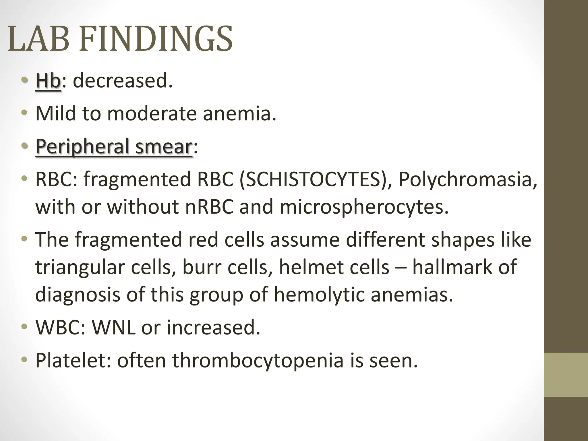
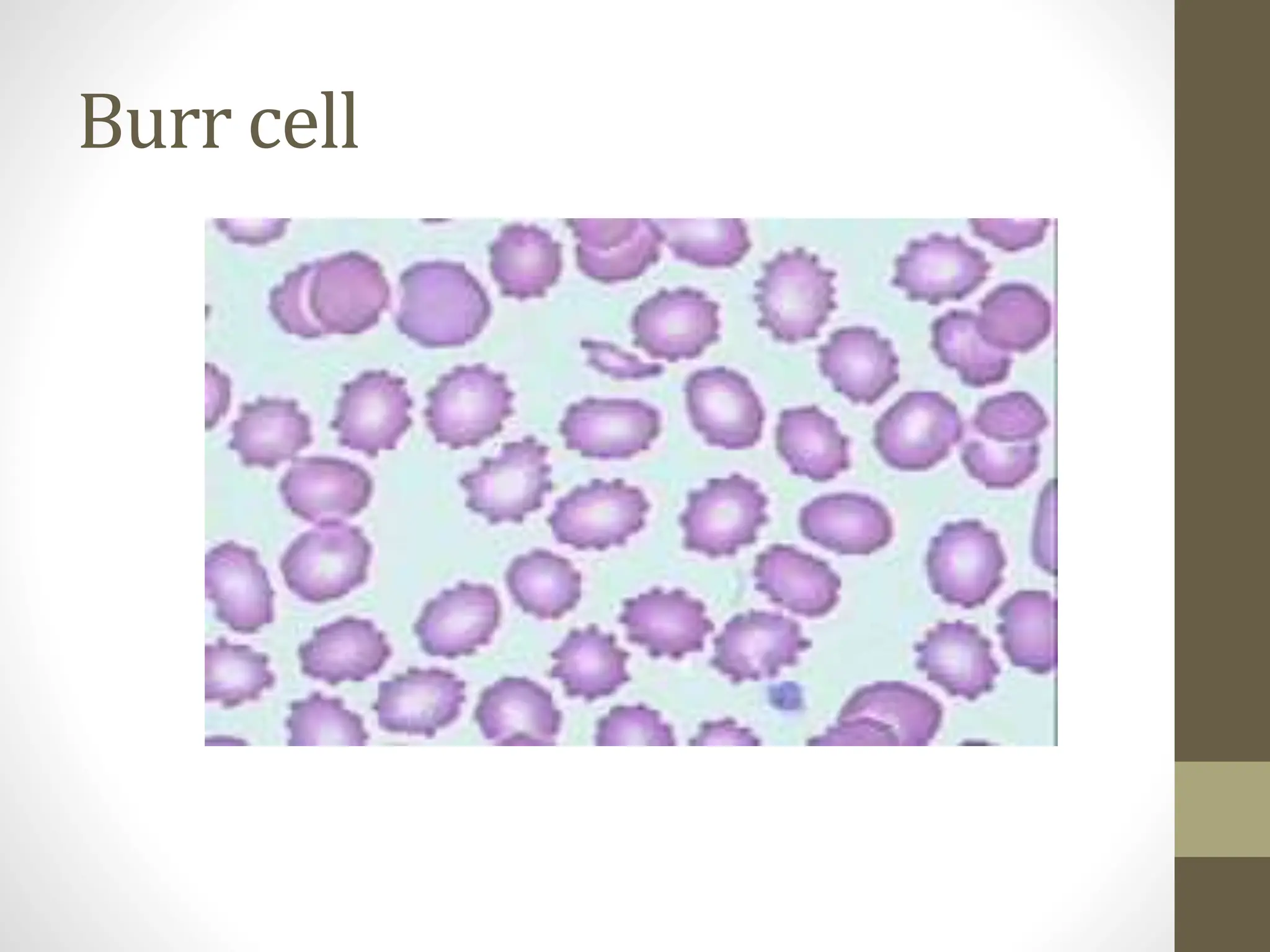
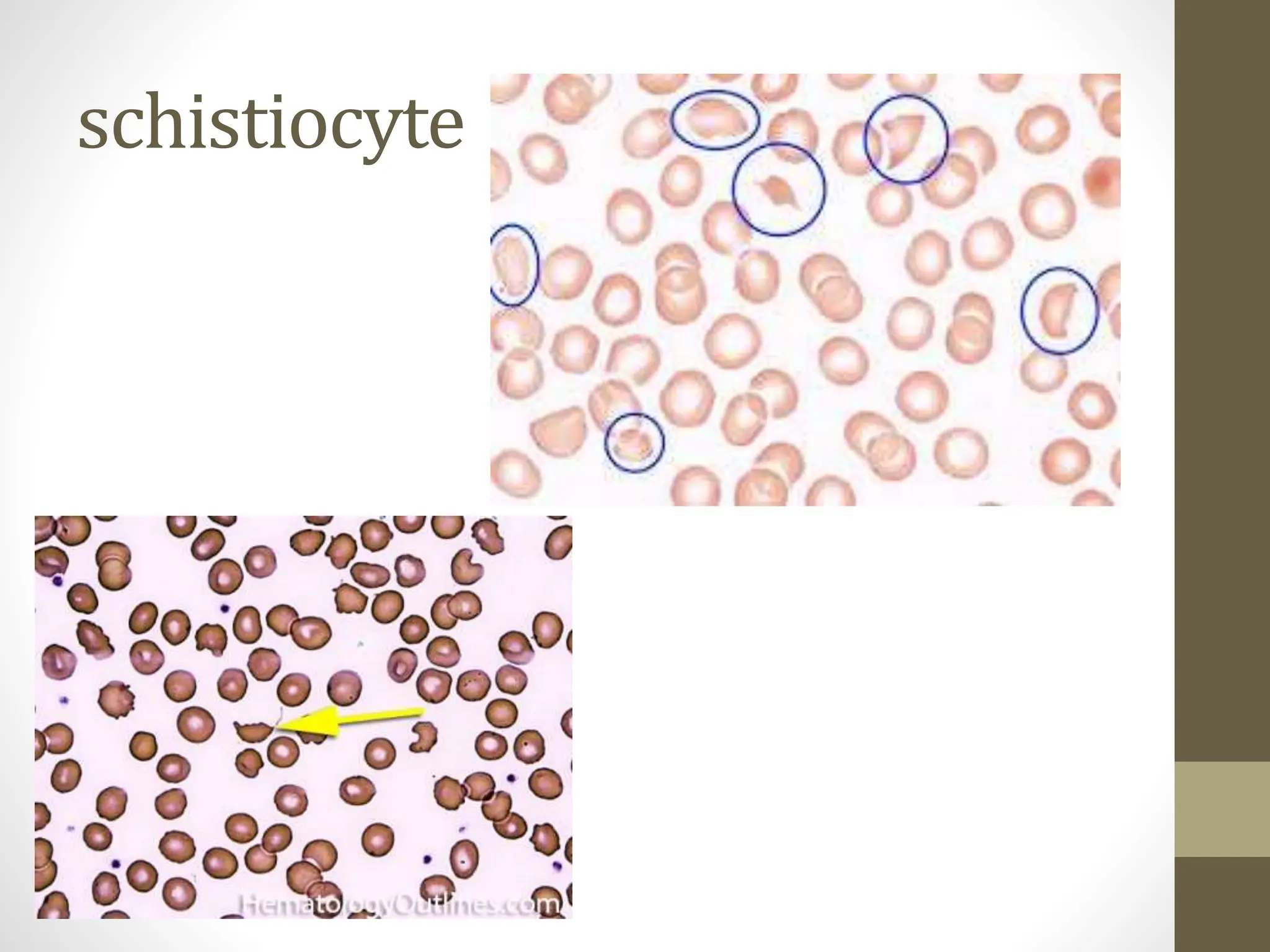
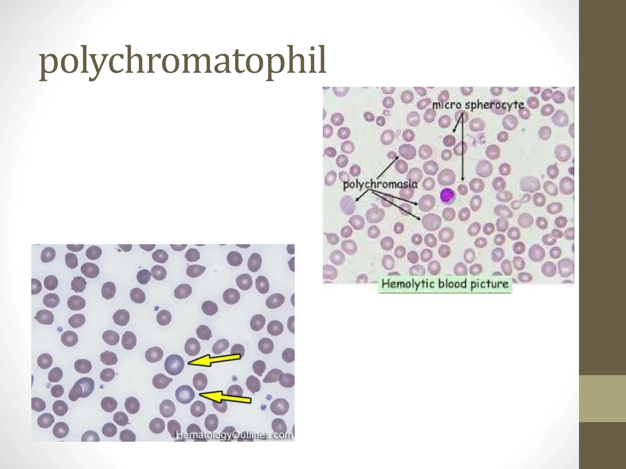
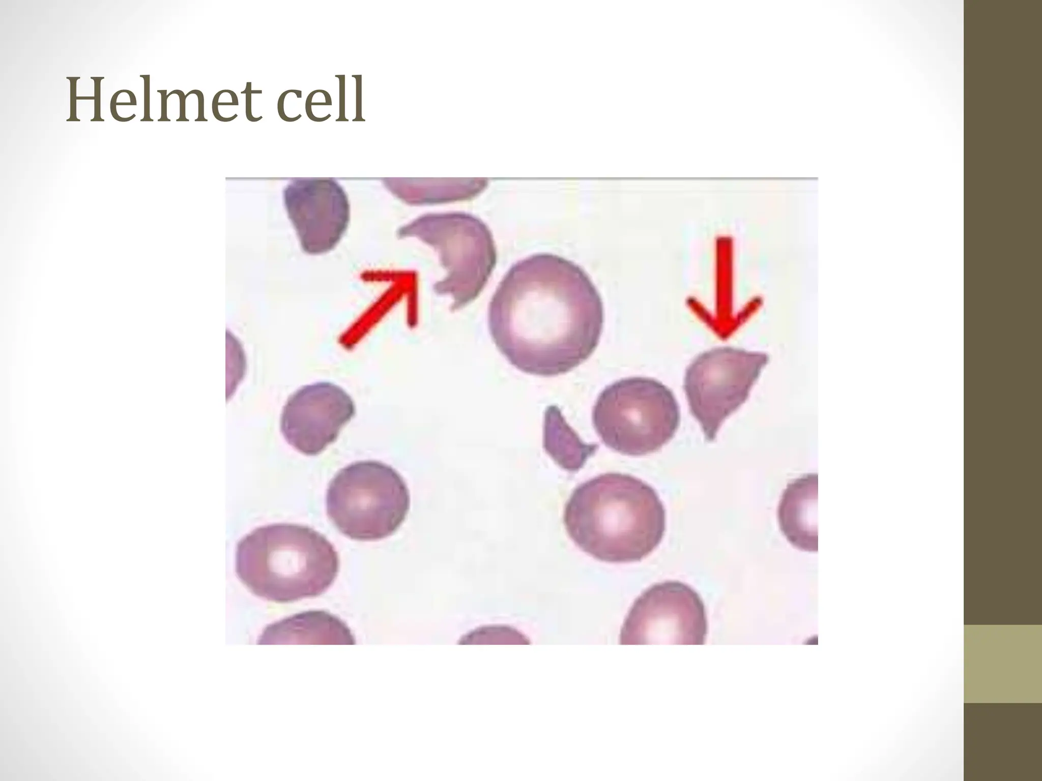
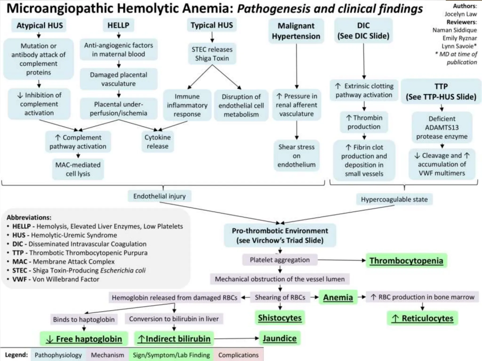
2. FRAGMENTATION SYNDROME
β’ Red cells when subjected to physical or
mechanical trauma in the circulation may
undergo fragmentation.
β’ This causes intravascular hemolysis and results in
hemolytic anemia.
β’ These are known as FRAGMENTATION
SYNDROME.
3. CLASSIFICATION
β’ Hemolytic anemia resulting from trauma to RBC
is classified according to the site of hemolysis:
β’ MACROANGIOPATHIC (large vessels)
β’ MICROANGIOPATHIC (capillaries)
4. MACROANGIOPATHIC
HEMOLYTIC ANEMIA
β’ Results most often due to direct red cell trauma
from abnormal vascular surface.
β’ Prosthetic heart valve, synthetic vascular graft.
β’ Known as CARDIAC HEMOLYTIC ANEMIA.
5. MIROANGIOPATHIC
HEMOLYTIC ANEMIA
β’ Results from abnormal luminal narrowing in
microcirculation, most often due to deposition of
fibrin and platelets that cause turbulent blood
flow.
6. CAUSES OF MAHA
β’ DISSEMINATED INTRAVASCULAR COAGULATION
β’ THROMBOTIC THROMBOCYTOPENIC PURPURA
β’ HEMOLYTIC UREMIC SYNDROME
β’ MALIGNANT HYPERTENSION
β’ SYSTEMIC LUPUS ERYTHEMATOSUS
β’ DISSEMINATED CANCER
β’ HELLP SYNDROME β HEMOLYSIS with ELEVATED
LIVER enzymes and LOW PLATELET
7. LAB FINDINGS
β’ Hb: decreased.
β’ Mild to moderate anemia.
β’ Peripheral smear:
β’ RBC: fragmented RBC (SCHISTOCYTES), Polychromasia,
with or without nRBC and microspherocytes.
β’ The fragmented red cells assume different shapes like
triangular cells, burr cells, helmet cells β hallmark of
diagnosis of this group of hemolytic anemias.
β’ WBC: WNL or increased.
β’ Platelet: often thrombocytopenia is seen.
8. Usually refers to
the polychromatophilic RBC which is less mature
non-nucleated red blood cell with a light
basophilic (light blue-purple) color on peripheral
smear which is due to the presence of cytoplasmic
RNA. Increased numbers may be seen in certain
anemias such as hemolytic anemia in which earlier
RBCs are being released by the reactive bone
marrow in response to the anemia with the earlier
release of these less mature RBCs into circulation.
Special stain can highlight the RNA molecules in
these cells and on the special stain these cell are
known as reticulocytes.
9. These are irregularly shaped assymetrical
fragmented RBCs that may have several
morphologic forms (e.g. Helmet cells, etc.). It is
usually the result of mechanical disruption of the
RBCs and many of which are generated by getting
stuck to fibrin strands within the vessel wall
(intravascular destruction) which may be
secondary to a Microangiopathic
hemolytic anemia such as HUS, TTP and DIC or a
fibrin-independent destructive path such as being
broken up by a mechanical heart valve.
10. Small round RBCs with no
central pallor that may be
seen in in blood smears from
patients with immune-
mediated hemolytic anemia,
burn injuries, and hereditary
spherocytosis.
15. MARCH HEMOGLOBINURIA
β’ This hemolytic disorder develops in athelets and
soldiers following long distance running or
marching, prolonged walking and vigorous
exercise.
β’ Mechanical destruction of red cells occurs in the
capillaries of the feet and results in transient
hemoglobinemia and hemoglobinuria.