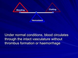
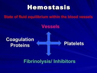
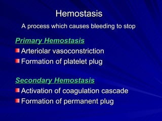
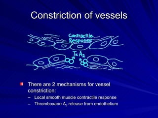
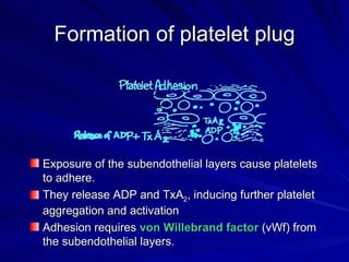
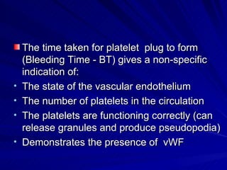
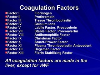
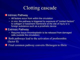
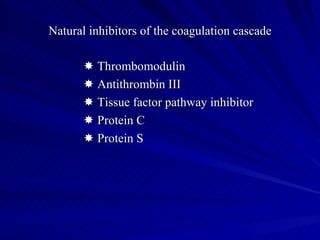
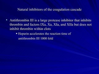
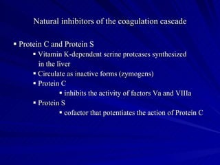
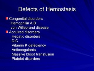
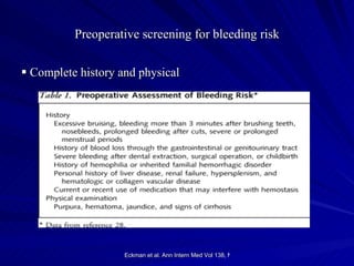
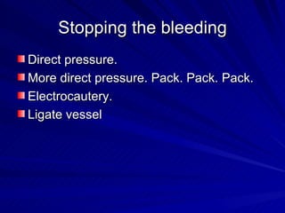
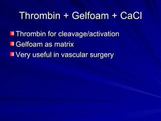
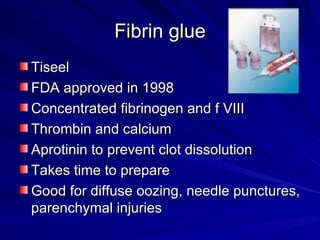
Hemostasis in the Surgical
        Patient

    Amr Aborahma , MD
  Lecturer of Vascular Surgery
WHAT ARE YOUR EXPECTATIONS ?
Hemostasis in the Surgical Patient
   Is It An Important Topic ?
From your point of view !!
Does All bleeding (eventually)
           stops ?
Bleeding                Clotting


                  Hemostasis


Under normal conditions, blood circulates
through the intact vasculature without
thrombus formation or haemorrhage
Hemostasis
State of fluid equilibrium within the blood vessels

                     Vessels

 Coagulation
  Proteins                        Platelets


           Fibrinolysis/ Inhibitors
Hemostasis
 A process which causes bleeding to stop

Primary Hemostasis
 Arteriolar vasoconstriction
 Formation of platelet plug

Secondary Hemostasis
 Activation of coagulation cascade
 Formation of permanent plug
Constriction of vessels


There are 2 mechanisms for vessel
constriction:
– Local smooth muscle contractile response
– Thromboxane A2 release from endothelium
Formation of platelet plug


Exposure of the subendothelial layers cause platelets
to adhere.
They release ADP and TxA2, inducing further platelet
aggregation and activation
Adhesion requires von Willebrand factor (vWf) from
the subendothelial layers.
The time taken for platelet plug to form
    (Bleeding Time - BT) gives a non-specific
    indication of:
•   The state of the vascular endothelium
•   The number of platelets in the circulation
•   The platelets are functioning correctly (can
    release granules and produce pseudopodia)
•   Demonstrates the presence of vWF
Coagulation Factors
 Factor I      Fibrinogen
 Factor II     Prothrombin
 Factor III    Tissue Thromboplastin
 Factor IV     Calcium Ions
 Factor V      Labile Factor, Proaccelerin
 Factor VII    Stable Factor, Proconvertin
 Factor VIII   Antihemophilic Factor
 Factor IX     Christmas Factor
 Factor X      Stuart-Prower Factor
 Factor XI     Plasma Thromboplastin Antecedent
 Factor XII    Hageman Factor
 Factor XIII   Fibrin Stabilizing Factor

All coagulation factors are made in the
liver, except for vWF
Clotting cascade
Intrinsic Pathway
– All factors occur from within the circulation
– in vivo, the pathway is triggered by exposure of "contact factors"
  to collagen or basement membrane at the site of injury or a
  foreign substance such as a prosthetic device
Extrinsic Pathway
– Requires tissue thromboplastin to be released from damaged
  cells (outside the circulation)
 Both pathways lead to the activation of prothrombin
(factor II)
 Final common pathway converts fibrinogen to fibrin
Clotting cascade
Natural inhibitors of the coagulation cascade

       Thrombomodulin
       Antithrombin III
       Tissue factor pathway inhibitor
       Protein C
       Protein S
Which is moving?
Natural inhibitors of the coagulation cascade

 Antithrombin III is a large protease inhibitor that inhibits
  thrombin and factors IXa, Xa, XIa, and XIIa but does not
  inhibit thrombin within clots
        Heparin accelerates the reaction time of
         antithrombin III 1000 fold
Natural inhibitors of the coagulation cascade

 Protein C and Protein S
       Vitamin K-dependent serine proteases synthesized
        in the liver
       Circulate as inactive forms (zymogens)
       Protein C
                 inhibits the activity of factors Va and VIIIa
       Protein S
                 cofactor that potentiates the action of Protein C
Take A Break !!!
Watch this video
Defects of Hemostasis
Congenital disorders
 Hemophilia A,B
 von Willebrand disease
Acquired disorders
  Hepatic disorders
  DIC
  Vitamin K defeciency
  Anticoagulants
  Massive blood transfusion
  Platelet disorders
Preoperative screening for bleeding risk

 Complete history and physical


                    Eckman et al. Ann Intern Med Vol 138, No 5
Preoperative screening for bleeding risk

 Incidence of a significant hereditary deficiency of a
   coagulation factor is low (1 per 10,000-40,000)
         approximately 1/3 of these are asymptomatic

 Acquired deficiencies of factors should be suspected in the
   presence of advance hepatic disease, malabsorption, or
   malnutrition
Hemostasis Screening Tests

•    Bleeding Time
•    Clotting Time
                              -Vascular
     Platelet Count           -Platelet
     PT                       -Coagulation factors
                              -Fibrinolysis
     APTT
     TT
     Euglobulin Clot Lysis Time
     D = Dimer
Clinical testing and preoperative screening

 Prothrombin time (PT)
    measure extrinsic and common pathways
    affected by low concentrations of fibrinogen, prothrombin
   and factors II, V, VII, X

 Activated partial thomboplastin time (aPTT)
    measures intrinsic and common pathways
    deficiencies in all clotting factors except factors VII and
   XIII may prolong the aPTT
Surgical hemostasis
Stopping the bleeding
Direct pressure.
More direct pressure. Pack. Pack. Pack.
Electrocautery.
Ligate vessel
Methylcellulose

Gelfoam
– Absorbable
– Liquefies in 2-5
  days
– Serves as a
  scaffold for
  coagulation
Oxidized regenerated cellulose
Surgicel
 – Binds platelets and chemically precipitates
   fibrin
Microfibrillar collagen
Decellularized bovine source
Stimulates latelet adhesion
Stops venous ooze
Absorbed in 90 days
Thrombin + Gelfoam + CaCl
Thrombin for cleavage/activation
Gelfoam as matrix
Very useful in vascular surgery
Fibrin glue
Tiseel
FDA approved in 1998
Concentrated fibrinogen and f VIII
Thrombin and calcium
Aprotinin to prevent clot dissolution
Takes time to prepare
Good for diffuse oozing, needle punctures,
parenchymal injuries
Questions
What is your feedback?
Thank You


Email: amr.aborahma@gmail.com

02a Surgical hemostasis

  • 1.
    Hemostasis in theSurgical Patient Amr Aborahma , MD Lecturer of Vascular Surgery
  • 2.
    WHAT ARE YOUREXPECTATIONS ?
  • 4.
    Hemostasis in theSurgical Patient Is It An Important Topic ?
  • 5.
    From your pointof view !!
  • 6.
    Does All bleeding(eventually) stops ?
  • 7.
    Bleeding Clotting Hemostasis Under normal conditions, blood circulates through the intact vasculature without thrombus formation or haemorrhage
  • 8.
    Hemostasis State of fluidequilibrium within the blood vessels Vessels Coagulation Proteins Platelets Fibrinolysis/ Inhibitors
  • 9.
    Hemostasis A processwhich causes bleeding to stop Primary Hemostasis Arteriolar vasoconstriction Formation of platelet plug Secondary Hemostasis Activation of coagulation cascade Formation of permanent plug
  • 11.
    Constriction of vessels Thereare 2 mechanisms for vessel constriction: – Local smooth muscle contractile response – Thromboxane A2 release from endothelium
  • 12.
    Formation of plateletplug Exposure of the subendothelial layers cause platelets to adhere. They release ADP and TxA2, inducing further platelet aggregation and activation Adhesion requires von Willebrand factor (vWf) from the subendothelial layers.
  • 13.
    The time takenfor platelet plug to form (Bleeding Time - BT) gives a non-specific indication of: • The state of the vascular endothelium • The number of platelets in the circulation • The platelets are functioning correctly (can release granules and produce pseudopodia) • Demonstrates the presence of vWF
  • 14.
    Coagulation Factors FactorI Fibrinogen Factor II Prothrombin Factor III Tissue Thromboplastin Factor IV Calcium Ions Factor V Labile Factor, Proaccelerin Factor VII Stable Factor, Proconvertin Factor VIII Antihemophilic Factor Factor IX Christmas Factor Factor X Stuart-Prower Factor Factor XI Plasma Thromboplastin Antecedent Factor XII Hageman Factor Factor XIII Fibrin Stabilizing Factor All coagulation factors are made in the liver, except for vWF
  • 15.
    Clotting cascade Intrinsic Pathway –All factors occur from within the circulation – in vivo, the pathway is triggered by exposure of "contact factors" to collagen or basement membrane at the site of injury or a foreign substance such as a prosthetic device Extrinsic Pathway – Requires tissue thromboplastin to be released from damaged cells (outside the circulation) Both pathways lead to the activation of prothrombin (factor II) Final common pathway converts fibrinogen to fibrin
  • 16.
  • 17.
    Natural inhibitors ofthe coagulation cascade  Thrombomodulin  Antithrombin III  Tissue factor pathway inhibitor  Protein C  Protein S
  • 18.
  • 19.
    Natural inhibitors ofthe coagulation cascade  Antithrombin III is a large protease inhibitor that inhibits thrombin and factors IXa, Xa, XIa, and XIIa but does not inhibit thrombin within clots  Heparin accelerates the reaction time of antithrombin III 1000 fold
  • 20.
    Natural inhibitors ofthe coagulation cascade  Protein C and Protein S  Vitamin K-dependent serine proteases synthesized in the liver  Circulate as inactive forms (zymogens)  Protein C  inhibits the activity of factors Va and VIIIa  Protein S  cofactor that potentiates the action of Protein C
  • 21.
    Take A Break!!! Watch this video
  • 23.
    Defects of Hemostasis Congenitaldisorders Hemophilia A,B von Willebrand disease Acquired disorders Hepatic disorders DIC Vitamin K defeciency Anticoagulants Massive blood transfusion Platelet disorders
  • 25.
    Preoperative screening forbleeding risk  Complete history and physical Eckman et al. Ann Intern Med Vol 138, No 5
  • 26.
    Preoperative screening forbleeding risk  Incidence of a significant hereditary deficiency of a coagulation factor is low (1 per 10,000-40,000)  approximately 1/3 of these are asymptomatic  Acquired deficiencies of factors should be suspected in the presence of advance hepatic disease, malabsorption, or malnutrition
  • 27.
    Hemostasis Screening Tests • Bleeding Time • Clotting Time -Vascular Platelet Count -Platelet PT -Coagulation factors -Fibrinolysis APTT TT Euglobulin Clot Lysis Time D = Dimer
  • 28.
    Clinical testing andpreoperative screening  Prothrombin time (PT)  measure extrinsic and common pathways  affected by low concentrations of fibrinogen, prothrombin and factors II, V, VII, X  Activated partial thomboplastin time (aPTT)  measures intrinsic and common pathways  deficiencies in all clotting factors except factors VII and XIII may prolong the aPTT
  • 29.
  • 30.
    Stopping the bleeding Directpressure. More direct pressure. Pack. Pack. Pack. Electrocautery. Ligate vessel
  • 31.
    Methylcellulose Gelfoam – Absorbable – Liquefiesin 2-5 days – Serves as a scaffold for coagulation
  • 32.
    Oxidized regenerated cellulose Surgicel – Binds platelets and chemically precipitates fibrin
  • 33.
    Microfibrillar collagen Decellularized bovinesource Stimulates latelet adhesion Stops venous ooze Absorbed in 90 days
  • 34.
    Thrombin + Gelfoam+ CaCl Thrombin for cleavage/activation Gelfoam as matrix Very useful in vascular surgery
  • 35.
    Fibrin glue Tiseel FDA approvedin 1998 Concentrated fibrinogen and f VIII Thrombin and calcium Aprotinin to prevent clot dissolution Takes time to prepare Good for diffuse oozing, needle punctures, parenchymal injuries
  • 36.
  • 37.
    What is yourfeedback?
  • 39.