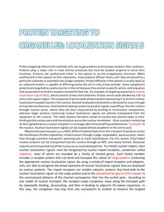
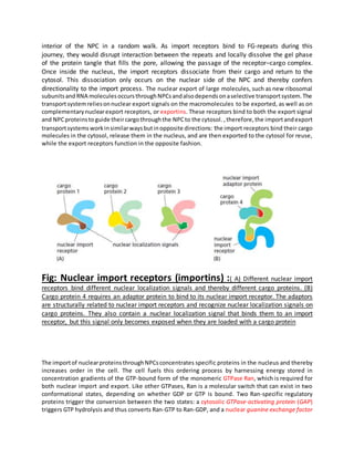
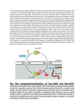
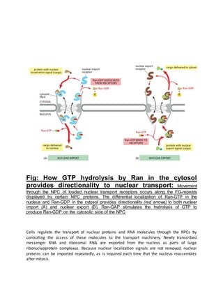
Proteins must be properly located within cells to carry out their functions. Protein targeting refers to how cells transport proteins to the correct locations after synthesis. There are several mechanisms for protein targeting. Some proteins diffuse through the cytosol and bind to receptors at their destination site, while others contain targeting sequences like nuclear localization signals that bind nuclear transport receptors to be actively transported into the nucleus. The import and export of proteins between the cytosol and nucleus is directed by gradients of Ran-GTP and Ran-GDP concentrations established by regulatory proteins localized to different cellular compartments. This compartmentalization of Ran states provides directionality to nuclear transport.
CLONAL SELECTION THEORY IS AN SCIENTIFIC THEORY IN IMMUNOLOGY THAT EXPALINS THE FUNCTION OF CELLS OF THE IMMUNE SYSTEM IN RESPONSE TO SPECIFIC ANTIGEN INVADING THE BODY.
I have tried to make a precise presentation on protein transport, targeting and sorting into organelle's other than nucleus. Hope this might help you. Comments are welcome.
Protein targeting or protein sorting is the biological mechanism by which proteins are transported to their appropriate destinations in the cell or outside it. Proteins can be targeted to the inner space of an organelle, different intracellular membranes, plasma membrane, or to exterior of the cell via secretion.
Protein targeting or protein sorting is the mechanism by which a cell transports to the appropriate positions in the cell or outside of it. Both in prokaryotes and eukaryotes, newly synthesized proteins must be delivered to a specific sub-cellular location or exported from the cell for correct activity. This phenomenon is called protein targeting. Protein targeting is necessary for proteins that are destined to work outside the cytoplasm.This delivery process is carried out based on information contained in the protein itself. Correct sorting is crucial for the cell; errors can lead to diseases. In 1970, Günter Blobel conducted experiments on the translocation of proteins across membranes. He was awarded the 1999 Nobel Prize for his findings. He discovered that many proteins have a signal sequence, that is, a short amino acid sequence at one end that functions like a postal code for the target organelle.
HAPTENS ARE LOW MOLECULAR WEIGHT COMPOUND, MOSTLY SMALL ORGANIC MOLECULES THAT ARE ANTIGENIC BUT NOT IMMUNOGENIC. THEY CAN BIND TO ANTIBODIES BY THEMSELVES BUT THEY ARE NOT RECOGNISED BY THE IMMUNE CELLS
How are macromolecules transported across the nuclear envelope Desc.pdfkellenaowardstrigl34
How are macromolecules transported across the nuclear envelope? Describe the structure that
regulates nuclear membrane permeability.
Solution
The transportation of molecules in and out of the nucleus is controlled by the nuclear pore
complex(NPC). Small molecules move through the nuclear membrane with ease but for
macromolecules like proteins, RNA etc, they require an association with transportation factors
like importins, karyopherins and exportins.
Proteins that need to be carried into the nucleus have nuclear localization signals (NLS) which
bind to the importins. NLS is a sequence of Amino acids which act as a tag. They are mostly
hydrophilic but hydrophobic sequences have also been documented.
Nuclear Import:
Importins bind to the macromolecule to be transported in the cytoplasm itself. The complex then
interacts with the NPC and passes through the channel. Once inside the nucleus, it interacts with
Ran-GTP and the importin dissociates from the macromolecule it was carrying. Then the
importin-Ran-GTP complex is transported to the cytoplasm where it is separated from importin
by the Ran Binding Protein (RanBP). After being separated from importin, GTPase activating
protein(GAP) binds with Ran-GTP and induces hydrolysis of GTP to GDP. The Ran-GDP
produced binds with the nuclear transport factor(NUTF2) and then returns to the nucleoplasm.
Inside the nucleus, Ran-GDP interacts with guanine nucleotide exchange factor(GEF) which
replaces GDP with GTP and Ran-GTP is born again to repeat the cycle.
Nuclear Export:
In the nucleoplasm, exportin binds the macromolecule to be transported outside the nucleus and
Ran-GTP. The complex diffuses through the pore to the cytoplasm where it dissociates. Ran-
GTP binds to GAP and hydrolysis resulting in Ran-GDP. Ran-GDP is returned to the nucleus
where it exchanges its ligand for GTP.
Nuclear Pore:
The nuclear pore complex is made of proteins called nucleoporins. Around 50% of nucleoporins
contains solenoid protein domains (alpha or beta-propeller fold) and the rest might have both as
separate structural domains. Each NPS has about 456 individual protein molecules with 30
distinct nucleoproteins. These are highly flexible proteins that lack an ordered secondary
structure..
Protein targeting or protein sorting is the biological mechanism by which proteins are transported to their appropriate destinations in the cell or outside it. Proteins can be targeted to the inner space of an organelle, different intracellular membranes, plasma membrane, or to exterior of the cell via secretion.
Ran is Ras-related nuclear protein or GTP-binding nuclear protein. I.pdfanand1213
Ran is Ras-related nuclear protein or GTP-binding nuclear protein. It is a small protein that is
involved in transport of large molecules in and out of the cell nucleus. Ran can exist in two
states, either bound to GDP or bound to GTP. When bound to GTP, it is capable of binding
karyopherins; the transport channels importins and exportins of the nuclear membrane.
The proteins importins bind molecules to be transported into the nucleus. These molecules have
nuclear localization signals exposed on them. Importin- binds to NLS sequence, which then
binds to importin-. The complex thus formed passes through the nuclear pore. Inside the nucleus,
RanGTP binds to importin- and displaced it. Another protein called CAS which is an exportin
binds RanGTP and displaces importin-, releasing the macromolecules.
The importin-RanGTP and importin – CAS-RanGTP complexes diffuse back to cytoplasm. The
GTP is hydrolyzed to GDP, which causes the release of importin- and importin- which
participates in another cycle of import around.
Solution
Ran is Ras-related nuclear protein or GTP-binding nuclear protein. It is a small protein that is
involved in transport of large molecules in and out of the cell nucleus. Ran can exist in two
states, either bound to GDP or bound to GTP. When bound to GTP, it is capable of binding
karyopherins; the transport channels importins and exportins of the nuclear membrane.
The proteins importins bind molecules to be transported into the nucleus. These molecules have
nuclear localization signals exposed on them. Importin- binds to NLS sequence, which then
binds to importin-. The complex thus formed passes through the nuclear pore. Inside the nucleus,
RanGTP binds to importin- and displaced it. Another protein called CAS which is an exportin
binds RanGTP and displaces importin-, releasing the macromolecules.
The importin-RanGTP and importin – CAS-RanGTP complexes diffuse back to cytoplasm. The
GTP is hydrolyzed to GDP, which causes the release of importin- and importin- which
participates in another cycle of import around..
Describe the role signal sequences play in protein localization. NES.pdffippsximenaal85949
Describe the role signal sequences play in protein localization. NES, NJS, Stop start, ER signal
sequences Describe the process of events involved in ligand production to exocytosis.
PowerPointslide 6 plus written details Post transcriptional modifications. Discuss 5\'capping,
splicing, poly A tail Describe three different mechanisms of signal termination Inhibitors -
extracellular, receptor inhibitors, internalization phosphorylation/dephosphorylation Give an
account of the role vesicle tethering plays in endocytosis. SNARES V and T, Rab effectors,
tethering docking fusion Protein glycosylation, discuss, its importance and the process of
glycosylation Occurs in ER, modified in Golgi, etc... Autocrine, paracrine and endocrine.
Discuss the differences between these signaling mechanisms. Write an account how signaling
can be terminated at the level of receptors Different cells can react differently to the same signal.
Discuss Example acetylcholine Outline the process of plasma membrane receptor production to
its final destination Two major populations of ribosome exist in our cells. Discuss
Solution
Ques-1:
The role of signal seqeunecs in protein localization:
ER signal sequences and stop-start:
Signal peptidase normally localized in the “endoplasmic reticulum” of eukaryotic cell. These
serine proteases digest the “nascent secretory polypeptides” after translation & post & co-
translational modifications. These signal peptidase enzymes are going to cleave signal peptides
of membrane proteins at their N-terminals.
Signal sequence of \"6 to 10 amino acid length\" normally cleaved by signal peptidases during or
after completion of translocation from ribosome & before entering into the endoplasmic
reticulum
Cotranslational translocation is mediated by signal recognition particle (SRP) & it is a 11S
ribonucleoprotien essential for translocation of nascent polypeptide to the “ER lumen”. This SRP
is formed of a nucleoprotein with 7S RNA, 11S & combines with several proteins to form the
eukaryotic signal recognition particle. The nascent polypeptide is emerged from the ribosomes,
followed by “recognition of signal sequence & bound by signal recognition particle(SRP), a
hydrophobic residue with 6 polypeptides 7SL RNA.
Therefore, when this SRP sequence exits from the ribosome, the nascent polypeptide stops being
translocated into the ER lumen. Signal recognition particle has bound by a signal sequence and
flowed by slow protein synthesis in the translation. This SRP further is going to bind to the
location near by endoplasmic reticulum followed by release of signal recognition particle but the
growing polypeptide is in the form of protein-ribosome complex that is at the exact location for
further protein translocation through Golgi-vesicle translocation channel.
The nuclear localization signal sequence (NLS) is mainly associated with “tags” in which amino
acids or proteins are imported into the nucleus using Ran-GTPases activity by nuclear transport.
So .
Proteins destined for secretion, integration in the plasma membrane, or inclusion in lysosomes generally share pathway that begins in the endoplasmic reticulum. Proteins destined for mitochondria, chloroplasts, or the nucleus use three separate mechanisms. And proteins destined for the cytosol simply remain where they are synthesized.
Introduction
Protein synthesis
Synthesis of secretory proteins on membrane-bound ribosomes
Processing of newly synthesized proteins in the ER
Synthesis of integral membrane protein on membrane bound ribosomes
Maintenance of membrane asymmetry
Conclusion
Reference
Professional air quality monitoring systems provide immediate, on-site data for analysis, compliance, and decision-making.
Monitor common gases, weather parameters, particulates.
Slide 1: Title Slide
Extrachromosomal Inheritance
Slide 2: Introduction to Extrachromosomal Inheritance
Definition: Extrachromosomal inheritance refers to the transmission of genetic material that is not found within the nucleus.
Key Components: Involves genes located in mitochondria, chloroplasts, and plasmids.
Slide 3: Mitochondrial Inheritance
Mitochondria: Organelles responsible for energy production.
Mitochondrial DNA (mtDNA): Circular DNA molecule found in mitochondria.
Inheritance Pattern: Maternally inherited, meaning it is passed from mothers to all their offspring.
Diseases: Examples include Leber’s hereditary optic neuropathy (LHON) and mitochondrial myopathy.
Slide 4: Chloroplast Inheritance
Chloroplasts: Organelles responsible for photosynthesis in plants.
Chloroplast DNA (cpDNA): Circular DNA molecule found in chloroplasts.
Inheritance Pattern: Often maternally inherited in most plants, but can vary in some species.
Examples: Variegation in plants, where leaf color patterns are determined by chloroplast DNA.
Slide 5: Plasmid Inheritance
Plasmids: Small, circular DNA molecules found in bacteria and some eukaryotes.
Features: Can carry antibiotic resistance genes and can be transferred between cells through processes like conjugation.
Significance: Important in biotechnology for gene cloning and genetic engineering.
Slide 6: Mechanisms of Extrachromosomal Inheritance
Non-Mendelian Patterns: Do not follow Mendel’s laws of inheritance.
Cytoplasmic Segregation: During cell division, organelles like mitochondria and chloroplasts are randomly distributed to daughter cells.
Heteroplasmy: Presence of more than one type of organellar genome within a cell, leading to variation in expression.
Slide 7: Examples of Extrachromosomal Inheritance
Four O’clock Plant (Mirabilis jalapa): Shows variegated leaves due to different cpDNA in leaf cells.
Petite Mutants in Yeast: Result from mutations in mitochondrial DNA affecting respiration.
Slide 8: Importance of Extrachromosomal Inheritance
Evolution: Provides insight into the evolution of eukaryotic cells.
Medicine: Understanding mitochondrial inheritance helps in diagnosing and treating mitochondrial diseases.
Agriculture: Chloroplast inheritance can be used in plant breeding and genetic modification.
Slide 9: Recent Research and Advances
Gene Editing: Techniques like CRISPR-Cas9 are being used to edit mitochondrial and chloroplast DNA.
Therapies: Development of mitochondrial replacement therapy (MRT) for preventing mitochondrial diseases.
Slide 10: Conclusion
Summary: Extrachromosomal inheritance involves the transmission of genetic material outside the nucleus and plays a crucial role in genetics, medicine, and biotechnology.
Future Directions: Continued research and technological advancements hold promise for new treatments and applications.
Slide 11: Questions and Discussion
Invite Audience: Open the floor for any questions or further discussion on the topic.
Deep Behavioral Phenotyping in Systems Neuroscience for Functional Atlasing a...Ana Luísa Pinho
Functional Magnetic Resonance Imaging (fMRI) provides means to characterize brain activations in response to behavior. However, cognitive neuroscience has been limited to group-level effects referring to the performance of specific tasks. To obtain the functional profile of elementary cognitive mechanisms, the combination of brain responses to many tasks is required. Yet, to date, both structural atlases and parcellation-based activations do not fully account for cognitive function and still present several limitations. Further, they do not adapt overall to individual characteristics. In this talk, I will give an account of deep-behavioral phenotyping strategies, namely data-driven methods in large task-fMRI datasets, to optimize functional brain-data collection and improve inference of effects-of-interest related to mental processes. Key to this approach is the employment of fast multi-functional paradigms rich on features that can be well parametrized and, consequently, facilitate the creation of psycho-physiological constructs to be modelled with imaging data. Particular emphasis will be given to music stimuli when studying high-order cognitive mechanisms, due to their ecological nature and quality to enable complex behavior compounded by discrete entities. I will also discuss how deep-behavioral phenotyping and individualized models applied to neuroimaging data can better account for the subject-specific organization of domain-general cognitive systems in the human brain. Finally, the accumulation of functional brain signatures brings the possibility to clarify relationships among tasks and create a univocal link between brain systems and mental functions through: (1) the development of ontologies proposing an organization of cognitive processes; and (2) brain-network taxonomies describing functional specialization. To this end, tools to improve commensurability in cognitive science are necessary, such as public repositories, ontology-based platforms and automated meta-analysis tools. I will thus discuss some brain-atlasing resources currently under development, and their applicability in cognitive as well as clinical neuroscience.
Seminar of U.V. Spectroscopy by SAMIR PANDASAMIR PANDA
Spectroscopy is a branch of science dealing the study of interaction of electromagnetic radiation with matter.
Ultraviolet-visible spectroscopy refers to absorption spectroscopy or reflect spectroscopy in the UV-VIS spectral region.
Ultraviolet-visible spectroscopy is an analytical method that can measure the amount of light received by the analyte.
(May 29th, 2024) Advancements in Intravital Microscopy- Insights for Preclini...Scintica Instrumentation
Intravital microscopy (IVM) is a powerful tool utilized to study cellular behavior over time and space in vivo. Much of our understanding of cell biology has been accomplished using various in vitro and ex vivo methods; however, these studies do not necessarily reflect the natural dynamics of biological processes. Unlike traditional cell culture or fixed tissue imaging, IVM allows for the ultra-fast high-resolution imaging of cellular processes over time and space and were studied in its natural environment. Real-time visualization of biological processes in the context of an intact organism helps maintain physiological relevance and provide insights into the progression of disease, response to treatments or developmental processes.
In this webinar we give an overview of advanced applications of the IVM system in preclinical research. IVIM technology is a provider of all-in-one intravital microscopy systems and solutions optimized for in vivo imaging of live animal models at sub-micron resolution. The system’s unique features and user-friendly software enables researchers to probe fast dynamic biological processes such as immune cell tracking, cell-cell interaction as well as vascularization and tumor metastasis with exceptional detail. This webinar will also give an overview of IVM being utilized in drug development, offering a view into the intricate interaction between drugs/nanoparticles and tissues in vivo and allows for the evaluation of therapeutic intervention in a variety of tissues and organs. This interdisciplinary collaboration continues to drive the advancements of novel therapeutic strategies.
2. Proteintargetingreferstothe methods cells use to get proteins to the proper location after synthesis.
Proteins play a major role in most cellular processes but must be located properly to serve their
functions. Proteins are synthesized either in the cytosol or on the endoplasmic reticulum. When
synthesized in the cytosol on free ribosomes, most proteins diffuse freely until they are bound to a
particularsubstrate or assemble intoalarger complex.Proteindiffusioninthe cytosol isusuallyrapid,so
an unbound protein is capable of diffusing across the cell in only a few seconds. Some cytoplasmic
proteinsare targetedtoa particularsite inthe cell because theycontainaspecific amino acid sequence
that causesthemto bindto receptorslocatedat that site. An example of targeting sequence is nuclear
localization signal (NLS), which consists of two short stretches of basic amino acids divided by a 10–11
aminoacid spacerregion.Thissequence of aminoacidsallowsaproteinpossessing it to bind to nuclear
localization receptorsfoundinthe nucleus.Nuclearlocalizationof proteins is believed to occur through
at leasttwomechanisms:small proteinslackingnuclearlocalizationsignals maydiffuse into the nucleus
through nuclear pores, where they are then sequestered by binding to intranuclear components,
whereas larger proteins containing nuclear localization signals are actively transported from the
cytoplasm to the nucleus. The latter process has been shown to involve two distinct steps in vitro:
bindingtothe nuclearpore and translocationacrossthe nuclear membrane. Once a protein containing
an NLS signal bindstoa nuclearreceptoritis nolongerable tofreelydiffuse andbecomes "localized" to
the nucleus. Nuclear localization signals can be located almost anywhere in the amino acid.
Macromoleculartransportacross NPCs differs fundamentally from the transport of proteins across
the membranesof otherorganelles,inthatitoccurs through a large, expandable, aqueous pore, rather
than through a protein transporter spanning one or more lipid bilayers. For this reason, fully folded
nuclear proteins can be transported into the nucleus through an NPC, and newly formed ribosomal
subunitsare transportedoutof the nucleusas an assembledparticle. To initiate nuclear import, most
nuclear localization signals must be recognized by nuclear import receptors, sometimes called
importins, most of which are encoded by a family of related genes. Each family member
encodes a receptor protein that can bind and transport the subset of cargo proteins containing
the appropriate nuclear localization signal .By using a variety of import receptors and adaptors,
cells are able to recognize the broad repertoire of nuclear localization signals that are displayed
on nuclear proteins. The import receptors are soluble cytosolic proteins that bind both to the
nuclear localization signal on the cargo protein and to the phenylalanine-glycine (FG) repeats in
the unstructured domains of the channel nucleoporins that line the central pore . According to
one model of nuclear transport, the receptor–cargo complexes move along the transport path
by repeatedly binding, dissociating, and then re-binding to adjacent FG-repeat sequences. In
this way, the complexes may hop from one nucleoporin to another to traverse the tangled
3. interior of the NPC in a random walk. As import receptors bind to FG-repeats during this
journey, they would disrupt interaction between the repeats and locally dissolve the gel phase
of the protein tangle that fills the pore, allowing the passage of the receptor–cargo complex.
Once inside the nucleus, the import receptors dissociate from their cargo and return to the
cytosol. This dissociation only occurs on the nuclear side of the NPC and thereby confers
directionality to the import process. The nuclear export of large molecules, such as new ribosomal
subunitsand RNA molecules occursthroughNPCsandalsodependsonaselective transportsystem.The
transportsystemreliesonnuclear export signals on the macromolecules to be exported, as well as on
complementarynuclearexport receptors, or exportins. These receptors bind to both the export signal
and NPCproteinsto guide theircargothroughthe NPCto the cytosol. ,therefore,the importandexport
transportsystems workinsimilarwaysbutinopposite directions: the import receptors bind their cargo
molecules in the cytosol, release them in the nucleus, and are then exported to the cytosol for reuse,
while the export receptors function in the opposite fashion.
Fig: Nuclear import receptors (importins) :( A) Different nuclear import
receptors bind different nuclear localization signals and thereby different cargo proteins. (B)
Cargo protein 4 requires an adaptor protein to bind to its nuclear import receptor. The adaptors
are structurally related to nuclear import receptors and recognize nuclear localization signals on
cargo proteins. They also contain a nuclear localization signal that binds them to an import
receptor, but this signal only becomes exposed when they are loaded with a cargo protein
The importof nuclearproteinsthroughNPCsconcentrates specific proteins in the nucleus and thereby
increases order in the cell. The cell fuels this ordering process by harnessing energy stored in
concentration gradients of the GTP-bound form of the monomeric GTPase Ran, which is required for
both nuclear import and export. Like other GTPases, Ran is a molecular switch that can exist in two
conformational states, depending on whether GDP or GTP is bound. Two Ran-specific regulatory
proteins trigger the conversion between the two states: a cytosolic GTPase-activating protein (GAP)
triggers GTP hydrolysis and thus converts Ran-GTP to Ran-GDP, and a nuclear guanine exchange factor
4. (GEF) promotesthe exchange of GDPfor GTP and thus convertsRan-GDP to Ran-GTP. Because Ran-GAP
is located in the cytosol and Ran-GEF is located in the nucleus where it is anchored to chromatin, the
cytosol contains mainly Ran-GDP, and the nucleus contains mainly Ran-GTP. This gradient of the two
conformational forms of Ran drives nuclear transport in the appropriate direction. Docking of nuclear
import receptors to FG-repeats on the cytosolic side of the NPC, for example, occurs whether or not
these receptors are loaded with appropriate cargo. Import receptors, facilitated by FG-repeat binding,
thenenterthe channel.If theyreachthe nuclearside of the pore complex, Ran-GTP binds to them, and,
if the receptorsarrive loadedwithcargomolecules, the Ran-GTPbindingcausesthe receptorstorelease
theircargo Because the Ran-GDPin the cytosol doesnotbindto import(orexport) receptors,unloading
occurs onlyon the nuclear side of the NPC. In this way, the nuclear localization of Ran-GTP creates the
directionality of the import process. Having discharged its cargo in the nucleus, the empty import
receptorwith Ran-GTPboundistransportedback throughthe pore complex to the cytosol. There, Ran-
GAP triggers Ran-GTP to hydrolyze its bound GTP, thereby converting it to Ran-GDP, which dissociates
fromthe receptor.The receptoristhenreadyfor anothercycle of nuclearimport. Nuclearexport occurs
by a similar mechanism, except that Ran-GTP in the nucleus promotes cargo binding to the export
receptor,rather than promoting cargo dissociation. Once the export receptor moves through the pore
to the cytosol, it encounters Ran-GAP, which induces the receptor to hydrolyze its GTP to GDP. As a
result,the exportreceptorreleasesbothitscargoand Ran-GDPinthe cytosol.Free exportreceptors are
then returned to the nucleus to complete the cycle.
Fig: The compartmentalization of Ran-GDP and Ran-GTP:
Localization of Ran-GDP in the cytosol and Ran-GTP in the nucleus results from the localization
of two Ran regulatory proteins: Ran GTPase activating protein (Ran-GAP) is located in the
cytosol, and Ran guanine nucleotide exchange factor (Ran-GEF) binds to chromatin and is
therefore located in the nucleus. Ran-GDP is imported into the nucleus by its own import
receptor, which is specific for the GDP-bound conformation of Ran. The Ran-GDP receptor is
structurally unrelated to the main family of nuclear transport receptors. However, it also binds
to FG-repeats in NPC channel nucleoporins.
5. Fig: How GTP hydrolysis by Ran in the cytosol
provides directionality to nuclear transport: Movement
through the NPC of loaded nuclear transport receptors occurs along the FG-repeats
displayed by certain NPC proteins. The differential localization of Ran-GTP in the
nucleus and Ran-GDP in the cytosol provides directionality (red arrows) to both nuclear
import (A) and nuclear export (B). Ran-GAP stimulates the hydrolysis of GTP to
produce Ran-GDP on the cytosolic side of the NPC
Cells regulate the transport of nuclear proteins and RNA molecules through the NPCs by
controlling the access of these molecules to the transport machinery. Newly transcribed
messenger RNA and ribosomal RNA are exported from the nucleus as parts of large
ribonucleoprotein complexes. Because nuclear localization signals are not removed, nuclear
proteins can be imported repeatedly, as is required each time that the nucleus reassembles
after mitosis.
6. -Molecular Biology of the Cell-Alberts, 6th edition
-MIN GAO AND DAVID M. KNIPE (1991), Distal Protein Sequences Can Affect the Function of a
Nuclear Localization Signal, MOLECULAR AND CELLULAR BIOLOGY, Vol. 12, No. 3