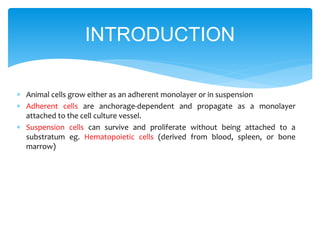
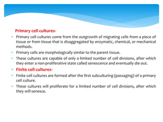
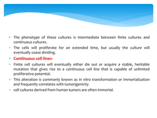
SlideShare a Scribd company logo
1 of 34
 Animal cells grow either as an adherent monolayer or in suspension
 Adherent cells are anchorage-dependent and propagate as a monolayer
attached to the cell culture vessel.
 Suspension cells can survive and proliferate without being attached to a
substratum eg. Hematopoietic cells (derived from blood, spleen, or bone
marrow)
INTRODUCTION
Primary cell cultures-
 Primary cell cultures come from the outgrowth of migrating cells from a piece of
tissue or from tissue that is disaggregated by enzymatic, chemical, or mechanical
methods.
 Primary cells are morphologically similar to the parent tissue.
 These cultures are capable of only a limited number of cell divisions, after which
they enter a non-proliferative state called senescence and eventually die out.
 Finite cell cultures-
 Finite cell cultures are formed after the first subculturing (passaging) of a primary
cell culture.
 These cultures will proliferate for a limited number of cell divisions, after which
they will senesce.
. The phenotype of these cultures is intermediate between finite cultures and
continuous cultures.
 The cells will proliferate for an extended time, but usually the culture will
eventually cease dividing.
 Continuous cell lines-
 Finite cell cultures will eventually either die out or acquire a stable, heritable
mutation that gives rise to a continuous cell line that is capable of unlimited
proliferative potential.
 This alteration is commonly known as in vitro transformation or immortalization
and frequently correlates with tumorigenicity
 cell cultures derived from human tumors are often immortal.
 Cell culture - Maintenance of dissociated cells in culture
 Tissue culture - Maintenance of tissue explants in culture
 Cell line - A culture that is subcultured beyond the initial primary culture phase
 Finite cell line - A cell line with a limited lifespan that eventually undergoes
senescence
 Continuous cell line - A cell line that is essentially immortal and continues
indefinitely
 Primary culture - The initial culture derived from in vivo material
 Clone - The progeny isolated from a single cell
 Immortalization - Enabling of cells to extend their life in culture
 Lag phase of growth - Initial phase of growth when cells are subcultured
GENERAL TERMS
 Log or exponential phase - The cells enter a period of exponential growth that
lasts until the entire growth surface is occupied or the cell concentration exceeds
the capacity of the medium.
 Stationary phase - Cell proliferation slows and stops.
 Decline phase - If the culture medium is not replaced and the cell number is not
reduced, the cells lose viability and their number decreases.
 Population doubling time - Time for cell number to double
 Cell banks - Repositories of cancer cell lines and related materials
 Substrate- The matrix on which a culture is grown
 Passage -Subculture of cells from one container to another
 Confluent - Situation wherein cells completely cover the substrate
 To ensure viability, genetic stability, and phenotypic stability, cell lines need to be
maintained in the exponential phase. This means that they need to be subcultured
on a regular basis before they enter the stationary growth phase, before a
monolayer becomes 100% confluent or before a suspension reaches its maximum
recommended cell density.
Laminar-Flow Hood-
• It is used to provide sterile environment and protect the laboratory worker
from exposure to aerosols from cell culture.
• Air is filtered through a HEPA (high efficiency particulate air) filter before
exiting the cabinet. They are classified at levels I, II, and III.
• Class I cabinets are the simplest and easiest to maintain but offer least sterile
protection to the cell culture.
• Class II cabinets are the most widely used for cell culture work and offer good
protection to both the operator and cell cultures since air passing over the
working area is HEPA filtered.
• Class III cabinets are completely sealed units and are used for more hazardous
types of work.
• Cabinet’s surface should be wiped with ethanol both before and after use.
• Cabinets are also equipped with a UV light that can be used to sterilize the
surfaces of the cabinet.
Class two type laminar flow
Inverted Microscope:
A simple inverted microscope is essential to look at cultures regularly to
detect morphological changes and the possibility of microbiological
contamination.
Humid CO2 Incubator
• A controlled atmosphere is achieved by
using a humidifying tray .
• It draws air from the incubator into a
sample chamber, determines the
concentration of CO2, and injects pure
CO2 into the incubator to make up any
deficiency.
• Air is circulated around the incubator by
natural convection or by using a fan to
keep both the CO2 level and the
temperature uniform.
Culture Vessels-
The choice of culture vessels is determined by
• the yield (number of cells) required
• whether the cell is grown in monolayer or suspension
• the sampling regime (i.e., are the samples to be collected simultaneously or at
intervals over a period of time)
Filter sterilization-
• Media that cannot be autoclaved must
be sterilized through a 0.22 μm pore
size membrane filter.
• Culture media, enzymes, hormones,
cofactors and bicarbonate buffers are
examples of non- autoclavable
substances.
• Cell Culture Media:
• Cell culture media are complex mixtures of salts, carbohydrates, vitamins,
amino acids, metabolic precursors, growth factors, hormones, and trace
elements.
• Carbohydrates are supplied in the form of glucose.
• The pH is maintained by one or more buffering systems; CO₂/sodium
bicarbonate, phosphate, and HEPES are the most common.
• Commonly used culture media include the following:
• Eagle’s basal medium (BME) has salt solution, non- essential amino acids, and
sodium pyruvate. It is formulated with a reduced sodium bicarbonate
concentration (1,500 mg/l) for use with 5% CO₂
• Dulbecco’s Modified Eagle’s Medium (DMEM) has twice the concentration of
amino acids and four times the amount of vitamins as EMEM.
• The original formulation contained 1,000 mg/L of glucose, but in the more
commonly used variations this amount was increased to 4,500 mg/L.
• Iscove’s Modified Dulbecco’s Medium (IMDM) was formulated for growth of
lymphocytes and hybridomas.
• Compared to DMEM, it has additional amino acids, vitamins and inorganic salts.
Potassium nitrate was substituted for ferric nitrate.
• It also contains HEPES and selenium, a reduced sodium bicarbonate
concentration (1,500 mg/L) for use with 5% CO₂.
• McCoy’s 5A and RPMI-1640 was originally used to grow Novikoff hepatoma cells
and will support the growth of primary cultures.
• RPMI-1640 is a modification of McCoy’s 5A and was developed for the long-term
culture of peripheral blood lymphocytes.
• Leibovitz’s L-15 is formulated for use without CO₂ incubation .
• The standard sodium bicarbonate/CO₂ buffering system is replaced by a
combination of phosphate buffers, free-base amino acids, higher levels of
sodium pyruvate, and galactose.
Media Formulations-
• Sodium bicarbonate and buffering
• Cells produce and require small amounts of carbon dioxide for growth and
survival.
• CO₂ dissolves freely into the medium and reacts with water to form carbonic
acid. As the cells metabolize and produce more CO₂, the pH of the medium
decreases as the chemical reaction below is driven to the right:
H₂O + CO₂ H₂CO₃ H+ + HCO₃–
• The optimal pH range of 7.2 to 7.4 can be maintained by supplementing the
medium with sodium bicarbonate and regulating the level of CO₂ in the
atmosphere above the medium as shown by the reaction below:
H₂O + CO₂ + NaHCO₃ H+ + Na+ + 2HCO₃–
• In tissue culture, cells are grown either in open systems (where there is free
exchange of the atmosphere immediately above the medium with the
atmosphere of the incubator) or in closed systems (where the two atmospheres
are kept separate).
• In closed systems the level of CO₂ is regulated by the metabolism of the cells.
The culture vessel must be sealed to retain any CO₂ generated by the cells.
• Consequently, closed systems provide additional protection against
contamination and have simpler incubator requirements than open systems.
• Closed systems usually require media with buffers based on Hanks’ balanced salt
solution having relatively low levels of sodium bicarbonate.
• In open systems, humidity (to reduce evaporation) and a means of regulating
CO₂ levels (if the culture medium contains sodium bicarbonate) are required
during incubation to maintain the pH of the culture medium.
• HEPES buffer
• HEPES and other organic buffers can be used with many cell lines to
effectively buffer the pH of the medium.
• HEPES increases the sensitivity of media to the phototoxic effects induced by
exposure to fluorescent Light.
• Phenol red
• Phenol red is used to monitor the pH of media.
• During cell growth, the medium changes color as it changes pH due to
metabolites released by the cells.
• At low pH levels, phenol red turns the medium yellow, while at higher pH
levels it turns the medium purple.
• For most tissue culture work (pH 7.4), the medium should be bright red.
• L-Glutamine
• L-Glutamine is an essential amino acid required by virtually all mammalian
and insect cells grown in culture.
• It is used for protein production, as an energy source, and in nucleic acid
metabolism.
Nonessential amino acids
• The inclusion of the other non-essential amino acids (alanine, asparagine, aspartic
acid, glycine, glutamic acid, proline, and serine) reduces the metabolic burden on
the cells allowing for an increase in cellular proliferation.
Sodium pyruvate
• Pyruvate may help in maintaining certain specialized cells, in clonal selection, in
reducing the serum concentration of the medium, and in reducing fluorescent
light-induced phototoxicity.
• Cellular metabolism of pyruvate produces carbon dioxide which is given off into
the atmosphere and becomes bicarbonate in the medium.
Antibiotics and Antimycotics
• Antibiotics and/or antimycotic agents are added to prevent contamination, as a
cure once contamination is found, to induce the expression of recombinant
proteins, or to maintain selective pressure on transfected cells.
• Routine use of antibiotics or antimycotics for cell culture is not recommended
unless they are specifically required.
• Antibiotics can mask contamination by mycoplasma and resistant bacteria.
Further, they can interfere with the metabolism of sensitive cells.
Serum
• Sera serve as a source for amino acids, proteins, vitamins , carbohydrates, lipids,
hormones, growth factors, minerals, and trace elements.
• Serum buffers the culture medium, inactivates proteolytic enzymes, increases
medium viscosity, and conditions the growth surface of the culture vessel.
• Sera from fetal and calf bovine sources are commonly used to support the
growth of cells in culture.
• Fetal serum is a rich source of growth factors and is appropriate for cell cloning
and for the growth of fastidious cells.
• Calf serum, because of its lower growth-promoting properties, is used in
contact-inhibition
• Attachment and Spreading Factor:
• Animal cells and extracellular matrix used in labs, like glass and plastic are both
negatively charged.
• Cross-linking with glycoproteins/ fibronectin or divalent cations Ca++ and Mg++
provide necessary electrostatic attraction for cells to bind to the matrix.
Disadvantages of serum
• Foetal calf serum (FCS) contains high level of arginase which depletes the medium
arginine an essential amino acid in the medium.
• The serum itself may be cytotoxic e.g., the enzyme polyamine oxidase in serum
may react with polyamine such as spermine/spermidine secreted by highly
proliferative cells and form cytotoxic polyaminoaldehyde.
• Serum varies from batch to batch, and at best a batch will last one year, It must
then be replaced with another batch that may be selected as similar, but will never
be identical, to the first batch.
• It is highly risky to use serum as it is very likely to carry contaminating
microorganisms into laboratory.
• The presence of serum creates a major obstacle to product purification and may
even limit the pharmaceutical acceptance of the product.
Serum free media
The development of a defined cell culture environment often termed serum free or
chemically defined media.
Advantages:
• Avoidance of serum toxicity.
• Less protein interference in bioassay.
• Improved reproducibility between cultures and avoidance of batch to batch
variation of sera.
• No serum proteases to degrade sensitive proteins.
• Selective cultures of differentiated and functional cell types from heterogeneous
population of primary cultures.
• In absence of serum, the phenotype of cell can be well controlled.
Passage Number and Population Doubling Level-
• Primary cultures are generally subcultured at a 1:2 ratio (they are split in half
with each passage).
• Passage number is generally the number of times the cells have been
subcultured into a new vessel.
• For diploid cultures, passage number is roughly equal to the number of
population doublings (or population doubling level, PDL) since the culture
was started.
Temperature-
• Most animal cell lines require 37°C for optimum growth.
• Insect and amphibian cells require lower temperatures (such as 28°C) as do
some animal cell lines which are temperature sensitive for their phenotypic
characteristics.
Examination of Cultures-
• Morphology and viability of cultures must be observed regularly and carefully.
• Medium must be examined for macroscopic evidence of microbial
contamination. This includes unusual pH shifts (yellow or purple color from the
phenol red), turbidity, or particles.
• Bacterial contamination will appear as small, shimmering black dots within the
spaces between the cells.
• Yeast contamination will appear as rounded or budding particles, while fungi
will have thin filamentous mycelia.
• Viable cells appear round and refractile whereas dead cells appear smaller and
darker.
• It is a fairly thick glass slide with two
counting chambers, one on each side. Each
counting chamber has a mirrored surface
with a 3 × 3 mm grid of 9 counting squares.
(See Figure 2) The chambers have raised
sides that will hold a coverslip exactly 0.1
mm above the chamber floor
• Take average of cells in four quadrants and
multiply it by 104 cells/ml
Cell Counting-
• Cell counts are necessary in order to establish or monitor growth rates as well
as to set up new cultures with known cell numbers.
• Hemocytometers are commonly used to estimate cell number and determine
cell viability.
Cell Viability-
• Viability assays measure the number of viable cells in a population.
• When combined with the total number of cells, the number of viable cells
provides an accurate indication of the health of the cell culture.
• Trypan blue and erythrosin B stains are actively excluded by viable cells but
are taken up and retained by dead cells, which lack an intact membrane.
Subculturing Monolayer Cells-
• Anchorage-dependent cell lines growing in monolayers need to be
subcultured at regular intervals to maintain them in exponential growth.
When the cells are near the end of exponential growth (roughly 70% to 90%
confluent), they are ready to be subcultured.
• Trypsinization is done to detach adhered cells from the surface.
• It involves the breakage of both intercellular and intracellular cell-to-surface
bonds digestion of their protein attachment bonds with proteolytic enzymes
such as trypsin/EDTA.
• For cells growing in suspension, subculturing does not require the
trypsinization steps needed for harvesting adherent cells.
• The preservation of cell stocks at temperatures below –130°C has allowed
the long-term storage of cells for periods of at least 2–3 decades.
• Several features are important for optimizing the viability of cells.
• The use of cryoprotective agents that prevent ice crystals forming and the
fragmenting of membranes is essential.
• The most commonly used cryoprotective agent is dimethylsulfoxide
(DMSO), but glycerol is an alternative.
• The rates of freezing and thawing also influence viability, and a freezing
rate of approximately 1°C/min is considered optimal.
• In contrast, thawing should be rapid and this is most easily achieved by
placing ampules in a water bath at 37°C.
• Cells are generally stored in liquid nitrogen at –196°C, but can remain viable
for short periods of time at –80°C.
Contamination of cells in culture can arise from many sources including other cell
lines, reagents, supplies such as pipettes and culture vessels, equipment such as
tissue culture hoods and incubators, and laboratory personnel.
• Microbial Contamination-
• Distinct changes to the medium such as turbidity, presence of particles visible in
suspension, and a rapid decline in pH (yellow color, indicating acidity) are all
indicators of bacterial contamination.
• Yeast cells are larger than bacteria, but may not appreciably change the pH of the
medium, and will appear as separate round or ovoid particles
• Mycoplasma Contamination-
• Cell lines are screened for mycoplasma contamination by direct (agarose and
broth culture) and indirect (Hoechst) methods.
• Mycoplasmas are small, slow-growing prokaryotes that lack a cell wall and
commonly infect cell cultures. They are generally unaffected by the antibiotics
commonly used against bacteria and fungi.
• Mycoplasma do not overgrow cell cultures and typically do not cause turbidity,
they can go undetected for long periods of time and can easily spread to other
cell cultures.
• The negative effects of mycoplasmal contamination include inhibition of
metabolism and growth, as well as interference with nucleic acid synthesis and
cell antigenicity.
• Mycoplasmal infection — eradication
• Elimination of mycoplasma is commonly achieved by treatment with various
commercially available antibiotics such as a quinolone derivative (Mycoplasma
Removal Agent), ciprofolxacin (Ciprobay), enrofloxacin (Baytril), and a
combination of tiamulin and minocycline (BM-Cyclin).
Characteristic Bacteria Yeast Fungi
Change in pH pH drop with most
infections
pH change with
Heavy infections
pH changes
sometimes
Cloudy medium:
Under microscope
(100–400x)
Shimmering in
spaces between
cells; rods or cocci
may be observed
Round or ovoid
particles that bud
off smaller particles
Thin filamentous
mycelia; sometimes
clumps of spores
• Potential hazards may be harbored by cancer cells both within cultures or
within sera.
• Cancer cells may carry viruses while serum may carry not only viruses but
other microorganisms as well..
• Blood borne pathogens such as hepatitis B virus (HBV) and the human
immunodeficiency virus (HIV) are perhaps the most common risk although
cells transformed with viral agents, such as SV-40, Epstein-Barr virus (EBV),
and HBV should also be treated with caution
• Viral infection is also possible from the culture of animal material .A reported
incident of cancer developing from a needle-stick injury highlights the risk of
possible transfer and infection but good practice should make this risk
extremely low.
Biosafety
MDA -MB -231
• Derived from breast cancer patient
• MDA-MB-231 is a breast cancer cell line appears phenotypically as spindle shaped
cells.
• In nude mice It produces metastasis in bone, Brain, adrenal gland and other
orgamisms.
• Medium
1.Culture Medium:
L-15
• 2.Freeze Medium:
• 70% DMEM, 20% FBS, 10% DMSO.
MCF-7
• MCF-7 is a cell line that was first isolated in 1970 from the breast tissue of a 69-year
old Caucasian woman.
• MCF-7 cells process estrogen, in the form of estradiol, via estrogen receptors in the
cell cytoplasm.
• MCF-7 cell line an estrogen receptor (ER) positive control cell line.
• When grown in vitro, the cell line is capable of forming domes and the epithelial like
cells grow in monolayers.
• Growth can be inhibited using tumor necrosis factor alpha (TNF alpha).
• Treatment of MCF-7 cancer cells with anti-estrogens can modulate insulin-like
growth factor finding protein’s, which
ultimately have the effect of a reduction
in cell growth.
T47D
It was isolated in 1979 from a pleural effusion obtained from a 54 year old female
patient with an infiltrating ductal carcinoma of the breast.
This differentiated epithelial cell line was found to contain
cytoplasmic junctions and receptors to 17 beta estradiol, other steroids and
calcitonin.
These cells exhibit epithelial morphology and form monolayers in culture

More Related Content

What's hot

Principles of cell culture
Principles of cell culturePrinciples of cell culture
Principles of cell cultureMegha Bedekar
 
Cell cloning, animal cell culture
Cell cloning, animal cell cultureCell cloning, animal cell culture
Cell cloning, animal cell cultureKAUSHAL SAHU
 
Viability testing of cells
Viability testing of cellsViability testing of cells
Viability testing of cellsIrene Daniel
 
Cell culture technique
Cell culture techniqueCell culture technique
Cell culture techniqueirtizaashaq
 
Cell line development ol
Cell line development olCell line development ol
Cell line development olAnkita Gurao
 
Cell line development
Cell line development Cell line development
Cell line development SumedhaBobade
 
Animal cell culture media
Animal cell culture mediaAnimal cell culture media
Animal cell culture mediaPankaj Nerkar
 
Basic of animal cell culture part I
Basic of animal cell culture part IBasic of animal cell culture part I
Basic of animal cell culture part IFahad Khan
 
8. Biology and characterization of cultured cells
8. Biology and characterization of cultured cells8. Biology and characterization of cultured cells
8. Biology and characterization of cultured cellsShailendra shera
 
Mammalian cell culture, basic techniques
Mammalian cell culture, basic techniquesMammalian cell culture, basic techniques
Mammalian cell culture, basic techniquesKAUSHAL SAHU
 
Maintenance of cell lines
Maintenance of cell linesMaintenance of cell lines
Maintenance of cell linesMariaKJohn
 
Cell synchronization
Cell synchronizationCell synchronization
Cell synchronizationKAUSHAL SAHU
 
Role of serum and supplements in culture medium k.s
Role of serum and supplements in culture medium k.sRole of serum and supplements in culture medium k.s
Role of serum and supplements in culture medium k.skailash saini
 
12. Cell synchronization and Immortalization
12. Cell synchronization and Immortalization12. Cell synchronization and Immortalization
12. Cell synchronization and ImmortalizationShailendra shera
 
Notes for Cell Culture Basic Techniques
Notes for Cell Culture Basic TechniquesNotes for Cell Culture Basic Techniques
Notes for Cell Culture Basic Techniquesimprovemed
 

What's hot (20)

Principles of cell culture
Principles of cell culturePrinciples of cell culture
Principles of cell culture
 
Cell cloning, animal cell culture
Cell cloning, animal cell cultureCell cloning, animal cell culture
Cell cloning, animal cell culture
 
Viability testing of cells
Viability testing of cellsViability testing of cells
Viability testing of cells
 
Cell culture technique
Cell culture techniqueCell culture technique
Cell culture technique
 
3D cell cultures
3D cell cultures3D cell cultures
3D cell cultures
 
Cell viability assays
Cell viability assaysCell viability assays
Cell viability assays
 
Various types of cell cultures
Various types of cell culturesVarious types of cell cultures
Various types of cell cultures
 
Cell culture media
Cell culture mediaCell culture media
Cell culture media
 
Cell line development ol
Cell line development olCell line development ol
Cell line development ol
 
Cell line development
Cell line development Cell line development
Cell line development
 
Animal cell culture media
Animal cell culture mediaAnimal cell culture media
Animal cell culture media
 
Basic of animal cell culture part I
Basic of animal cell culture part IBasic of animal cell culture part I
Basic of animal cell culture part I
 
8. Biology and characterization of cultured cells
8. Biology and characterization of cultured cells8. Biology and characterization of cultured cells
8. Biology and characterization of cultured cells
 
Stem cell culture
Stem cell cultureStem cell culture
Stem cell culture
 
Mammalian cell culture, basic techniques
Mammalian cell culture, basic techniquesMammalian cell culture, basic techniques
Mammalian cell culture, basic techniques
 
Maintenance of cell lines
Maintenance of cell linesMaintenance of cell lines
Maintenance of cell lines
 
Cell synchronization
Cell synchronizationCell synchronization
Cell synchronization
 
Role of serum and supplements in culture medium k.s
Role of serum and supplements in culture medium k.sRole of serum and supplements in culture medium k.s
Role of serum and supplements in culture medium k.s
 
12. Cell synchronization and Immortalization
12. Cell synchronization and Immortalization12. Cell synchronization and Immortalization
12. Cell synchronization and Immortalization
 
Notes for Cell Culture Basic Techniques
Notes for Cell Culture Basic TechniquesNotes for Cell Culture Basic Techniques
Notes for Cell Culture Basic Techniques
 

Similar to Cell culture

Physiology of microorganism 1
Physiology of microorganism 1Physiology of microorganism 1
Physiology of microorganism 1Cavoy Calvert
 
Basic of Animal cell culture part II
Basic of Animal cell culture part IIBasic of Animal cell culture part II
Basic of Animal cell culture part IIFahad Khan
 
Dr. Saraswathi_ Assistant professor, Kristujayanti College_Animal Cell Cultur...
Dr. Saraswathi_ Assistant professor, Kristujayanti College_Animal Cell Cultur...Dr. Saraswathi_ Assistant professor, Kristujayanti College_Animal Cell Cultur...
Dr. Saraswathi_ Assistant professor, Kristujayanti College_Animal Cell Cultur...Saraswathi29574
 
Animalcellculturetechniques
AnimalcellculturetechniquesAnimalcellculturetechniques
AnimalcellculturetechniquesANU RAJ
 
Animal cell culture
Animal cell cultureAnimal cell culture
Animal cell cultureAfrinshaik1
 
Tissue culture
Tissue cultureTissue culture
Tissue culturerubina1000
 
Presentation on cell line by Shakira 2
Presentation on cell line by Shakira 2Presentation on cell line by Shakira 2
Presentation on cell line by Shakira 2Shakira Sulehri
 
Chapter 6 microbial growth partial
Chapter 6 microbial growth partialChapter 6 microbial growth partial
Chapter 6 microbial growth partialBilalHoushaymi
 
Fermentation
FermentationFermentation
FermentationROHIT PAL
 
animalcellculture-191120055700.pdf
animalcellculture-191120055700.pdfanimalcellculture-191120055700.pdf
animalcellculture-191120055700.pdfRafiaRayanabtbc
 
Cultivating organisms
Cultivating organismsCultivating organisms
Cultivating organismsweeklymedic
 
Cell culture techniques
Cell culture  techniquesCell culture  techniques
Cell culture techniquesNivedhitha S
 
Virus Isolation II.pdf
Virus Isolation II.pdfVirus Isolation II.pdf
Virus Isolation II.pdfnezifzenu2023
 
Tissue culture for natural product
Tissue culture for natural productTissue culture for natural product
Tissue culture for natural productIjazAhmad120
 
Microbial metabolites
Microbial metabolitesMicrobial metabolites
Microbial metabolitesRinaldo John
 

Similar to Cell culture (20)

Cell culture
Cell cultureCell culture
Cell culture
 
Physiology of microorganism 1
Physiology of microorganism 1Physiology of microorganism 1
Physiology of microorganism 1
 
Basic of Animal cell culture part II
Basic of Animal cell culture part IIBasic of Animal cell culture part II
Basic of Animal cell culture part II
 
Dr. Saraswathi_ Assistant professor, Kristujayanti College_Animal Cell Cultur...
Dr. Saraswathi_ Assistant professor, Kristujayanti College_Animal Cell Cultur...Dr. Saraswathi_ Assistant professor, Kristujayanti College_Animal Cell Cultur...
Dr. Saraswathi_ Assistant professor, Kristujayanti College_Animal Cell Cultur...
 
Animalcellculturetechniques
AnimalcellculturetechniquesAnimalcellculturetechniques
Animalcellculturetechniques
 
Animal cell culture
Animal cell cultureAnimal cell culture
Animal cell culture
 
Tissue culture
Tissue cultureTissue culture
Tissue culture
 
cell_cutlure_0.pptx
cell_cutlure_0.pptxcell_cutlure_0.pptx
cell_cutlure_0.pptx
 
Presentation on cell line by Shakira 2
Presentation on cell line by Shakira 2Presentation on cell line by Shakira 2
Presentation on cell line by Shakira 2
 
Chapter 6 microbial growth partial
Chapter 6 microbial growth partialChapter 6 microbial growth partial
Chapter 6 microbial growth partial
 
Fermentation
FermentationFermentation
Fermentation
 
Animal cell culture
Animal cell cultureAnimal cell culture
Animal cell culture
 
animalcellculture-191120055700.pdf
animalcellculture-191120055700.pdfanimalcellculture-191120055700.pdf
animalcellculture-191120055700.pdf
 
Tissue culture
Tissue cultureTissue culture
Tissue culture
 
Cultivating organisms
Cultivating organismsCultivating organisms
Cultivating organisms
 
5273239.ppt
5273239.ppt5273239.ppt
5273239.ppt
 
Cell culture techniques
Cell culture  techniquesCell culture  techniques
Cell culture techniques
 
Virus Isolation II.pdf
Virus Isolation II.pdfVirus Isolation II.pdf
Virus Isolation II.pdf
 
Tissue culture for natural product
Tissue culture for natural productTissue culture for natural product
Tissue culture for natural product
 
Microbial metabolites
Microbial metabolitesMicrobial metabolites
Microbial metabolites
 

More from Kanchan Rawat

More from Kanchan Rawat (7)

plastome engineering
plastome engineeringplastome engineering
plastome engineering
 
transgene silencing
transgene silencingtransgene silencing
transgene silencing
 
Protein targeting
Protein targetingProtein targeting
Protein targeting
 
Expression vectors
Expression vectorsExpression vectors
Expression vectors
 
Lipids
LipidsLipids
Lipids
 
Lysosomes
LysosomesLysosomes
Lysosomes
 
Natural killer cells
Natural killer cellsNatural killer cells
Natural killer cells
 

Recently uploaded

All-domain Anomaly Resolution Office U.S. Department of Defense (U) Case: “Eg...
All-domain Anomaly Resolution Office U.S. Department of Defense (U) Case: “Eg...All-domain Anomaly Resolution Office U.S. Department of Defense (U) Case: “Eg...
All-domain Anomaly Resolution Office U.S. Department of Defense (U) Case: “Eg...Sérgio Sacani
 
Boyles law module in the grade 10 science
Boyles law module in the grade 10 scienceBoyles law module in the grade 10 science
Boyles law module in the grade 10 sciencefloriejanemacaya1
 
Recombination DNA Technology (Nucleic Acid Hybridization )
Recombination DNA Technology (Nucleic Acid Hybridization )Recombination DNA Technology (Nucleic Acid Hybridization )
Recombination DNA Technology (Nucleic Acid Hybridization )aarthirajkumar25
 
G9 Science Q4- Week 1-2 Projectile Motion.ppt
G9 Science Q4- Week 1-2 Projectile Motion.pptG9 Science Q4- Week 1-2 Projectile Motion.ppt
G9 Science Q4- Week 1-2 Projectile Motion.pptMAESTRELLAMesa2
 
A relative description on Sonoporation.pdf
A relative description on Sonoporation.pdfA relative description on Sonoporation.pdf
A relative description on Sonoporation.pdfnehabiju2046
 
GFP in rDNA Technology (Biotechnology).pptx
GFP in rDNA Technology (Biotechnology).pptxGFP in rDNA Technology (Biotechnology).pptx
GFP in rDNA Technology (Biotechnology).pptxAleenaTreesaSaji
 
Artificial Intelligence In Microbiology by Dr. Prince C P
Artificial Intelligence In Microbiology by Dr. Prince C PArtificial Intelligence In Microbiology by Dr. Prince C P
Artificial Intelligence In Microbiology by Dr. Prince C PPRINCE C P
 
Call Us ≽ 9953322196 ≼ Call Girls In Mukherjee Nagar(Delhi) |
Call Us ≽ 9953322196 ≼ Call Girls In Mukherjee Nagar(Delhi) |Call Us ≽ 9953322196 ≼ Call Girls In Mukherjee Nagar(Delhi) |
Call Us ≽ 9953322196 ≼ Call Girls In Mukherjee Nagar(Delhi) |aasikanpl
 
VIRUSES structure and classification ppt by Dr.Prince C P
VIRUSES structure and classification ppt by Dr.Prince C PVIRUSES structure and classification ppt by Dr.Prince C P
VIRUSES structure and classification ppt by Dr.Prince C PPRINCE C P
 
Bentham & Hooker's Classification. along with the merits and demerits of the ...
Bentham & Hooker's Classification. along with the merits and demerits of the ...Bentham & Hooker's Classification. along with the merits and demerits of the ...
Bentham & Hooker's Classification. along with the merits and demerits of the ...Nistarini College, Purulia (W.B) India
 
Call Girls in Mayapuri Delhi 💯Call Us 🔝9953322196🔝 💯Escort.
Call Girls in Mayapuri Delhi 💯Call Us 🔝9953322196🔝 💯Escort.Call Girls in Mayapuri Delhi 💯Call Us 🔝9953322196🔝 💯Escort.
Call Girls in Mayapuri Delhi 💯Call Us 🔝9953322196🔝 💯Escort.aasikanpl
 
SOLUBLE PATTERN RECOGNITION RECEPTORS.pptx
SOLUBLE PATTERN RECOGNITION RECEPTORS.pptxSOLUBLE PATTERN RECOGNITION RECEPTORS.pptx
SOLUBLE PATTERN RECOGNITION RECEPTORS.pptxkessiyaTpeter
 
NAVSEA PEO USC - Unmanned & Small Combatants 26Oct23.pdf
NAVSEA PEO USC - Unmanned & Small Combatants 26Oct23.pdfNAVSEA PEO USC - Unmanned & Small Combatants 26Oct23.pdf
NAVSEA PEO USC - Unmanned & Small Combatants 26Oct23.pdfWadeK3
 
Analytical Profile of Coleus Forskohlii | Forskolin .pptx
Analytical Profile of Coleus Forskohlii | Forskolin .pptxAnalytical Profile of Coleus Forskohlii | Forskolin .pptx
Analytical Profile of Coleus Forskohlii | Forskolin .pptxSwapnil Therkar
 
Biopesticide (2).pptx .This slides helps to know the different types of biop...
Biopesticide (2).pptx  .This slides helps to know the different types of biop...Biopesticide (2).pptx  .This slides helps to know the different types of biop...
Biopesticide (2).pptx .This slides helps to know the different types of biop...RohitNehra6
 
Scheme-of-Work-Science-Stage-4 cambridge science.docx
Scheme-of-Work-Science-Stage-4 cambridge science.docxScheme-of-Work-Science-Stage-4 cambridge science.docx
Scheme-of-Work-Science-Stage-4 cambridge science.docxyaramohamed343013
 
Grafana in space: Monitoring Japan's SLIM moon lander in real time
Grafana in space: Monitoring Japan's SLIM moon lander  in real timeGrafana in space: Monitoring Japan's SLIM moon lander  in real time
Grafana in space: Monitoring Japan's SLIM moon lander in real timeSatoshi NAKAHIRA
 
Is RISC-V ready for HPC workload? Maybe?
Is RISC-V ready for HPC workload? Maybe?Is RISC-V ready for HPC workload? Maybe?
Is RISC-V ready for HPC workload? Maybe?Patrick Diehl
 

Recently uploaded (20)

All-domain Anomaly Resolution Office U.S. Department of Defense (U) Case: “Eg...
All-domain Anomaly Resolution Office U.S. Department of Defense (U) Case: “Eg...All-domain Anomaly Resolution Office U.S. Department of Defense (U) Case: “Eg...
All-domain Anomaly Resolution Office U.S. Department of Defense (U) Case: “Eg...
 
Boyles law module in the grade 10 science
Boyles law module in the grade 10 scienceBoyles law module in the grade 10 science
Boyles law module in the grade 10 science
 
Recombination DNA Technology (Nucleic Acid Hybridization )
Recombination DNA Technology (Nucleic Acid Hybridization )Recombination DNA Technology (Nucleic Acid Hybridization )
Recombination DNA Technology (Nucleic Acid Hybridization )
 
G9 Science Q4- Week 1-2 Projectile Motion.ppt
G9 Science Q4- Week 1-2 Projectile Motion.pptG9 Science Q4- Week 1-2 Projectile Motion.ppt
G9 Science Q4- Week 1-2 Projectile Motion.ppt
 
A relative description on Sonoporation.pdf
A relative description on Sonoporation.pdfA relative description on Sonoporation.pdf
A relative description on Sonoporation.pdf
 
GFP in rDNA Technology (Biotechnology).pptx
GFP in rDNA Technology (Biotechnology).pptxGFP in rDNA Technology (Biotechnology).pptx
GFP in rDNA Technology (Biotechnology).pptx
 
Artificial Intelligence In Microbiology by Dr. Prince C P
Artificial Intelligence In Microbiology by Dr. Prince C PArtificial Intelligence In Microbiology by Dr. Prince C P
Artificial Intelligence In Microbiology by Dr. Prince C P
 
Call Us ≽ 9953322196 ≼ Call Girls In Mukherjee Nagar(Delhi) |
Call Us ≽ 9953322196 ≼ Call Girls In Mukherjee Nagar(Delhi) |Call Us ≽ 9953322196 ≼ Call Girls In Mukherjee Nagar(Delhi) |
Call Us ≽ 9953322196 ≼ Call Girls In Mukherjee Nagar(Delhi) |
 
VIRUSES structure and classification ppt by Dr.Prince C P
VIRUSES structure and classification ppt by Dr.Prince C PVIRUSES structure and classification ppt by Dr.Prince C P
VIRUSES structure and classification ppt by Dr.Prince C P
 
Bentham & Hooker's Classification. along with the merits and demerits of the ...
Bentham & Hooker's Classification. along with the merits and demerits of the ...Bentham & Hooker's Classification. along with the merits and demerits of the ...
Bentham & Hooker's Classification. along with the merits and demerits of the ...
 
Call Girls in Mayapuri Delhi 💯Call Us 🔝9953322196🔝 💯Escort.
Call Girls in Mayapuri Delhi 💯Call Us 🔝9953322196🔝 💯Escort.Call Girls in Mayapuri Delhi 💯Call Us 🔝9953322196🔝 💯Escort.
Call Girls in Mayapuri Delhi 💯Call Us 🔝9953322196🔝 💯Escort.
 
SOLUBLE PATTERN RECOGNITION RECEPTORS.pptx
SOLUBLE PATTERN RECOGNITION RECEPTORS.pptxSOLUBLE PATTERN RECOGNITION RECEPTORS.pptx
SOLUBLE PATTERN RECOGNITION RECEPTORS.pptx
 
NAVSEA PEO USC - Unmanned & Small Combatants 26Oct23.pdf
NAVSEA PEO USC - Unmanned & Small Combatants 26Oct23.pdfNAVSEA PEO USC - Unmanned & Small Combatants 26Oct23.pdf
NAVSEA PEO USC - Unmanned & Small Combatants 26Oct23.pdf
 
Analytical Profile of Coleus Forskohlii | Forskolin .pptx
Analytical Profile of Coleus Forskohlii | Forskolin .pptxAnalytical Profile of Coleus Forskohlii | Forskolin .pptx
Analytical Profile of Coleus Forskohlii | Forskolin .pptx
 
Biopesticide (2).pptx .This slides helps to know the different types of biop...
Biopesticide (2).pptx  .This slides helps to know the different types of biop...Biopesticide (2).pptx  .This slides helps to know the different types of biop...
Biopesticide (2).pptx .This slides helps to know the different types of biop...
 
9953056974 Young Call Girls In Mahavir enclave Indian Quality Escort service
9953056974 Young Call Girls In Mahavir enclave Indian Quality Escort service9953056974 Young Call Girls In Mahavir enclave Indian Quality Escort service
9953056974 Young Call Girls In Mahavir enclave Indian Quality Escort service
 
The Philosophy of Science
The Philosophy of ScienceThe Philosophy of Science
The Philosophy of Science
 
Scheme-of-Work-Science-Stage-4 cambridge science.docx
Scheme-of-Work-Science-Stage-4 cambridge science.docxScheme-of-Work-Science-Stage-4 cambridge science.docx
Scheme-of-Work-Science-Stage-4 cambridge science.docx
 
Grafana in space: Monitoring Japan's SLIM moon lander in real time
Grafana in space: Monitoring Japan's SLIM moon lander  in real timeGrafana in space: Monitoring Japan's SLIM moon lander  in real time
Grafana in space: Monitoring Japan's SLIM moon lander in real time
 
Is RISC-V ready for HPC workload? Maybe?
Is RISC-V ready for HPC workload? Maybe?Is RISC-V ready for HPC workload? Maybe?
Is RISC-V ready for HPC workload? Maybe?
 

Cell culture

  • 1.
  • 2.  Animal cells grow either as an adherent monolayer or in suspension  Adherent cells are anchorage-dependent and propagate as a monolayer attached to the cell culture vessel.  Suspension cells can survive and proliferate without being attached to a substratum eg. Hematopoietic cells (derived from blood, spleen, or bone marrow) INTRODUCTION
  • 3. Primary cell cultures-  Primary cell cultures come from the outgrowth of migrating cells from a piece of tissue or from tissue that is disaggregated by enzymatic, chemical, or mechanical methods.  Primary cells are morphologically similar to the parent tissue.  These cultures are capable of only a limited number of cell divisions, after which they enter a non-proliferative state called senescence and eventually die out.  Finite cell cultures-  Finite cell cultures are formed after the first subculturing (passaging) of a primary cell culture.  These cultures will proliferate for a limited number of cell divisions, after which they will senesce.
  • 4. . The phenotype of these cultures is intermediate between finite cultures and continuous cultures.  The cells will proliferate for an extended time, but usually the culture will eventually cease dividing.  Continuous cell lines-  Finite cell cultures will eventually either die out or acquire a stable, heritable mutation that gives rise to a continuous cell line that is capable of unlimited proliferative potential.  This alteration is commonly known as in vitro transformation or immortalization and frequently correlates with tumorigenicity  cell cultures derived from human tumors are often immortal.
  • 5.  Cell culture - Maintenance of dissociated cells in culture  Tissue culture - Maintenance of tissue explants in culture  Cell line - A culture that is subcultured beyond the initial primary culture phase  Finite cell line - A cell line with a limited lifespan that eventually undergoes senescence  Continuous cell line - A cell line that is essentially immortal and continues indefinitely  Primary culture - The initial culture derived from in vivo material  Clone - The progeny isolated from a single cell  Immortalization - Enabling of cells to extend their life in culture  Lag phase of growth - Initial phase of growth when cells are subcultured GENERAL TERMS
  • 6.  Log or exponential phase - The cells enter a period of exponential growth that lasts until the entire growth surface is occupied or the cell concentration exceeds the capacity of the medium.  Stationary phase - Cell proliferation slows and stops.  Decline phase - If the culture medium is not replaced and the cell number is not reduced, the cells lose viability and their number decreases.  Population doubling time - Time for cell number to double  Cell banks - Repositories of cancer cell lines and related materials  Substrate- The matrix on which a culture is grown  Passage -Subculture of cells from one container to another  Confluent - Situation wherein cells completely cover the substrate  To ensure viability, genetic stability, and phenotypic stability, cell lines need to be maintained in the exponential phase. This means that they need to be subcultured on a regular basis before they enter the stationary growth phase, before a monolayer becomes 100% confluent or before a suspension reaches its maximum recommended cell density.
  • 7. Laminar-Flow Hood- • It is used to provide sterile environment and protect the laboratory worker from exposure to aerosols from cell culture. • Air is filtered through a HEPA (high efficiency particulate air) filter before exiting the cabinet. They are classified at levels I, II, and III. • Class I cabinets are the simplest and easiest to maintain but offer least sterile protection to the cell culture. • Class II cabinets are the most widely used for cell culture work and offer good protection to both the operator and cell cultures since air passing over the working area is HEPA filtered. • Class III cabinets are completely sealed units and are used for more hazardous types of work. • Cabinet’s surface should be wiped with ethanol both before and after use. • Cabinets are also equipped with a UV light that can be used to sterilize the surfaces of the cabinet.
  • 8. Class two type laminar flow
  • 9. Inverted Microscope: A simple inverted microscope is essential to look at cultures regularly to detect morphological changes and the possibility of microbiological contamination.
  • 10. Humid CO2 Incubator • A controlled atmosphere is achieved by using a humidifying tray . • It draws air from the incubator into a sample chamber, determines the concentration of CO2, and injects pure CO2 into the incubator to make up any deficiency. • Air is circulated around the incubator by natural convection or by using a fan to keep both the CO2 level and the temperature uniform.
  • 11. Culture Vessels- The choice of culture vessels is determined by • the yield (number of cells) required • whether the cell is grown in monolayer or suspension • the sampling regime (i.e., are the samples to be collected simultaneously or at intervals over a period of time)
  • 12. Filter sterilization- • Media that cannot be autoclaved must be sterilized through a 0.22 μm pore size membrane filter. • Culture media, enzymes, hormones, cofactors and bicarbonate buffers are examples of non- autoclavable substances.
  • 13. • Cell Culture Media: • Cell culture media are complex mixtures of salts, carbohydrates, vitamins, amino acids, metabolic precursors, growth factors, hormones, and trace elements. • Carbohydrates are supplied in the form of glucose. • The pH is maintained by one or more buffering systems; CO₂/sodium bicarbonate, phosphate, and HEPES are the most common. • Commonly used culture media include the following: • Eagle’s basal medium (BME) has salt solution, non- essential amino acids, and sodium pyruvate. It is formulated with a reduced sodium bicarbonate concentration (1,500 mg/l) for use with 5% CO₂ • Dulbecco’s Modified Eagle’s Medium (DMEM) has twice the concentration of amino acids and four times the amount of vitamins as EMEM. • The original formulation contained 1,000 mg/L of glucose, but in the more commonly used variations this amount was increased to 4,500 mg/L.
  • 14. • Iscove’s Modified Dulbecco’s Medium (IMDM) was formulated for growth of lymphocytes and hybridomas. • Compared to DMEM, it has additional amino acids, vitamins and inorganic salts. Potassium nitrate was substituted for ferric nitrate. • It also contains HEPES and selenium, a reduced sodium bicarbonate concentration (1,500 mg/L) for use with 5% CO₂. • McCoy’s 5A and RPMI-1640 was originally used to grow Novikoff hepatoma cells and will support the growth of primary cultures. • RPMI-1640 is a modification of McCoy’s 5A and was developed for the long-term culture of peripheral blood lymphocytes. • Leibovitz’s L-15 is formulated for use without CO₂ incubation . • The standard sodium bicarbonate/CO₂ buffering system is replaced by a combination of phosphate buffers, free-base amino acids, higher levels of sodium pyruvate, and galactose.
  • 15. Media Formulations- • Sodium bicarbonate and buffering • Cells produce and require small amounts of carbon dioxide for growth and survival. • CO₂ dissolves freely into the medium and reacts with water to form carbonic acid. As the cells metabolize and produce more CO₂, the pH of the medium decreases as the chemical reaction below is driven to the right: H₂O + CO₂ H₂CO₃ H+ + HCO₃– • The optimal pH range of 7.2 to 7.4 can be maintained by supplementing the medium with sodium bicarbonate and regulating the level of CO₂ in the atmosphere above the medium as shown by the reaction below: H₂O + CO₂ + NaHCO₃ H+ + Na+ + 2HCO₃–
  • 16. • In tissue culture, cells are grown either in open systems (where there is free exchange of the atmosphere immediately above the medium with the atmosphere of the incubator) or in closed systems (where the two atmospheres are kept separate). • In closed systems the level of CO₂ is regulated by the metabolism of the cells. The culture vessel must be sealed to retain any CO₂ generated by the cells. • Consequently, closed systems provide additional protection against contamination and have simpler incubator requirements than open systems. • Closed systems usually require media with buffers based on Hanks’ balanced salt solution having relatively low levels of sodium bicarbonate. • In open systems, humidity (to reduce evaporation) and a means of regulating CO₂ levels (if the culture medium contains sodium bicarbonate) are required during incubation to maintain the pH of the culture medium.
  • 17. • HEPES buffer • HEPES and other organic buffers can be used with many cell lines to effectively buffer the pH of the medium. • HEPES increases the sensitivity of media to the phototoxic effects induced by exposure to fluorescent Light. • Phenol red • Phenol red is used to monitor the pH of media. • During cell growth, the medium changes color as it changes pH due to metabolites released by the cells. • At low pH levels, phenol red turns the medium yellow, while at higher pH levels it turns the medium purple. • For most tissue culture work (pH 7.4), the medium should be bright red. • L-Glutamine • L-Glutamine is an essential amino acid required by virtually all mammalian and insect cells grown in culture. • It is used for protein production, as an energy source, and in nucleic acid metabolism.
  • 18. Nonessential amino acids • The inclusion of the other non-essential amino acids (alanine, asparagine, aspartic acid, glycine, glutamic acid, proline, and serine) reduces the metabolic burden on the cells allowing for an increase in cellular proliferation. Sodium pyruvate • Pyruvate may help in maintaining certain specialized cells, in clonal selection, in reducing the serum concentration of the medium, and in reducing fluorescent light-induced phototoxicity. • Cellular metabolism of pyruvate produces carbon dioxide which is given off into the atmosphere and becomes bicarbonate in the medium. Antibiotics and Antimycotics • Antibiotics and/or antimycotic agents are added to prevent contamination, as a cure once contamination is found, to induce the expression of recombinant proteins, or to maintain selective pressure on transfected cells. • Routine use of antibiotics or antimycotics for cell culture is not recommended unless they are specifically required. • Antibiotics can mask contamination by mycoplasma and resistant bacteria. Further, they can interfere with the metabolism of sensitive cells.
  • 19. Serum • Sera serve as a source for amino acids, proteins, vitamins , carbohydrates, lipids, hormones, growth factors, minerals, and trace elements. • Serum buffers the culture medium, inactivates proteolytic enzymes, increases medium viscosity, and conditions the growth surface of the culture vessel. • Sera from fetal and calf bovine sources are commonly used to support the growth of cells in culture. • Fetal serum is a rich source of growth factors and is appropriate for cell cloning and for the growth of fastidious cells. • Calf serum, because of its lower growth-promoting properties, is used in contact-inhibition • Attachment and Spreading Factor: • Animal cells and extracellular matrix used in labs, like glass and plastic are both negatively charged. • Cross-linking with glycoproteins/ fibronectin or divalent cations Ca++ and Mg++ provide necessary electrostatic attraction for cells to bind to the matrix.
  • 20. Disadvantages of serum • Foetal calf serum (FCS) contains high level of arginase which depletes the medium arginine an essential amino acid in the medium. • The serum itself may be cytotoxic e.g., the enzyme polyamine oxidase in serum may react with polyamine such as spermine/spermidine secreted by highly proliferative cells and form cytotoxic polyaminoaldehyde. • Serum varies from batch to batch, and at best a batch will last one year, It must then be replaced with another batch that may be selected as similar, but will never be identical, to the first batch. • It is highly risky to use serum as it is very likely to carry contaminating microorganisms into laboratory. • The presence of serum creates a major obstacle to product purification and may even limit the pharmaceutical acceptance of the product.
  • 21. Serum free media The development of a defined cell culture environment often termed serum free or chemically defined media. Advantages: • Avoidance of serum toxicity. • Less protein interference in bioassay. • Improved reproducibility between cultures and avoidance of batch to batch variation of sera. • No serum proteases to degrade sensitive proteins. • Selective cultures of differentiated and functional cell types from heterogeneous population of primary cultures. • In absence of serum, the phenotype of cell can be well controlled.
  • 22. Passage Number and Population Doubling Level- • Primary cultures are generally subcultured at a 1:2 ratio (they are split in half with each passage). • Passage number is generally the number of times the cells have been subcultured into a new vessel. • For diploid cultures, passage number is roughly equal to the number of population doublings (or population doubling level, PDL) since the culture was started. Temperature- • Most animal cell lines require 37°C for optimum growth. • Insect and amphibian cells require lower temperatures (such as 28°C) as do some animal cell lines which are temperature sensitive for their phenotypic characteristics.
  • 23. Examination of Cultures- • Morphology and viability of cultures must be observed regularly and carefully. • Medium must be examined for macroscopic evidence of microbial contamination. This includes unusual pH shifts (yellow or purple color from the phenol red), turbidity, or particles. • Bacterial contamination will appear as small, shimmering black dots within the spaces between the cells. • Yeast contamination will appear as rounded or budding particles, while fungi will have thin filamentous mycelia. • Viable cells appear round and refractile whereas dead cells appear smaller and darker.
  • 24. • It is a fairly thick glass slide with two counting chambers, one on each side. Each counting chamber has a mirrored surface with a 3 × 3 mm grid of 9 counting squares. (See Figure 2) The chambers have raised sides that will hold a coverslip exactly 0.1 mm above the chamber floor • Take average of cells in four quadrants and multiply it by 104 cells/ml Cell Counting- • Cell counts are necessary in order to establish or monitor growth rates as well as to set up new cultures with known cell numbers. • Hemocytometers are commonly used to estimate cell number and determine cell viability.
  • 25. Cell Viability- • Viability assays measure the number of viable cells in a population. • When combined with the total number of cells, the number of viable cells provides an accurate indication of the health of the cell culture. • Trypan blue and erythrosin B stains are actively excluded by viable cells but are taken up and retained by dead cells, which lack an intact membrane. Subculturing Monolayer Cells- • Anchorage-dependent cell lines growing in monolayers need to be subcultured at regular intervals to maintain them in exponential growth. When the cells are near the end of exponential growth (roughly 70% to 90% confluent), they are ready to be subcultured.
  • 26. • Trypsinization is done to detach adhered cells from the surface. • It involves the breakage of both intercellular and intracellular cell-to-surface bonds digestion of their protein attachment bonds with proteolytic enzymes such as trypsin/EDTA. • For cells growing in suspension, subculturing does not require the trypsinization steps needed for harvesting adherent cells.
  • 27. • The preservation of cell stocks at temperatures below –130°C has allowed the long-term storage of cells for periods of at least 2–3 decades. • Several features are important for optimizing the viability of cells. • The use of cryoprotective agents that prevent ice crystals forming and the fragmenting of membranes is essential. • The most commonly used cryoprotective agent is dimethylsulfoxide (DMSO), but glycerol is an alternative. • The rates of freezing and thawing also influence viability, and a freezing rate of approximately 1°C/min is considered optimal. • In contrast, thawing should be rapid and this is most easily achieved by placing ampules in a water bath at 37°C. • Cells are generally stored in liquid nitrogen at –196°C, but can remain viable for short periods of time at –80°C.
  • 28. Contamination of cells in culture can arise from many sources including other cell lines, reagents, supplies such as pipettes and culture vessels, equipment such as tissue culture hoods and incubators, and laboratory personnel. • Microbial Contamination- • Distinct changes to the medium such as turbidity, presence of particles visible in suspension, and a rapid decline in pH (yellow color, indicating acidity) are all indicators of bacterial contamination. • Yeast cells are larger than bacteria, but may not appreciably change the pH of the medium, and will appear as separate round or ovoid particles • Mycoplasma Contamination- • Cell lines are screened for mycoplasma contamination by direct (agarose and broth culture) and indirect (Hoechst) methods. • Mycoplasmas are small, slow-growing prokaryotes that lack a cell wall and commonly infect cell cultures. They are generally unaffected by the antibiotics commonly used against bacteria and fungi.
  • 29. • Mycoplasma do not overgrow cell cultures and typically do not cause turbidity, they can go undetected for long periods of time and can easily spread to other cell cultures. • The negative effects of mycoplasmal contamination include inhibition of metabolism and growth, as well as interference with nucleic acid synthesis and cell antigenicity. • Mycoplasmal infection — eradication • Elimination of mycoplasma is commonly achieved by treatment with various commercially available antibiotics such as a quinolone derivative (Mycoplasma Removal Agent), ciprofolxacin (Ciprobay), enrofloxacin (Baytril), and a combination of tiamulin and minocycline (BM-Cyclin).
  • 30. Characteristic Bacteria Yeast Fungi Change in pH pH drop with most infections pH change with Heavy infections pH changes sometimes Cloudy medium: Under microscope (100–400x) Shimmering in spaces between cells; rods or cocci may be observed Round or ovoid particles that bud off smaller particles Thin filamentous mycelia; sometimes clumps of spores
  • 31. • Potential hazards may be harbored by cancer cells both within cultures or within sera. • Cancer cells may carry viruses while serum may carry not only viruses but other microorganisms as well.. • Blood borne pathogens such as hepatitis B virus (HBV) and the human immunodeficiency virus (HIV) are perhaps the most common risk although cells transformed with viral agents, such as SV-40, Epstein-Barr virus (EBV), and HBV should also be treated with caution • Viral infection is also possible from the culture of animal material .A reported incident of cancer developing from a needle-stick injury highlights the risk of possible transfer and infection but good practice should make this risk extremely low. Biosafety
  • 32. MDA -MB -231 • Derived from breast cancer patient • MDA-MB-231 is a breast cancer cell line appears phenotypically as spindle shaped cells. • In nude mice It produces metastasis in bone, Brain, adrenal gland and other orgamisms. • Medium 1.Culture Medium: L-15 • 2.Freeze Medium: • 70% DMEM, 20% FBS, 10% DMSO.
  • 33. MCF-7 • MCF-7 is a cell line that was first isolated in 1970 from the breast tissue of a 69-year old Caucasian woman. • MCF-7 cells process estrogen, in the form of estradiol, via estrogen receptors in the cell cytoplasm. • MCF-7 cell line an estrogen receptor (ER) positive control cell line. • When grown in vitro, the cell line is capable of forming domes and the epithelial like cells grow in monolayers. • Growth can be inhibited using tumor necrosis factor alpha (TNF alpha). • Treatment of MCF-7 cancer cells with anti-estrogens can modulate insulin-like growth factor finding protein’s, which ultimately have the effect of a reduction in cell growth.
  • 34. T47D It was isolated in 1979 from a pleural effusion obtained from a 54 year old female patient with an infiltrating ductal carcinoma of the breast. This differentiated epithelial cell line was found to contain cytoplasmic junctions and receptors to 17 beta estradiol, other steroids and calcitonin. These cells exhibit epithelial morphology and form monolayers in culture